
Article Figures & Data
Figures
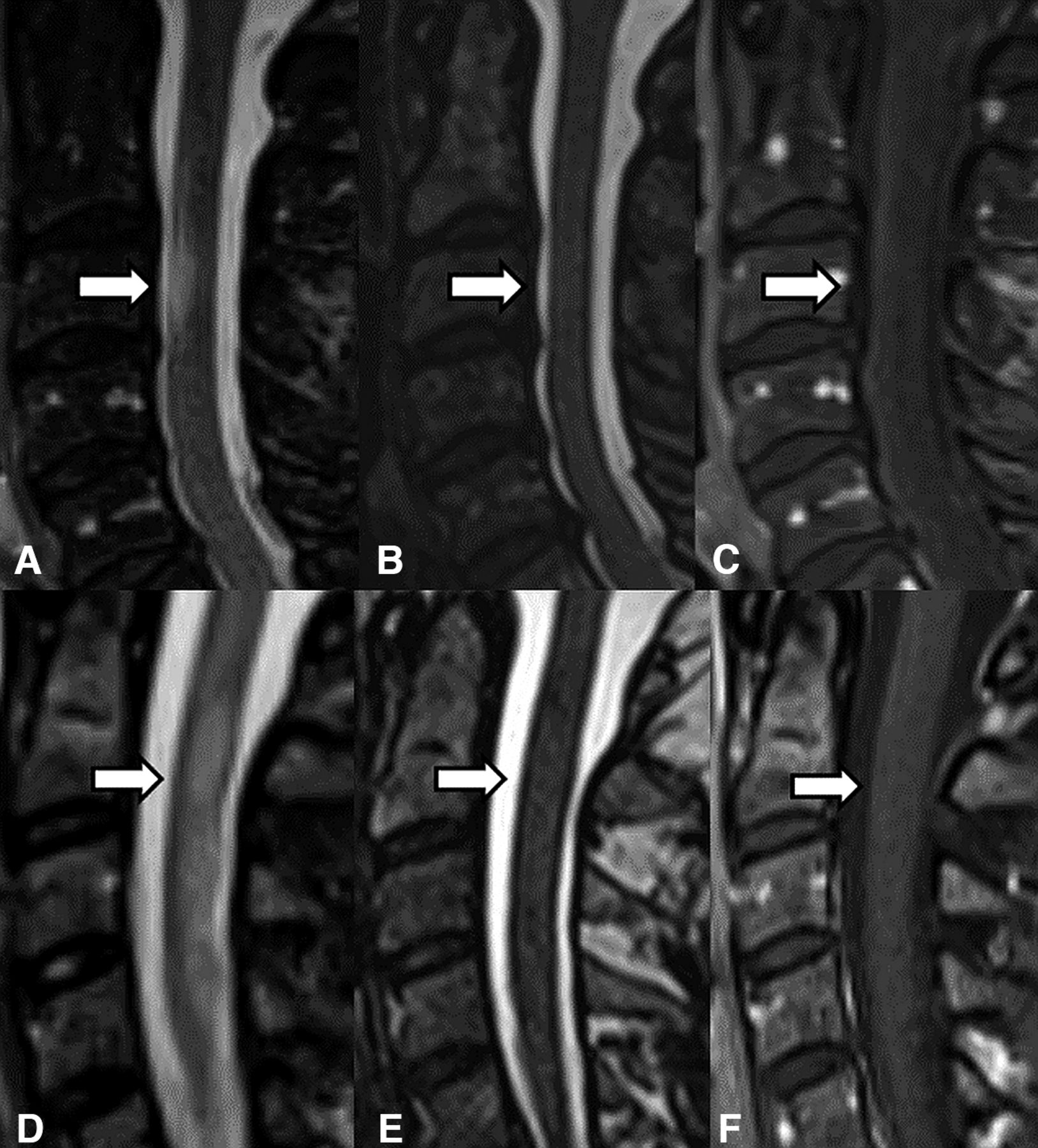
- FIG 1.
MR imaging of 2 patients with MS. A 62-year-old woman in the top row (A–C) and a 52-year-old woman in the bottom row (D–F). The first panel (A and D) shows a sagittal STIR image of an MS lesion (arrow) in the cervical spine in both patients, which is not conspicuous on sagittal T2-SPACE (B and E) or sagittal postcontrast T1WI (C and F).
- FIG 2.
MR imaging of a 38-year-old woman with MS. Sagittal T2-SPACE (left) shows focal T1–T3 hyperintensity (arrow) with focally increased volume corresponding to an active cord lesion on sagittal T1 postcontrast (right).
- FIG 3.
MR imaging of a 25-year-old man with relapsing-remitting MS. Sagittal T2-SPACE (left) shows focal C2–C3 hyperintensity (arrow), with focal increased cord volume corresponding to an enhancing, active lesion on sagittal T1 postcontrast (right) image.
- FIG 4.
MR imaging of a 32-year-old woman with relapsing-remitting MS. Sagittal T2-SPACE (left) shows focal T2 hyperintensity (arrow) with normal/decreased cord volume. No active lesion/enhancement is seen on the sagittal T1 postcontrast image (right).
- FIG 5.
MR imaging of a 51-year-old woman with MS. Sagittal T1 postcontrast imaging shows an enhancing lesion (arrow) in the C3-C4 cord (left) without associated hyperintensity on T2-SPACE (right).
- FIG 6.
Observational imaging progression of an active MS lesion in the C6 spinal cord across time to a chronic lesion in a 36-year-old woman with MS. Sagittal T2-SPACE (A) and sagittal postcontrast T1WI (B) show a T2-hyperintense lesion with cord expansion and enhancement compatible with an active lesion. Follow-up T2-SPACE (C) and postcontrast T1 (D) after 2 years demonstrate no T2 hyperintensity at the same site, with mild cord volume loss, and no enhancement, suggestive of a chronic lesion.


Jump to section
Related Articles
Cited By...
- No citing articles found.