
Article Figures & Data
Figures
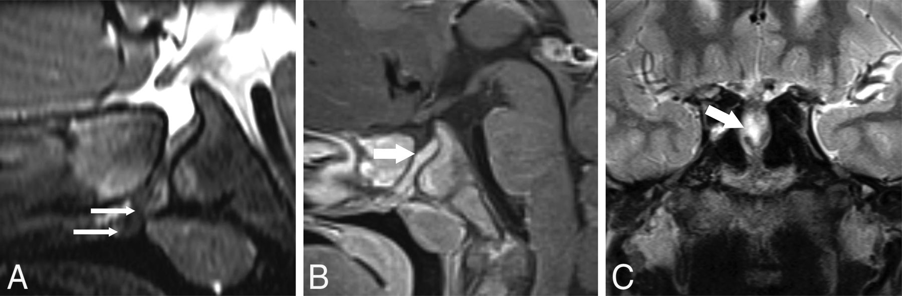
- FIG 1.
ON and chiasmatic findings on MR imaging. A–C, P5; An 11-month-old girl. A, Axial T2 sampling perfection with application-optimized contrasts by using different flip angle evolution (SPACE sequence; Siemens) image shows a right MGDA with mild asymmetric right ON thickening (long arrow) without signal abnormality. B, Reformatted coronal T2 SPACE image reveals a chiasmatic deformity, asymmetrically thickened on the right (short arrow). C, Coronal contrast-enhanced fat-suppressed T1-weighted image shows minimal peripheral enhancement around the right side of the chiasm (short arrow).
- FIG 2.
PCPC and pituitary fossa anomalies on MR imaging and CT. P3; A 14-month-old girl with bilateral MGDA. A, Sagittal T2-weighted MR imaging shows a PCPC with apposed margins (long, wide arrow) and minimal inferior pituitary fossa beaking. B and C, P25; A male patient with bilateral MGDA. B, Sagittal T1 MPRAGE MR imaging in a patient 5 years of age shows partial sphenoid pneumatization and a partially empty sella. The PCPC is difficult to appreciate (long, wide arrow) but is readily seen at 4 years of age on sagittal reformatted CT (long, wide arrow, C). Note the normal spheno-occipital synchondrosis dorsal to the PCPC.
- FIG 3.
PCPC with a sphenoid cephalocele on MR imaging. A and B, P1; A 16-month-old boy with a right MGDA. A, Reformatted sagittal T2 SPACE sequence. B, Sagittal contrast-enhanced T1 SPACE images show a deep-set and angulated pituitary fossa associated with a wide PCPC containing pituitary tissue (wide white arrow). Note that in A, there is also tubular hypointense tissue that differs in signal intensity from pituitary tissue projecting into the nasopharynx inferior to the pituitary tissue (thin white arrow). C, P6; A 14-year-old boy with a right MGDA. Coronal T2-weighted image shows a sphenoid cephalocele containing CSF and pituitary gland (wide white arrow).
- FIG 4.
PCPC, tubular nasopharyngeal hamartoma, and oculomotor nerve abnormality on MR imaging. A–C, P7; A 15-year-old girl with a left MGDA. A, Sagittal T1-weighted image shows a deep-set pituitary fossa and PCPC (long, wide white arrow) and a heterogeneous part-fatty, tubular lesion protruding into the nasopharynx (long, thin white arrows). B, Axial T2 SPACE sequence MR imaging shows asymmetric thickening of the cisternal segment of the left oculomotor nerve (black arrow), ipsilateral to the MGDA. C, Axial contrast-enhanced fat-suppressed T1-weighted MR imaging shows avid oculomotor nerve enhancement (black arrow) that appeared stable at 2-year follow-up.
- FIG 5.
Off-midline left sphenoid bone cleft and oculomotor nerve abnormality on CT and MR imaging. A–C, P31; A 5-year-old girl with a left MGDA. A, 2D reformatted contrast-enhanced coronal CT image shows a sloping pituitary fossa with a left-sided sphenoid bone cleft (wide white arrow). B, Axial T2 SPACE sequence MR imaging shows a proximally thickened left oculomotor nerve that originates anomalously from the left side of the midbrain (black arrow) with a prominent tortuous vessel (thin white arrow) originating near the origin of the left superior cerebellar and posterior cerebral arteries (corroborated on MRA; not shown). C, Axial fat-suppressed contrast-enhanced T1-weighted MR imaging shows the tortuous vessel (thin white arrow) with surrounding abnormal enhancement encompassing the left oculomotor nerve root entry zone with enhancement of the proximal cisternal oculomotor nerve segment (black arrow). These findings appeared stable during a 6-year period.


Jump to section
Related Articles
Cited By...
- No citing articles found.