Advertisement

Main menu

  • Home
  • Content
    • Current Issue
    • Accepted Manuscripts
    • Article Preview
    • Past Issue Archive
    • Video Articles
    • AJNR Case Collection
    • Case of the Week Archive
    • Case of the Month Archive
    • Classic Case Archive
  • Special Collections
    • AJNR Awards
    • Low-Field MRI
    • Alzheimer Disease
    • ASNR Foundation Special Collection
    • Photon-Counting CT
    • View All
  • Multimedia
    • AJNR Podcasts
    • AJNR SCANtastic
    • Trainee Corner
    • MRI Safety Corner
    • Imaging Protocols
  • For Authors
    • Submit a Manuscript
    • Submit a Video Article
    • Submit an eLetter to the Editor/Response
    • Manuscript Submission Guidelines
    • Statistical Tips
    • Fast Publishing of Accepted Manuscripts
    • Graphical Abstract Preparation
    • Imaging Protocol Submission
    • Author Policies
  • About Us
    • About AJNR
    • Editorial Board
    • Editorial Board Alumni
  • More
    • Become a Reviewer/Academy of Reviewers
    • Subscribers
    • Permissions
    • Alerts
    • Feedback
    • Advertisers
    • ASNR Home

User menu

  • Alerts
  • Log in

Search

  • Advanced search
American Journal of Neuroradiology

American Journal of Neuroradiology

ASHNR American Society of Functional Neuroradiology ASHNR American Society of Pediatric Neuroradiology ASSR
  • Alerts
  • Log in

Advanced Search

  • Home
  • Content
    • Current Issue
    • Accepted Manuscripts
    • Article Preview
    • Past Issue Archive
    • Video Articles
    • AJNR Case Collection
    • Case of the Week Archive
    • Case of the Month Archive
    • Classic Case Archive
  • Special Collections
    • AJNR Awards
    • Low-Field MRI
    • Alzheimer Disease
    • ASNR Foundation Special Collection
    • Photon-Counting CT
    • View All
  • Multimedia
    • AJNR Podcasts
    • AJNR SCANtastic
    • Trainee Corner
    • MRI Safety Corner
    • Imaging Protocols
  • For Authors
    • Submit a Manuscript
    • Submit a Video Article
    • Submit an eLetter to the Editor/Response
    • Manuscript Submission Guidelines
    • Statistical Tips
    • Fast Publishing of Accepted Manuscripts
    • Graphical Abstract Preparation
    • Imaging Protocol Submission
    • Author Policies
  • About Us
    • About AJNR
    • Editorial Board
    • Editorial Board Alumni
  • More
    • Become a Reviewer/Academy of Reviewers
    • Subscribers
    • Permissions
    • Alerts
    • Feedback
    • Advertisers
    • ASNR Home
  • Follow AJNR on Twitter
  • Visit AJNR on Facebook
  • Follow AJNR on Instagram
  • Join AJNR on LinkedIn
  • RSS Feeds

AJNR Awards, New Junior Editors, and more. Read the latest AJNR updates

Research ArticleNEUROIMAGING PHYSICS/FUNCTIONAL NEUROIMAGING/CT AND MRI TECHNOLOGY
Open Access

Imaging and Anesthesia Protocol Optimization in Sedated Clinical Resting-State fMRI

Elmira Hassanzadeh, Alyssa Ailion, Masoud Hassanzadeh, Alena Hornak, Noam Peled, Dana Martino, Simon K. Warfield, Zhou Lan, Taha Gholipour and Steven M. Stufflebeam
American Journal of Neuroradiology December 2024, DOI: https://doi.org/10.3174/ajnr.A8438
Elmira Hassanzadeh
aFrom the Department of Radiology (E.H., Z.L.), Brigham and Women’s Hospital, Harvard Medical School, Boston, Massachusetts
  • Find this author on Google Scholar
  • Find this author on PubMed
  • Search for this author on this site
  • ORCID record for Elmira Hassanzadeh
Alyssa Ailion
bDepartments of Neurology (A.A., A.H., D.M.), Boston Children’s Hospital, Harvard Medical School, Boston, Massachusetts
  • Find this author on Google Scholar
  • Find this author on PubMed
  • Search for this author on this site
Masoud Hassanzadeh
dDepartments of Surgery (M.H.), Massachusetts General Hospital, Harvard Medical School, Boston, Massachusetts
  • Find this author on Google Scholar
  • Find this author on PubMed
  • Search for this author on this site
  • ORCID record for Masoud Hassanzadeh
Alena Hornak
bDepartments of Neurology (A.A., A.H., D.M.), Boston Children’s Hospital, Harvard Medical School, Boston, Massachusetts
  • Find this author on Google Scholar
  • Find this author on PubMed
  • Search for this author on this site
Noam Peled
eDepartments of Radiology (N.P., S.M.S.), Massachusetts General Hospital, Harvard Medical School, Boston, Massachusetts
  • Find this author on Google Scholar
  • Find this author on PubMed
  • Search for this author on this site
  • ORCID record for Noam Peled
Dana Martino
bDepartments of Neurology (A.A., A.H., D.M.), Boston Children’s Hospital, Harvard Medical School, Boston, Massachusetts
  • Find this author on Google Scholar
  • Find this author on PubMed
  • Search for this author on this site
Simon K. Warfield
cDepartments of Radiology (S.K.W.), Boston Children’s Hospital, Harvard Medical School, Boston, Massachusetts
  • Find this author on Google Scholar
  • Find this author on PubMed
  • Search for this author on this site
  • ORCID record for Simon K. Warfield
Zhou Lan
aFrom the Department of Radiology (E.H., Z.L.), Brigham and Women’s Hospital, Harvard Medical School, Boston, Massachusetts
  • Find this author on Google Scholar
  • Find this author on PubMed
  • Search for this author on this site
Taha Gholipour
fComprehensive Epilepsy Center (T.G.), University of California San Diego, San Diego, California
  • Find this author on Google Scholar
  • Find this author on PubMed
  • Search for this author on this site
  • ORCID record for Taha Gholipour
Steven M. Stufflebeam
eDepartments of Radiology (N.P., S.M.S.), Massachusetts General Hospital, Harvard Medical School, Boston, Massachusetts
  • Find this author on Google Scholar
  • Find this author on PubMed
  • Search for this author on this site
  • Article
  • Figures & Data
  • Supplemental
  • Info & Metrics
  • Responses
  • References
  • PDF
Loading

Article Figures & Data

Figures

  • Tables
  • FIG 1.
    • Download figure
    • Open in new tab
    • Download powerpoint
    FIG 1.

    Flow diagram of patient selection.
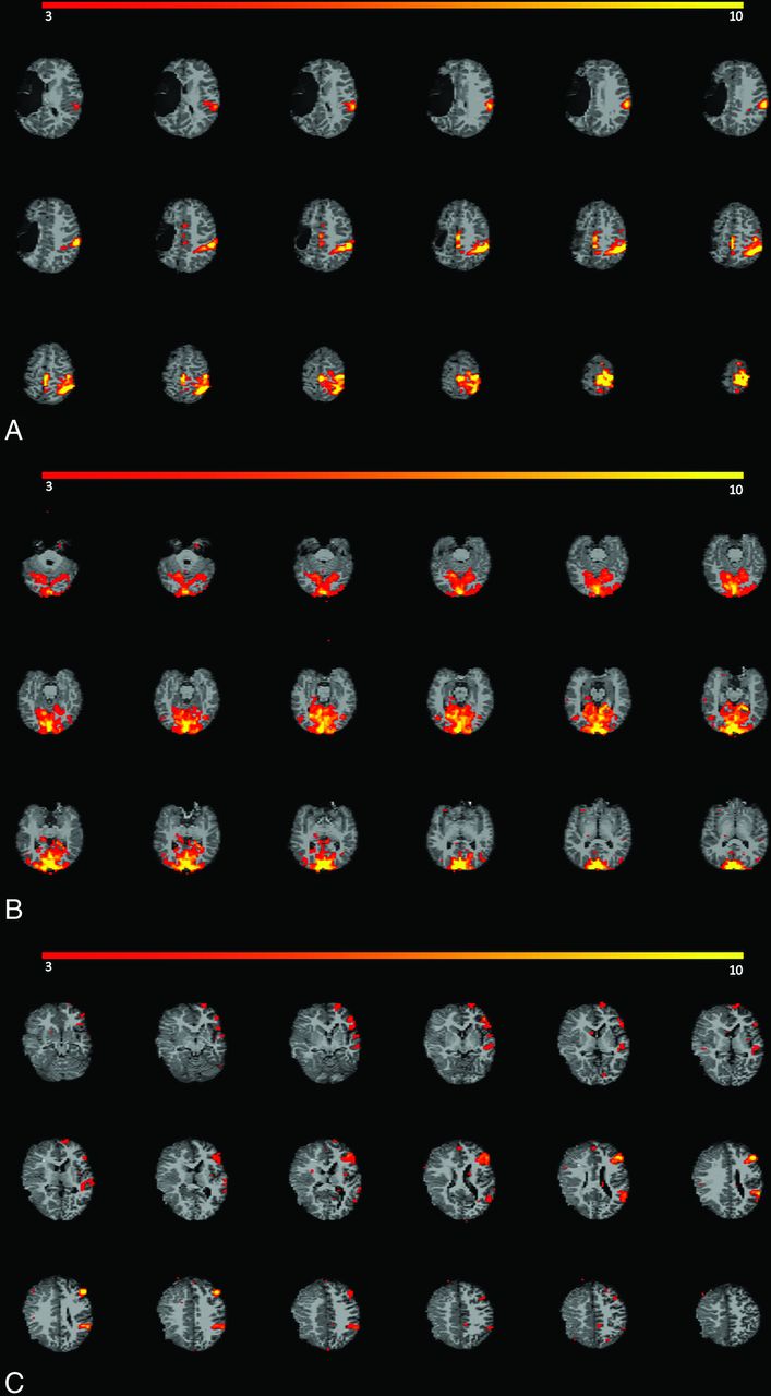
  • FIG 2.
    • Download figure
    • Open in new tab
    • Download powerpoint
    FIG 2.

    Examples of robust RSNs in our study population. Network activations are depicted in red-yellow spectrum. A, Robust motor network activation is seen (both visually and quantitatively) in an 8-year-old boy with epilepsy who underwent a 10.4-minute rs-fMRI with acceleration under propofol anesthesia. Structural images showed chronic right MCA infarction. B, Robust visual network activation is seen in a 9-year-old boy with epilepsy who underwent a 7.25-minute rs-fMRI without acceleration under propofol anesthesia. No structural abnormality was detected. C, Robust language network activation is seen (both visually and quantitatively) in a 5-year-old girl with seizure who underwent a 6.75-minute rs-fMRI without acceleration under propofol, sevoflurane, and fentanyl anesthesia. Subacute infarction in the right MCA territory was seen on structural images.
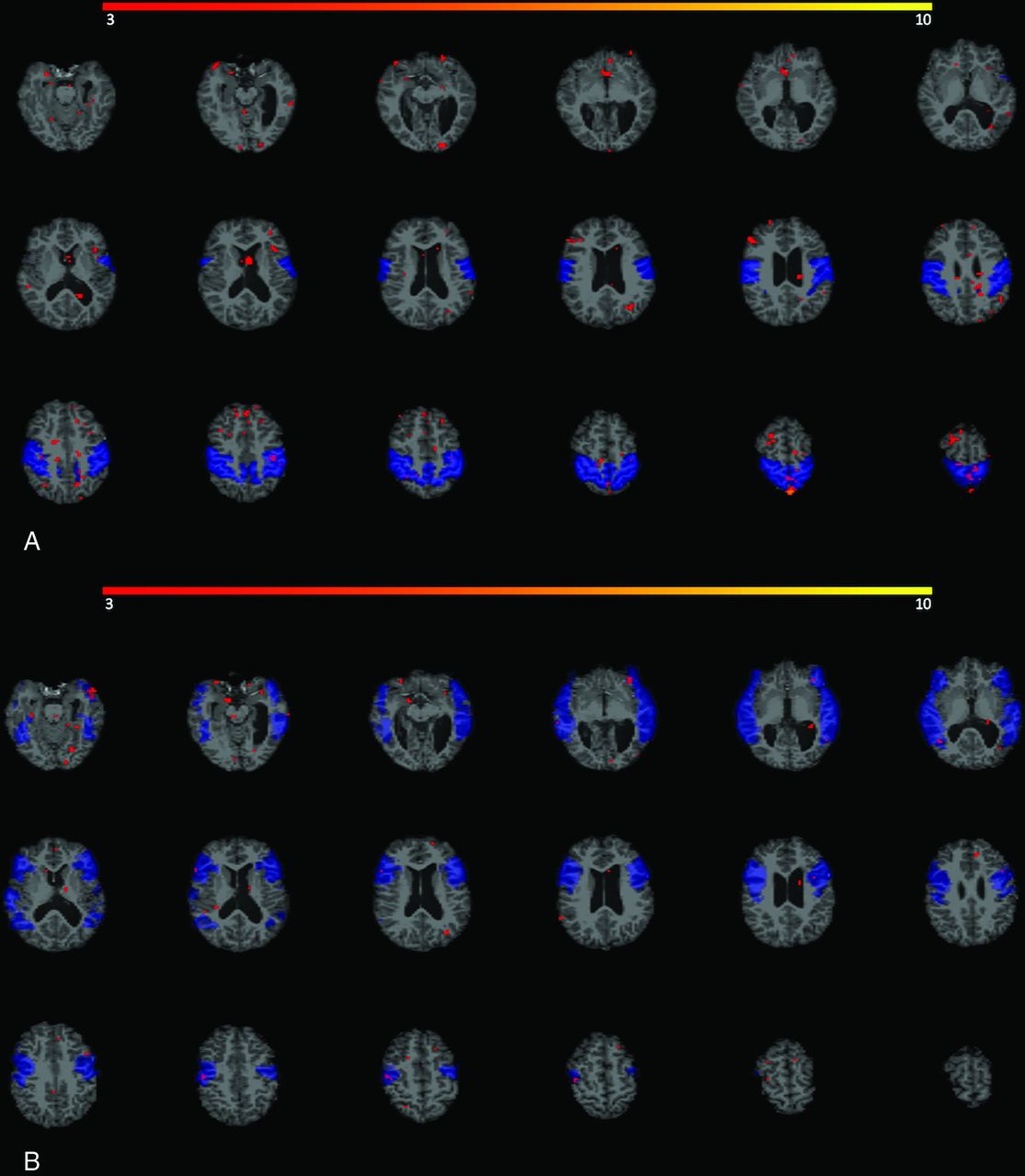
  • FIG 3.
    • Download figure
    • Open in new tab
    • Download powerpoint
    FIG 3.

    Example of weak RSNs in a 9-year-old boy with seizure who underwent 7-minute rs-fMRI under propofol, sevoflurane, and N2O anesthesia. Structural images showed mesial temporal sclerosis, and chronic left germinal matrix hemorrhage. No meaningful activation (depicted in red) is seen in motor network (A) or language network (B) boundaries, shown as blue template masks.

Tables

  • Figures
    • View popup
    Table 1:

    Demographics, clinical, and imaging characteristics of study population

    Variablesn = 69
    Age (yr)
     Mean (SD)6.9 (4.26)
     Range1–17
    Sex
     Girl30 (43%)
    Indication for the examination
     Seizure35 (51%)
     Tumor7 (10%)
     Other27 (39%)
    rs-fMRI with multiband28 (41%)
    fMRI scan length (minute)
     Mean (SD)7.7 (2.01)
     Range4.5–10.4
    Number of BOLD volumes
     Mean (SD)271.3 (167.8)
     Range90–550
    Framewise displacement (mm)
     Mean (SD)0.05 (0.04)
     Range0.02–0.2
    MRI with structural abnormality39 (57%)
    Anesthesia with propofol58 (84%)
    Anesthesia with sevoflurane34 (49%)
    Anesthesia with N2O12 (17%)
    Anesthesia with other agents15 (22%)
    • View popup
    Table 2:

    ORs and CIs from multivariable logistic regression analysis of predictive factors in robust rs-fMRI overall networks with qualitative assessment

    VariableOR (Exp (B))95% CI for Exp (B)P Value
    rs-fMRI with multiband2.370.44–12.65.31
    Anesthesia with propofol1.410.27–7.29.68
    Anesthesia with sevoflurane0.200.05–0.79.02a
    Anesthesia with N2O0.800.16–4.11.79
    Anesthesia with other agents1.420.32–6.42.65
    Abnormality on structural MRI1.020.34–3.09.97
    Sex = boy0.970.32–2.91.96
    Age0.930.81–1.06.27
    Scan length in minutes1.060.73–1.53.76
    • ↵a Indicates statistical significance at the .05 level.

    • View popup
    Table 3:

    ORs and CIs from multivariable logistic regression analysis of predictive factors in robust rs-fMRI motor-language networks with quantitative assessment

    VariableOR (Exp (B))95% CI for Exp (B)P Value
    rs-fMRI with multiband1.110.21–5.97.90
    Anesthesia with propofol2.510.44–14.32.30
    Anesthesia with sevoflurane0.180.04–0.80.02a
    Anesthesia with N2O0.410.08–2.13.29
    Anesthesia with other agents2.150.40–11.52.37
    Abnormality on structural MRI1.570.47–5.26.46
    Sex = boy1.960.55–6.92.29
    Age0.930.81–1.07.31
    Scan length in minutes1.150.79–1.67.47
    • ↵a Indicates statistical significance at the .05 level.

    • View popup
    Table 4:

    ORs and CIs from multivariable logistic regression analysis of predictive factors in robust rs-fMRI motor network with quantitative assessment

    VariableOR (Exp (B))95% CI for Exp (B)P Value
    rs-fMRI with multiband3.290.41–26.63.26
    Anesthesia with propofol5.390.84–34.75.08
    Anesthesia with sevoflurane0.100.02–0.64.02a
    Anesthesia with N2O1.040.16–6.54.97
    Anesthesia with other agents1.720.26–11.14.57
    Abnormality on structural MRI1.540.38–6.23.55
    Sex = boy2.830.63–12.70.17
    Age0.940.81–1.10.47
    Scan length in minutes1.060.68–1.65.81
    • ↵a Indicates statistical significance at the .05 level.

    • View popup
    Table 5:

    ORs and CIs from multivariable logistic regression analysis of predictive factors in robust rs-fMRI language network with quantitative assessment

    VariableOR (Exp (B))95% CI for Exp (B)P Value
    rs-fMRI with multiband0.970.13–7.49.98
    Anesthesia with propofol0.410.05–3.40.41
    Anesthesia with sevoflurane0.340.06–2.01.23
    Anesthesia with N2O0.220.03–1.35.10
    Anesthesia with other agents1.680.26–11.03.59
    Abnormality on structural MRI0.910.21–3.98.89
    Sex = boy1.720.38–7.69.48
    Age1.000.85–1.19.97
    Scan length in minute1.410.91–2.20.13
PreviousNext
Back to top
Advertisement
Print
Download PDF
Email Article

Thank you for your interest in spreading the word on American Journal of Neuroradiology.

NOTE: We only request your email address so that the person you are recommending the page to knows that you wanted them to see it, and that it is not junk mail. We do not capture any email address.

Enter multiple addresses on separate lines or separate them with commas.
Imaging and Anesthesia Protocol Optimization in Sedated Clinical Resting-State fMRI
(Your Name) has sent you a message from American Journal of Neuroradiology
(Your Name) thought you would like to see the American Journal of Neuroradiology web site.
CAPTCHA
This question is for testing whether or not you are a human visitor and to prevent automated spam submissions.
Cite this article
Elmira Hassanzadeh, Alyssa Ailion, Masoud Hassanzadeh, Alena Hornak, Noam Peled, Dana Martino, Simon K. Warfield, Zhou Lan, Taha Gholipour, Steven M. Stufflebeam
Imaging and Anesthesia Protocol Optimization in Sedated Clinical Resting-State fMRI
American Journal of Neuroradiology Dec 2024, DOI: 10.3174/ajnr.A8438

Citation Manager Formats

  • BibTeX
  • Bookends
  • EasyBib
  • EndNote (tagged)
  • EndNote 8 (xml)
  • Medlars
  • Mendeley
  • Papers
  • RefWorks Tagged
  • Ref Manager
  • RIS
  • Zotero
0 Responses
Respond to this article
Share
Bookmark this article
Imaging and Anesthesia Protocol Optimization in Sedated Clinical Resting-State fMRI
Elmira Hassanzadeh, Alyssa Ailion, Masoud Hassanzadeh, Alena Hornak, Noam Peled, Dana Martino, Simon K. Warfield, Zhou Lan, Taha Gholipour, Steven M. Stufflebeam
American Journal of Neuroradiology Dec 2024, DOI: 10.3174/ajnr.A8438
del.icio.us logo Twitter logo Facebook logo Mendeley logo
  • Tweet Widget
  • Facebook Like
  • Google Plus One
Purchase

Jump to section

  • Article
    • Abstract
    • ABBREVIATIONS:
    • MATERIALS AND METHODS
    • RESULTS
    • DISCUSSION
    • CONCLUSIONS
    • Footnotes
    • References
  • Figures & Data
  • Supplemental
  • Info & Metrics
  • Responses
  • References
  • PDF

Related Articles

  • PubMed
  • Google Scholar

Cited By...

  • No citing articles found.
  • Crossref
  • Google Scholar

This article has not yet been cited by articles in journals that are participating in Crossref Cited-by Linking.

More in this TOC Section

  • A Comprehensive Review of IA Imaging Modalities
  • Synthetic MRI based on 3D-QALAS MR Quantification
  • Delta wave MRI
Show more Neuroimaging Physics/Functional Neuroimaging/CT and MRI Technology

Similar Articles

Advertisement

Indexed Content

  • Current Issue
  • Accepted Manuscripts
  • Article Preview
  • Past Issues
  • Editorials
  • Editor's Choice
  • Fellows' Journal Club
  • Letters to the Editor
  • Video Articles

Cases

  • Case Collection
  • Archive - Case of the Week
  • Archive - Case of the Month
  • Archive - Classic Case

More from AJNR

  • Trainee Corner
  • Imaging Protocols
  • MRI Safety Corner
  • Book Reviews

Multimedia

  • AJNR Podcasts
  • AJNR Scantastics

Resources

  • Turnaround Time
  • Submit a Manuscript
  • Submit a Video Article
  • Submit an eLetter to the Editor/Response
  • Manuscript Submission Guidelines
  • Statistical Tips
  • Fast Publishing of Accepted Manuscripts
  • Graphical Abstract Preparation
  • Imaging Protocol Submission
  • Evidence-Based Medicine Level Guide
  • Publishing Checklists
  • Author Policies
  • Become a Reviewer/Academy of Reviewers
  • News and Updates

About Us

  • About AJNR
  • Editorial Board
  • Editorial Board Alumni
  • Alerts
  • Permissions
  • Not an AJNR Subscriber? Join Now
  • Advertise with Us
  • Librarian Resources
  • Feedback
  • Terms and Conditions
  • AJNR Editorial Board Alumni

American Society of Neuroradiology

  • Not an ASNR Member? Join Now

© 2025 by the American Society of Neuroradiology All rights, including for text and data mining, AI training, and similar technologies, are reserved.
Print ISSN: 0195-6108 Online ISSN: 1936-959X

Powered by HighWire