Advertisement

Main menu

  • Home
  • Content
    • Current Issue
    • Accepted Manuscripts
    • Article Preview
    • Past Issue Archive
    • Video Articles
    • AJNR Case Collection
    • Case of the Week Archive
    • Case of the Month Archive
    • Classic Case Archive
  • Special Collections
    • AJNR Awards
    • Low-Field MRI
    • Alzheimer Disease
    • ASNR Foundation Special Collection
    • Photon-Counting CT
    • View All
  • Multimedia
    • AJNR Podcasts
    • AJNR SCANtastic
    • Trainee Corner
    • MRI Safety Corner
    • Imaging Protocols
  • For Authors
    • Submit a Manuscript
    • Submit a Video Article
    • Submit an eLetter to the Editor/Response
    • Manuscript Submission Guidelines
    • Statistical Tips
    • Fast Publishing of Accepted Manuscripts
    • Graphical Abstract Preparation
    • Imaging Protocol Submission
    • Author Policies
  • About Us
    • About AJNR
    • Editorial Board
    • Editorial Board Alumni
  • More
    • Become a Reviewer/Academy of Reviewers
    • Subscribers
    • Permissions
    • Alerts
    • Feedback
    • Advertisers
    • ASNR Home

User menu

  • Alerts
  • Log in

Search

  • Advanced search
American Journal of Neuroradiology

American Journal of Neuroradiology

ASHNR American Society of Functional Neuroradiology ASHNR American Society of Pediatric Neuroradiology ASSR
  • Alerts
  • Log in

Advanced Search

  • Home
  • Content
    • Current Issue
    • Accepted Manuscripts
    • Article Preview
    • Past Issue Archive
    • Video Articles
    • AJNR Case Collection
    • Case of the Week Archive
    • Case of the Month Archive
    • Classic Case Archive
  • Special Collections
    • AJNR Awards
    • Low-Field MRI
    • Alzheimer Disease
    • ASNR Foundation Special Collection
    • Photon-Counting CT
    • View All
  • Multimedia
    • AJNR Podcasts
    • AJNR SCANtastic
    • Trainee Corner
    • MRI Safety Corner
    • Imaging Protocols
  • For Authors
    • Submit a Manuscript
    • Submit a Video Article
    • Submit an eLetter to the Editor/Response
    • Manuscript Submission Guidelines
    • Statistical Tips
    • Fast Publishing of Accepted Manuscripts
    • Graphical Abstract Preparation
    • Imaging Protocol Submission
    • Author Policies
  • About Us
    • About AJNR
    • Editorial Board
    • Editorial Board Alumni
  • More
    • Become a Reviewer/Academy of Reviewers
    • Subscribers
    • Permissions
    • Alerts
    • Feedback
    • Advertisers
    • ASNR Home
  • Follow AJNR on Twitter
  • Visit AJNR on Facebook
  • Follow AJNR on Instagram
  • Join AJNR on LinkedIn
  • RSS Feeds

AJNR Awards, New Junior Editors, and more. Read the latest AJNR updates

Review ArticleBRAIN TUMOR IMAGING

Mesenchymal Nonmeningothelial Tumors of the CNS: Evolving Molecular Landscape and Implications for Neuroradiologists

Neetu Soni, Manish Ora, Denes Szekeres, Girish Bathla, Amit Desai, Vivek Gupta, Aparna Singhal and Amit Agarwal
American Journal of Neuroradiology May 2025, DOI: https://doi.org/10.3174/ajnr.A8519
Neetu Soni
aFrom the Department of Radiology (N.S., A.D., V.G., A.A.), Mayo Clinic, Jacksonville, Florida
  • Find this author on Google Scholar
  • Find this author on PubMed
  • Search for this author on this site
  • ORCID record for Neetu Soni
Manish Ora
bDepartment of Nuclear Medicine (M.O.), Sanjay Gandhi Post Graduate Institute of Medical Science, Lucknow, India
  • Find this author on Google Scholar
  • Find this author on PubMed
  • Search for this author on this site
  • ORCID record for Manish Ora
Denes Szekeres
cUniversity of Rochester (D.S.), School of Medicine and Dentistry, Rochester, New York
  • Find this author on Google Scholar
  • Find this author on PubMed
  • Search for this author on this site
Girish Bathla
dDepartment of Radiology (G.B.), Mayo Clinic, Rochester, Minnesota
  • Find this author on Google Scholar
  • Find this author on PubMed
  • Search for this author on this site
  • ORCID record for Girish Bathla
Amit Desai
aFrom the Department of Radiology (N.S., A.D., V.G., A.A.), Mayo Clinic, Jacksonville, Florida
  • Find this author on Google Scholar
  • Find this author on PubMed
  • Search for this author on this site
  • ORCID record for Amit Desai
Vivek Gupta
aFrom the Department of Radiology (N.S., A.D., V.G., A.A.), Mayo Clinic, Jacksonville, Florida
  • Find this author on Google Scholar
  • Find this author on PubMed
  • Search for this author on this site
  • ORCID record for Vivek Gupta
Aparna Singhal
eDepartment of Radiology (A.S.), UAB Hospital, Birmingham, Alabama
  • Find this author on Google Scholar
  • Find this author on PubMed
  • Search for this author on this site
  • ORCID record for Aparna Singhal
Amit Agarwal
aFrom the Department of Radiology (N.S., A.D., V.G., A.A.), Mayo Clinic, Jacksonville, Florida
  • Find this author on Google Scholar
  • Find this author on PubMed
  • Search for this author on this site
  • ORCID record for Amit Agarwal
  • Article
  • Figures & Data
  • Supplemental
  • Info & Metrics
  • Responses
  • References
  • PDF
Loading

Article Figures & Data

Figures

  • Tables
  • FIG 1.
    • Download figure
    • Open in new tab
    • Download powerpoint
    FIG 1.

    Spinal mesenchymal chondrosarcoma with HEY1-NCOA2 fusion gene in a 16-year-old girl. Sagittal and axial MR images (A–C) reveal a lobulated well-circumscribed mass in the extradural space of the cervical spine extending along the left neural foramen (arrow). Lesion shows an isointense signal on T1-weighted image (A), with homogeneous contrast enhancement (B) and areas of signal drop-out on axial gradient image (C). Amorphous calcification noted within the tumor on axial CT (D, arrow). H&E revealed spindle cell morphology with abrupt areas of cartilage formation (E, arrows). Tumor showed positive staining for CD99 and SATB2 (F) on immunohistochemistry. Molecular testing confirmed the presence of a HEY1-NCOA2 gene fusion event in the tumor, further confirming a diagnosis of mesenchymal chondrosarcoma. The HEY1-NCOA2 fusion is most frequently observed in mesenchymal chondrosarcomas.

  • FIG 2.
    • Download figure
    • Open in new tab
    • Download powerpoint
    FIG 2.

    Metastatic solitary fibrous tumor in a 69-year-old woman. MR images (A–D) depict a large multilobulated mass along the inferior tentorial surface (arrow) with avid contrast enhancement and adjacent cerebellar edema. Smaller similar nodules are noted along the superior tentorial surface and along right cerebellar convexity (B and D, arrows). Osseous and pulmonary metastatic deposits with increased uptake seen on FDG-PET study (E, arrows). The tumor cells show nuclear expression of STAT6 (F), compatible with the presence of NAB2-STAT6 fusion and diagnostic of solitary fibrous tumor.

  • FIG 3.
    • Download figure
    • Open in new tab
    • Download powerpoint
    FIG 3.

    Intracranial inflammatory myofibroblastic tumor in a 35-year-old man. Multiple axial MR images (A–C) reveal a lobulated mass (arrows) in the left basifrontal region with extensive vasogenic edema in the adjacent parenchyma, disproportionate to the size of the mass (A). Lesion shows foci of signal drop-out (hemorrhage) on SWI (B) and heterogeneous enhancement (C). Lesion shows increased uptake on FDG-PET examination (D). Rapid increase in the size of the mass was noted in the follow-up MRI after 3 months with clinical deterioration (Supplemental Data). Surgical resection was performed with histopathology revealing epithelioid inflammatory myofibroblastic tumor. Immunohistochemistry depicted perinuclear anaplastic lymphoma kinase staining, correlating with the presence of RANBP2-anaplastic lymphoma kinase gene fusion (Supplemental Data).
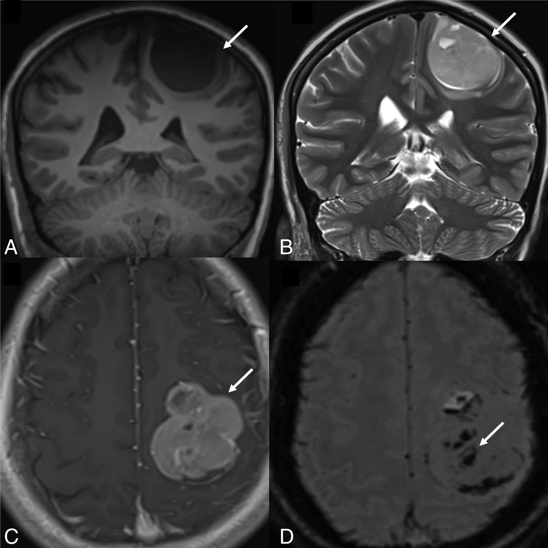
  • FIG 4.
    • Download figure
    • Open in new tab
    • Download powerpoint
    FIG 4.

    Intracranial mesenchymal tumor, FET::CREB fusion-positive in a 19-year-old man. Sagittal T1-weighted (A) and contrast-enhanced (B) images reveal a large extra-axial dural-based mass along the frontal convexity with heterogeneous enhancement. Lesion shows heterogeneous hyperintense signal on FLAIR (C) with areas of signal drop-out (calcification) on SWI (arrow) (D). Histopathology demonstrated a low-grade mesenchymal neoplasm with clear cell features and fibrillary deposits. Further molecular characterization by targeted next generation sequencing (sarcoma targeted gene fusion/rearrangement panel) revealed FET::CREB fusion.

  • FIG 5.
    • Download figure
    • Open in new tab
    • Download powerpoint
    FIG 5.

    Spinal CIC-rearranged sarcoma in a 14-year-old boy. MR images depict a small well-circumscribed peridural mass in the midthoracic region (arrows) with isointense T1 signal (A), hyperintense T2 signal (B), and avid contrast enhancement (C). The tumor extends through the left neural foramen into the paraspinal space with mass effect on the cord. Tumor shows increased uptake on FDG-PET study (arrow) (D). Histopathology (Supplemental Data) revealed sarcomatous cells with neoplastic cells positive for cyclin and calretinin immunohistochemical stains. Molecular cytogenetic studies (fluorescence in site hybridization) showed balanced rearrangement of the CIC locus in 91% of the nuclei (181/200).

  • FIG 6.
    • Download figure
    • Open in new tab
    • Download powerpoint
    FIG 6.

    Primary intracranial sarcoma, DICER1-mutant, in a 15-year-old girl. MR images depict a small well-circumscribed extra-axial dural-based mass along the left frontal convexity with buckling of the underlying cortex (arrows). Tumor shows hypointense T1 signal (A), hyperintense T2 signal (B), and avid contrast enhancement (C). Multiple foci of signal drop-out noted on SWI suggest hemorrhagic foci. Histopathology (Supplemental Data) demonstrates cellular composition of spindled and pleomorphic tumor cells with high mitotic activity. The tumor cells are positive for MyoD1 (patchy, focal), and negative for desmin on immunohistochemistry. “Somatic disease/germline comparator exome” sequencing panel showed pathogenic germline variant for DICER1. Overall, the histomorphologic, immunophenotypic, and genetic findings are diagnostic of primary intracranial sarcoma, DICER1-mutant.

Tables

  • Figures
  • New World Health Organization 2021 classification of CNS mesenchymal tumors and their molecular markers

    Mesenchymal, Non-Meningothelial Tumors Involving the CNS
    Soft tissue tumorsFibroblastic and myofibroblastic tumorsSolitary fibrous tumorPartially or completely lack STAT6 nuclear expression on immunohistochemistry
    Vascular tumorsHemangiomas and vascular malformationsDiagnostic molecular pathology is not clinically relevant
    Hemangioblastoma
    Skeletal muscle tumorsRhabdomyosarcomaPAX3::FOXO1 or PAX7::FOXO1 fusion in alveolar subtype, with a worse prognosis
    Tumors of uncertain differentiationIntracranial mesenchymal tumor, FET::CREB fusion-positiveDiagnostic FET::CREB family gene fusions
    CIC-rearranged sarcomaCIC gene fusion and up-regulation of ETV complements
    Primary intracranial sarcoma, DICER1-mutantDICER1 mutations with DNA-methylation profile distinct
    Ewing sarcomaDefinitive diagnosis requires confirmation of FET::ETS–type gene fusion
    Chondro-osseous tumorsChondrogenic tumorsMesenchymal chondrosarcomaHEY1::NCOA2 gene fusion
    ChondrosarcomaIDH mutation (approximately 60%)
    Notochordal tumorsChordomaSMARCB1 deletions in poorly differentiated chordomas
    Emerging entitiesSpindle cell carcinoma, NTRK-rearrangedFrequently having GJA4 p.Gly41Cys mutations
    Dural angioleiomyomaNTRK1/2/3 gene fusions with DNA-methylation profile distinct
    • Note:—ETV indicates ETS Variant Transcription Factor.

PreviousNext
Back to top
Advertisement
Print
Download PDF
Email Article

Thank you for your interest in spreading the word on American Journal of Neuroradiology.

NOTE: We only request your email address so that the person you are recommending the page to knows that you wanted them to see it, and that it is not junk mail. We do not capture any email address.

Enter multiple addresses on separate lines or separate them with commas.
Mesenchymal Nonmeningothelial Tumors of the CNS: Evolving Molecular Landscape and Implications for Neuroradiologists
(Your Name) has sent you a message from American Journal of Neuroradiology
(Your Name) thought you would like to see the American Journal of Neuroradiology web site.
CAPTCHA
This question is for testing whether or not you are a human visitor and to prevent automated spam submissions.
Cite this article
Neetu Soni, Manish Ora, Denes Szekeres, Girish Bathla, Amit Desai, Vivek Gupta, Aparna Singhal, Amit Agarwal
Mesenchymal Nonmeningothelial Tumors of the CNS: Evolving Molecular Landscape and Implications for Neuroradiologists
American Journal of Neuroradiology May 2025, DOI: 10.3174/ajnr.A8519

Citation Manager Formats

  • BibTeX
  • Bookends
  • EasyBib
  • EndNote (tagged)
  • EndNote 8 (xml)
  • Medlars
  • Mendeley
  • Papers
  • RefWorks Tagged
  • Ref Manager
  • RIS
  • Zotero
0 Responses
Respond to this article
Share
Bookmark this article
Mesenchymal Non-Meningothelial Tumors of the CNS
Neetu Soni, Manish Ora, Denes Szekeres, Girish Bathla, Amit Desai, Vivek Gupta, Aparna Singhal, Amit Agarwal
American Journal of Neuroradiology May 2025, DOI: 10.3174/ajnr.A8519
del.icio.us logo Twitter logo Facebook logo Mendeley logo
  • Tweet Widget
  • Facebook Like
  • Google Plus One
Purchase

Jump to section

  • Article
    • SUMMARY:
    • ABBREVIATIONS:
    • DISCUSSION
    • CONCLUSIONS
    • Footnotes
    • References
  • Figures & Data
  • Supplemental
  • Info & Metrics
  • Responses
  • References
  • PDF

Related Articles

  • PubMed
  • Google Scholar

Cited By...

  • No citing articles found.
  • Crossref
  • Google Scholar

This article has not yet been cited by articles in journals that are participating in Crossref Cited-by Linking.

More in this TOC Section

  • Brain Tumor Subtyping Image-Based search via MRI
  • Neuroimaging of Erdheim-Chester Disease
  • CE MRI for Brain Metastasis Detection
Show more Brain Tumor Imaging

Similar Articles

Advertisement

Indexed Content

  • Current Issue
  • Accepted Manuscripts
  • Article Preview
  • Past Issues
  • Editorials
  • Editor's Choice
  • Fellows' Journal Club
  • Letters to the Editor
  • Video Articles

Cases

  • Case Collection
  • Archive - Case of the Week
  • Archive - Case of the Month
  • Archive - Classic Case

More from AJNR

  • Trainee Corner
  • Imaging Protocols
  • MRI Safety Corner
  • Book Reviews

Multimedia

  • AJNR Podcasts
  • AJNR Scantastics

Resources

  • Turnaround Time
  • Submit a Manuscript
  • Submit a Video Article
  • Submit an eLetter to the Editor/Response
  • Manuscript Submission Guidelines
  • Statistical Tips
  • Fast Publishing of Accepted Manuscripts
  • Graphical Abstract Preparation
  • Imaging Protocol Submission
  • Evidence-Based Medicine Level Guide
  • Publishing Checklists
  • Author Policies
  • Become a Reviewer/Academy of Reviewers
  • News and Updates

About Us

  • About AJNR
  • Editorial Board
  • Editorial Board Alumni
  • Alerts
  • Permissions
  • Not an AJNR Subscriber? Join Now
  • Advertise with Us
  • Librarian Resources
  • Feedback
  • Terms and Conditions
  • AJNR Editorial Board Alumni

American Society of Neuroradiology

  • Not an ASNR Member? Join Now

© 2025 by the American Society of Neuroradiology All rights, including for text and data mining, AI training, and similar technologies, are reserved.
Print ISSN: 0195-6108 Online ISSN: 1936-959X

Powered by HighWire