
Article Figures & Data
Figures
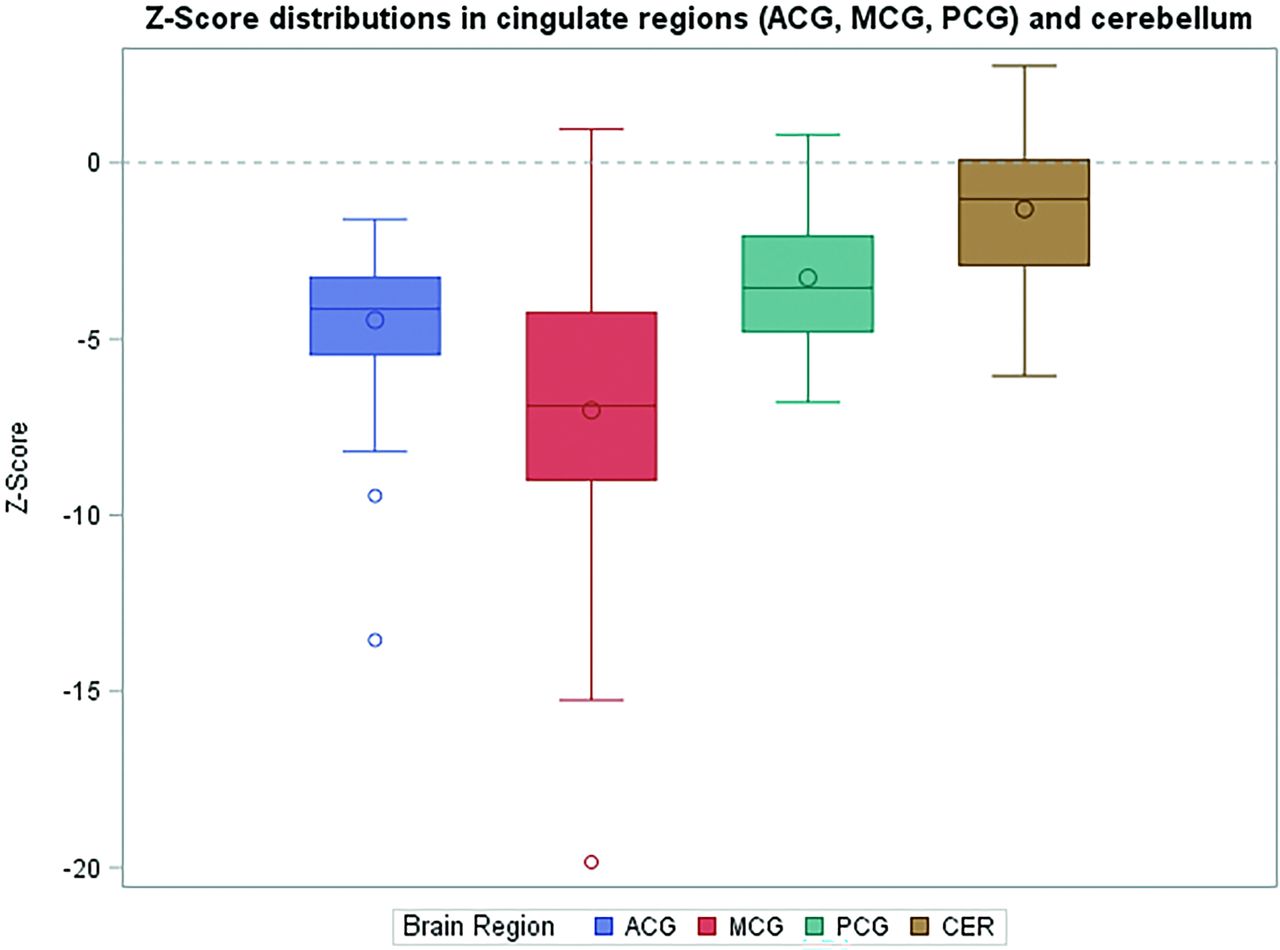
- FIG 1.
Z score distributions in cingulate regions (ACG, MCG, PCG) and CBL. Boxplots illustrating the z score distributions for the ACG, MCG, PCG, and CBL. The median z scores for the ACG, MCG, and PCG are notably lower than those of the CBL, which serves as the reference region. The CBL shows a narrow distribution centered near zero, confirming its role as a stable control. In contrast, the MCG demonstrates the widest range of z scores with the lowest median value, indicating greater variability in FDG uptake, while the ACG and PCG show intermediate distributions with less variability. The circles outside the boxes for ACG and MCG represent statistical outliers.
- FIG 2.
MRI findings in a representative subject. A 71-year-old man presenting with cognitive decline and gait impairment underwent MRI as part of his initial work-up. Axial (A and B) and coronal (C) reformats of 3D T1-weighted MPRAGE images are shown, demonstrating characteristic findings of communicating hydrocephalus. A, Crowding of sulci near the vertex. B, Disproportionate enlargement of the lateral ventricles. C, Narrowing of the callosal angle.
- FIG 3.
FDG-PET/MRI and statistical parametric maps highlighting the effects of hydrocephalus on SPM analysis. Shown is the same patient as in Fig 2. A, Axial attenuation-corrected PET image fused with 3D T2-FLAIR imaging shows enlarged lateral ventricles resulting in PCG displacement (red arrows). B, Unfused axial PET image (both A and B windowed to 0–15 maximum SUV). C and D, Corresponding axial SPM z score maps with D illustrating the outlined PCG region. The PCG is misplaced onto photopenic enlarged ventricles, leading to artifactually low z scores (left PCG score: −5.4; right PCG z score: −5.7). The color scale for C, D, G, and H indicates z scores. E–H, Analogous left parasagittal view shows PCG displacement and the artifacts induced by ventricular enlargement on SPM analysis, with the PCG region outlined in H.


Tables
Comparison of qualitative and quantitative abnormal FDG-PET findings across brain regionsa
Region Qualitative Results (Determined Visually) Quantitative Results (SPM Z Score ≤2) Right ACG 33/48 (68.7%) 47/48 (97.9%) Left ACG 34/48 (70.8%) 43/48 (89.6%) Right MCG 14/48 (29.1%) 47/48 (97.9%) Left MCG 16/48 (33.3%) 45/48 (93.8%) Right PCG 6/48 (12.5%) 35/48 (72.9%) Left PCG 6/48 (12.5%) 39/48 (81.3%) Right CBL 10.4 16/48 (33.3%) Left CBL 10.4 14/48 (29.2%) ↵a Shown are absolute numbers and percentages of subjects with abnormal findings in each brain region, as determined on visual assessment, as well as on quantitative analysis (SPM z scores below −2). These findings highlight discrepancies between qualitative and quantitative assessments across the ACG, MCG, PCG, and CBL, with a notably higher percentage of abnormalities identified quantitatively compared with the qualitative visual analysis.


Jump to section
Related Articles
Cited By...
- No citing articles found.