
Abstract
Summary: A patient with Goldenhar's syndrome (oculoauriculovertebral dysplasia) and unilateral aplasia of all semicircular canals is presented. This is the first report of such a finding and may support the hypothesis that Goldenhar's syndrome and the CHARGE association have a common pathogenetic mechanism.
Oculoauriculovertebral dysplasia, also referred to as Goldenhar's syndrome, was first described in 1952. The condition is a clinically heterogeneous disorder. In its most typical presentation, it is characterized by hemifacial microsomia in association with other anomalies, including vertebral defects and epibulbar dermoids. Bilateral involvement of the craniofacies is known to occur, however, with one side being more severely affected than the other side. Cardiac, renal, anal, and CNS malformations have been reported to occur in patients with Goldenhar's syndrome. The cause of the condition remains unclear (1). The cause of Goldenhar's syndrome seems to be heterogeneous (2). Most cases are sporadic. However, familial cases and concordant monozygotic twins have been reported, suggesting a genetic origin (3). In addition, several chromosomal abnormalities have been documented in affected individuals (4).
A wide variety of external and middle ear malformations have been described in association with the syndrome. These anomalies result from maldevelopment of structures derived from the first and second branchial arches. Inner ear malformations have also been reported in patients with oculoauriculovertebral dysplasia but are very rare (5, 6).
We present the CT and MR imaging findings of a patient with oculoauriculovertebral dysplasia. A unique malformation of the labyrinth with unilateral aplasia of the semicircular canals was observed in the proband.
Case Report
At the age of 3 months, a boy with Goldenhar's syndrome was referred for imaging studies of the temporal bone. The diagnosis of Goldenhar's syndrome was made based on the association of anal atresia, cervical vertebral fusion at the C6–C7 level, hemifacial microsomia with microtia, a preauricular tag, and narrowing of the external auditory canal on the right side. In addition, a solitary left kidney and ventricular septal defect were found.
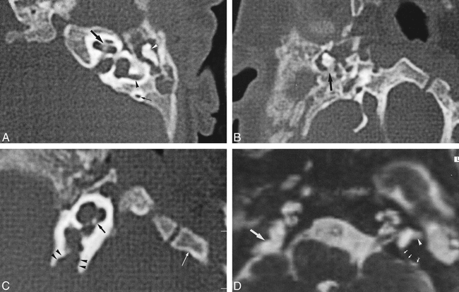
The patient underwent a temporal bone CT examination using contiguous 1-mm-thick axial sections made parallel to the infraorbital-meatal line. Images were reconstructed with a matrix size of 512 × 512 and a 10-cm field of view. Opacification of the middle ear and mastoid was noted on the left side, with preservation of the ossicular chain (Fig 1A). The inner ear was normal. The facial nerve canal showed a normal course. On the right side, multiple anomalies were detected. The external auditory canal was narrow, but no atretic plate was present. Malrotation of the ossicular chain with fixation to the walls of the tympanic cavity was present (Fig 1B). The cavity was also opacified. The stapes was not found. Enlargement of the vestibular aqueduct was noted, as was cochlear dysplasia, with modiolar deficiency (Fig 1C). No semicircular canals were present on the right side. The facial nerve canal was also absent.

Three-month-old boy with Goldenhar's syndrome.
A, Axial 1-mm CT section through the left temporal bone shows a normal cochlea (large arrow), with normal appearance of the modiolus and osseous spiral lamina, as well as normal posterior (small arrow) and lateral (black arrowhead) semicircular canals. Note also the normal incudomalleal articulation (white arrowhead) and the opacification of the middle ear.
B, Axial 1-mm CT section through the right middle ear shows fixation of the short process of the incus to the tympanic wall (arrow). The middle ear cavity is opacified.
C, Axial 1-mm CT section through the right inner ear shows deficiency of the cochlear modiolus (black arrow). Note the enlargement of the vestibular aqueduct (arrowheads). Because of hemifacial microsomia, extreme skull base asymmetry is present, with rotation of the clivus (white arrow). Note also the absence of semicircular canals.
D, Axial 1-mm 3D FT-CISS MR image through both temporal bones shows normal inner ear structures on the left side. Note the presence of normal lateral (arrowhead) and posterior (small arrows) semicircular canals on the left side, whereas these structures are absent on the right side. The vestibular aqueduct on the right side is abnormally enlarged (large arrow).
MR imaging of the temporal bone was performed with axial 1-mm 3D Fourier transform constructive interference in steady state (FT-CISS) images. Sequence parameters for the 3D FT-CISS sequence were 17/7 (TR/TE), with a flip angle of 50°, a field of view of 160 mm, and a matrix of 256 × 256. The MR examination confirmed the cochlear anomaly and clearly showed the enlargement of the vestibular aqueduct (Fig 1D). Aplasia of the semicircular canals and absence of the facial nerve were confirmed on the right side.
Discussion
In contrast to middle ear anomalies, inner ear anomalies are rarely observed in patients with Goldenhar's syndrome. In addition to minor deformities of the bony cochlea, Phelps et al (6) described an abnormal course of the internal auditory meatus. This abnormality could be secondary to the hemifacial microsomia, described as occurring in many patients with Goldenhar's syndrome. They also noted that the lateral semicircular canals were mildly dysplastic and dilated in nine of 29 malformed temporal bones of patients with the syndrome (6); however, they did not consider these anomalies to be significant. Another semicircular canal anomaly consisting of an absent common crus was reported by Manfré et al (5). A complex malformation was seen in which the anterior limb of the superior semicircular canal and the inferior limb of the posterior semicircular canal were joined together, resulting in a single posterosuperior canal, combined with a cystic lateral semicircular canal.
Bilateral absence of the semicircular canals has been reported previously to occur in patients with the CHARGE association (7–9) but not in association with Goldenhar's syndrome. CHARGE association is a disorder characterized by multiple congenital anomalies. An association is defined as a nonrandom occurrence, in two or more persons, of multiple anomalies not yet known to be a syndrome or a sequence. The mnemonic term CHARGE refers to the different possible anomalies: C-coloboma, H-heart defects, A-atresia of the choanae, R-retarded growth and development and/or CNS anomalies, G-genital hypoplasia, E-ear anomalies and/or deafness (8, 9). The CHARGE association is not a specific disorder sui generis nor is it a diagnosis. The cause of the CHARGE association remains unknown. Different chromosomal abnormalities have been reported in cases with the CHARGE association, suggesting that several genes may play a role in the pathogenesis of this entity (10–13).
In addition, two reports on four temporal bones are available in the literature that describe bilateral semicircular canal aplasia in patients who do not have the CHARGE association (14, 15). Two of these four patients also exhibited Kallmann's syndrome (8). Support for a possible link between the CHARGE association and Kallmann's syndrome can be found in the literature. Cortez et al (16) described the case of a 17-year-old boy with Kallmann's syndrome and a complex congenital heart malformation. He also had neurosensory hearing loss and mental retardation. They noted that seven previously reported patients with Kallmann's syndrome and cardiac abnormalities were short (heights more than 2 SD below the mean for their ages), lacked a family history of Kallmann's syndrome, and were mentally retarded (16). Dominant inheritance was postulated by Levy and Knudtzon (17) in a family with two sisters, ages 13 and 19 years, with classical anosmia and hypogonadotropic hypogonadism associated with Kallmann's syndrome. In addition, they had bilateral vesicoureteral reflux and unilateral hearing loss. One of the girls had unilateral coloboma of the optic nerve. The father had no clinical signs of either hypogonadism or anosmia. However, he had unilateral hearing loss and duplication of the left ureter and died suddenly at the age of 40 years from myocardial infarction and undiagnosed coarctation of the aorta (17). In addition, Klein et al (18) described the association of Kallmann's syndrome with choanal atresia.
The observation of unilateral semicircular canal aplasia in this patient is interesting because it supports the hypothesis presented by Van Meter and Weaver (19) that oculoauriculovertebral dysplasia and the CHARGE association may have a common pathogenetic mechanism. Van Meter and Weaver postulated this hypothesis after having examined two infants with features of both oculoauriculovertebral dysplasia and the CHARGE association.
In conclusion, this case of a patient with Goldenhar's syndrome and unilateral aplasia of all semicircular canals supports the hypothesis that Goldenhar's syndrome and the CHARGE association may have a common pathogenetic mechanism. More patients with Goldenhar's syndrome and other branchial arch disorders should be investigated by imaging studies of the temporal bone for better delineation of each disorder and understanding of the cause.
Footnotes
↵1 Address reprint requests to Marc M. Lemmerling, MD, PhD, University Hospital Gent, Department of Radiology, De Pintelaan 185, 9000 Gent, Belgium.
References
- Received May 12, 1999.
- Accepted after revision January 19, 2000.
- Copyright © American Society of Neuroradiology
In this issue


Jump to section
Related Articles
Cited By...
- No citing articles found.