Advertisement

Main menu

  • Home
  • Content
    • Current Issue
    • Accepted Manuscripts
    • Article Preview
    • Past Issue Archive
    • AJNR Case Collection
    • Case of the Week Archive
    • Classic Case Archive
    • Case of the Month Archive
  • Special Collections
    • Spinal CSF Leak Articles (Jan 2020-June 2024)
    • 2024 AJNR Journal Awards
    • Most Impactful AJNR Articles
  • Multimedia
    • AJNR Podcast
    • AJNR Scantastics
    • Video Articles
  • For Authors
    • Submit a Manuscript
    • Author Policies
    • Fast publishing of Accepted Manuscripts
    • Graphical Abstract Preparation
    • Manuscript Submission Guidelines
    • Imaging Protocol Submission
    • Submit a Case for the Case Collection
  • About Us
    • About AJNR
    • Editorial Board
  • More
    • Become a Reviewer/Academy of Reviewers
    • Subscribers
    • Permissions
    • Alerts
    • Feedback
    • Advertisers
    • ASNR Home
  • Other Publications
    • ajnr

User menu

  • Alerts
  • Log in

Search

  • Advanced search
American Journal of Neuroradiology

American Journal of Neuroradiology

ASHNR American Society of Functional Neuroradiology ASHNR American Society of Pediatric Neuroradiology ASSR
  • Alerts
  • Log in

Advanced Search

  • Home
  • Content
    • Current Issue
    • Accepted Manuscripts
    • Article Preview
    • Past Issue Archive
    • AJNR Case Collection
    • Case of the Week Archive
    • Classic Case Archive
    • Case of the Month Archive
  • Special Collections
    • Spinal CSF Leak Articles (Jan 2020-June 2024)
    • 2024 AJNR Journal Awards
    • Most Impactful AJNR Articles
  • Multimedia
    • AJNR Podcast
    • AJNR Scantastics
    • Video Articles
  • For Authors
    • Submit a Manuscript
    • Author Policies
    • Fast publishing of Accepted Manuscripts
    • Graphical Abstract Preparation
    • Manuscript Submission Guidelines
    • Imaging Protocol Submission
    • Submit a Case for the Case Collection
  • About Us
    • About AJNR
    • Editorial Board
  • More
    • Become a Reviewer/Academy of Reviewers
    • Subscribers
    • Permissions
    • Alerts
    • Feedback
    • Advertisers
    • ASNR Home
  • Follow AJNR on Twitter
  • Visit AJNR on Facebook
  • Follow AJNR on Instagram
  • Join AJNR on LinkedIn
  • RSS Feeds

Welcome to the new AJNR, Updated Hall of Fame, and more. Read the full announcements.


AJNR is seeking candidates for the position of Associate Section Editor, AJNR Case Collection. Read the full announcement.


Research ArticleHEAD AND NECK

Castleman's Disease in the Retropharyngeal Space: CT and MR Imaging Findings

Ji Hoon Shin, Ho Kyu Lee, Sang Yoon Kim, Shin Kwang Khang, Sung Hye Park, Choong Gon Choi and Dae Chul Suh
American Journal of Neuroradiology August 2000, 21 (7) 1337-1339;
Ji Hoon Shin
aFrom the Departments of Radiology (J.H.S., H.K.L., C.G.C., D.C.S.), Otolaryngology (S.Y.K.), and Pathology (S.K.K., S.H.P.), Asan Medical Center, University of Ulsan College of Medicine, Seoul, South Korea.
  • Find this author on Google Scholar
  • Find this author on PubMed
  • Search for this author on this site
Ho Kyu Lee
aFrom the Departments of Radiology (J.H.S., H.K.L., C.G.C., D.C.S.), Otolaryngology (S.Y.K.), and Pathology (S.K.K., S.H.P.), Asan Medical Center, University of Ulsan College of Medicine, Seoul, South Korea.
  • Find this author on Google Scholar
  • Find this author on PubMed
  • Search for this author on this site
Sang Yoon Kim
aFrom the Departments of Radiology (J.H.S., H.K.L., C.G.C., D.C.S.), Otolaryngology (S.Y.K.), and Pathology (S.K.K., S.H.P.), Asan Medical Center, University of Ulsan College of Medicine, Seoul, South Korea.
  • Find this author on Google Scholar
  • Find this author on PubMed
  • Search for this author on this site
Shin Kwang Khang
aFrom the Departments of Radiology (J.H.S., H.K.L., C.G.C., D.C.S.), Otolaryngology (S.Y.K.), and Pathology (S.K.K., S.H.P.), Asan Medical Center, University of Ulsan College of Medicine, Seoul, South Korea.
  • Find this author on Google Scholar
  • Find this author on PubMed
  • Search for this author on this site
Sung Hye Park
aFrom the Departments of Radiology (J.H.S., H.K.L., C.G.C., D.C.S.), Otolaryngology (S.Y.K.), and Pathology (S.K.K., S.H.P.), Asan Medical Center, University of Ulsan College of Medicine, Seoul, South Korea.
  • Find this author on Google Scholar
  • Find this author on PubMed
  • Search for this author on this site
Choong Gon Choi
aFrom the Departments of Radiology (J.H.S., H.K.L., C.G.C., D.C.S.), Otolaryngology (S.Y.K.), and Pathology (S.K.K., S.H.P.), Asan Medical Center, University of Ulsan College of Medicine, Seoul, South Korea.
  • Find this author on Google Scholar
  • Find this author on PubMed
  • Search for this author on this site
Dae Chul Suh
aFrom the Departments of Radiology (J.H.S., H.K.L., C.G.C., D.C.S.), Otolaryngology (S.Y.K.), and Pathology (S.K.K., S.H.P.), Asan Medical Center, University of Ulsan College of Medicine, Seoul, South Korea.
  • Find this author on Google Scholar
  • Find this author on PubMed
  • Search for this author on this site
  • Article
  • Figures & Data
  • Info & Metrics
  • Responses
  • References
  • PDF
Loading

Abstract

Summary: We report CT and MR imaging findings in a case of Castleman's disease involving the retropharyngeal space in a middle-aged woman. On CT scans, a well-marginated, homogeneous, and densely enhancing mass was detected in the right retropharyngeal space. The mass was isointense to the muscle on T1-weighted MR images, hyperintense to the muscle on T2-weighted MR images, and showed homogeneous, strong enhancement on contrast-enhanced T1-weighted MR images. The linear hypointense signal in an arborizing pattern was observed within the mass on all pulse sequences.

Castleman's disease is a rare lymphoproliferative disorder of uncertain cause characterized by a distinctive pattern of hypervascular lymphoid hyperplasia (1). Although it is usually reported as a solitary mediastinal mass, involvement of other anatomic sites has been reported, with the head and neck being the second most common area (2). We describe a case of hyaline-vascular-type Castleman's disease located in the retropharyngeal space.

Case Report

A 34-year-old woman initially presented with a 5-year history of painless swelling on the right side of her oropharynx and a 2-year history of a slightly enlarging mass on the right side of her neck. Her medical history was unremarkable.

Unenhanced CT scans showed a large, well-circumscribed, solid mass in the right retropharyngeal space, which was isodense to the muscle. The mass showed homogeneous, strong enhancement on the contrast-enhanced CT scans (Fig 1A).

fig 1.
  • Download figure
  • Open in new tab
  • Download powerpoint
fig 1.

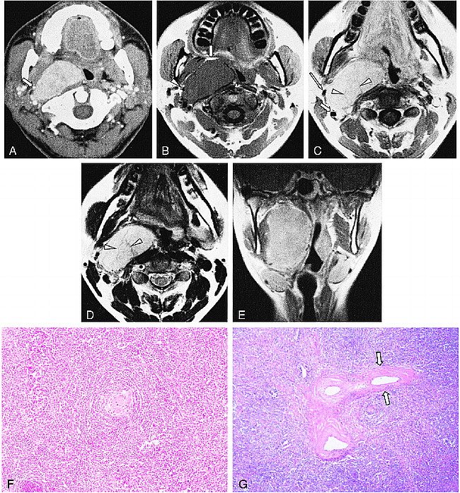
Images from the case of a 34-year-old woman with an enlarging mass on the right side of her neck.

A, Contrast-enhanced axial CT scan shows a well-marginated, intensely enhancing mass in the right retropharyngeal space. Note the styloid process, which is anterolaterally displaced by the mass (arrow).

B, T1-weighed (450/12) axial MR image shows a well-marginated mass isointense to the muscle in the right retropharyngeal space. The mass displaces the parapharyngeal fat anteriorly (arrow), pharyngeal mucosal space medially, styloid process anterolaterally (arrowhead), and internal carotid artery laterally.

C, Contrast-enhanced T1-weighed (450/12) axial MR image (obtained at a slightly lower level than that shown in panel B) reveals strong enhancement of the lesion. The displaced right internal carotid artery (short arrow) and external carotid artery (long arrow) are separated. Note the linear hypointense signal within the mass (arrowheads).

D, T2-weighted (3700/99) axial MR image (obtained at the same level as that shown in panel C) shows the lesion hyperintense to the muscle. The linear hypointense signal seen in panel C is also seen as a hypointense signal (arrowheads).

E, Contrast-enhanced T1-weighed (513/12) coronal MR image shows the mass occupying the entire right retropharyngeal and parapharyngeal space from below the skull base to the hyoid bone level.

F, Photomicrograph (original magnification, ×100; hematoxylin and eosin stain) of the right retropharyngeal lymph node shows a follicle with an onion skin–appearing, prominent mantle and a penetrating hyalinizing vessel into the germinal center, consistent with hyaline-vascular-type Castleman's disease.

G, Low-power photomicrograph (original magnification, ×40; hematoxylin and eosin stain) of the right retropharyngeal lymph node shows perivascular lamellar fibrosis (arrows), which corresponds to the linear hypointense signals in an arborizing pattern on the MR images.

One month later, MR imaging was performed. On the MR images, the mass was located within the right retropharyngeal space, displacing the parapharyngeal fat anteriorly, the pharyngeal mucosal space medially, the styloid process anterolaterally, and the carotid space laterally. It was isointense to the muscle on the T1-weighted images (Fig 1B). There was a nonenhancing linear hypointense signal in an arborizing pattern within the mass on T1- and T2-weighted images (Fig 1C and D). The mass occupied the right retropharyngeal and parapharyngeal space from below the skull base to the hyoid bone level on the contrast-enhanced T1-weighted coronal image (Fig 1E). There was a small lymph node with a 1-cm diameter in the right internal jugular chain just below the main mass.

Excision of the neck mass was performed via a transcervical approach. A 5 × 8-cm, well-encapsulated, ovoid mass was removed without much difficulty. Histopathologic evaluation revealed Castleman's disease (giant lymph node hyperplasia) of the hyaline-vascular type (Fig 1F). The mass was thought to arise from the lateral retropharyngeal lymph node. The small lymph node just below the main mass was proved reactive hyperplasia without tumor cells.

Discussion

In 1956, Castleman et al (3) described a group of patients with large thymoma-like masses in the anterior mediastinum, which they called mediastinal lymph node hyperplasia. The cause of Castleman's disease is uncertain; it is thought to be inflammatory or hamartomatous in nature (4, 5). Two distinct histologic variants are recognized (4). The most common is the hyaline-vascular type (more than 90% of the cases), which consists of small lymphoreticular follicles distributed within a hypervascular hyalinized stroma. The plasma cell type is less common (fewer than 10% of the cases) and consists of larger lymphoreticular nodules that are separated by sheets of plasma cells and a somewhat less vascular stroma. Patients with the hyaline-vascular-type disease are usually asymptomatic, but they may complain of symptoms caused by the compression of adjacent structures or may present with a palpable mass. Systemic manifestations are commonly seen in the plasma cell type and include fever, anemia, and hyperglobulinemia.

Castleman's disease is usually limited to one site, although an aggressive multicentric form has been described. The most common site is the mediastinum (approximately 70%). Additional sites of occurrence include the axilla, retroperitoneum, mesentery, vulva, pancreas, pelvis, and neck (1, 4). Fewer than 10% of cases arise in the head and neck (6−11). In our case, the tumor was located within the right retropharyngeal space. It was thought to arise from a retropharyngeal node, probably the lateral group, which is located at the anterolateral aspect of the prevertebral muscle, although a carotid space origin could not be completely excluded. Several cases of Castleman's disease involving the retropharyngeal or parapharyngeal space, medial to the carotid sheath, as in our case, have been reported (7−9).

The typical CT findings of Castleman's disease are a well-marginated, homogeneous, and densely enhancing mass (6, 7), although ring enhancement can also be seen (1, 10). The hyaline-vascular variant tends to show more prominent enhancement on CT scans, most likely because of its greater vascularity (1, 10).

Typically, Castleman's disease appears on MR images as low-to-intermediate signals compared with muscle on T1-weighted images and high signals on T2-weighted images (7, 9). In a few previous reports (12, 13), linear, arborizing, hypointense signals within the mass have been attributed to calcification, fibrous septations, vessels, or sinus histiocytosis. The linear hypointense signals seen on MR images in our case were proved perivascular lamellar fibrosis on pathologic correlation (Fig 1G). These linear hypointense signals could be an important clue to the diagnosis of Castleman's disease, although they occur infrequently.

The MR imaging differential diagnoses for Castleman's disease include paraganglioma and schwannoma. Paragangliomas in this area usually displace the internal carotid artery anteriorly because the tumor arises around the vagus nerve. Furthermore, paragangliomas have multiple focal and serpentine areas of low signal intensity, representing vascular flow voids within the mass, especially in large lesions. Schwannomas that arise from the vagus nerve and the sympathetic chain usually displace the internal carotid artery anteriorly because these nerves are posterior to this vessel. Frequently, schwannomas have inhomogeneous signal intensity due to cystic change. These features could be the clue to the differential diagnosis, with Castleman's disease in the retropharyngeal or parapharyngeal space.

Complete surgical excision is the treatment of choice for Castleman's disease in the head and neck, with a 100% control rate for the hyaline-vascular type. The plasma cell type, however, requires excision with close follow-up and possible systemic chemotherapy (14). Malignant transformation or association with other malignancies is rare and includes plasmacytoma, malignant lymphoma, and Kaposi's sarcoma (15). Its malignant potential, although rare, raises suggestions of a relationship with an immune dysfunction or even primary lymphoproliferative disorder (16).

Conclusion

Castleman's disease is a rare mass, which may involve the retropharyngeal space. On CT scans, it appears as a well-marginated, homogeneously enhancing mass. It has an isointense signal compared with muscle on T1-weighted MR images and a hyperintense signal compared with muscle on T2-weighted MR images, with homogeneous, strong enhancement. Linear hypointense signals in an arborizing pattern may be present within the mass. Castleman's disease should be considered in the differential diagnosis of masses involving the retropharyngeal space.

Footnotes

  • ↵1 Address reprint requests to Ho Kyu Lee, MD, Department of Radiology, Asan Medical Center, 388-1 Poongnap-Dong, Songpa-Ku, Seoul 138-736, South Korea.

References

  1. ↵
    Chaloupka JC, Castillo M, Hudgins P. Castleman disease in the neck: atypical appearance on CT. AJR Am J Radiol 1990;154:1051-1052
    PubMed
  2. ↵
    Frizzera G. Castleman's disease: more questions than answers. Hum Pathol 1985;16:202-205
    CrossRefPubMed
  3. ↵
    Castleman B, Iverson L, Menendez VP. Localized mediasinal lymph node hyperplasia resembling thymoma. Cancer 1956;9:822-830
    CrossRefPubMed
  4. ↵
    Keller AR, Hochholzer L, Castleman B. Hyaline-vascular and plasma-cell types of giant lymph node hyperplasia of the mediastinum and other locations. Cancer 1972;29:670-683
    CrossRefPubMed
  5. Johnson JT, Oral A, Nalesnik M, Roscoe GJ, Whiteside TL. Giant lymph node hyperplasia: clinical and immunohistologic correlation of an intermediate variant. Ear Nose Throat J 1985;64:249-254
    PubMed
  6. ↵
    Koslin DB, Berland LL, Sekar BC. Cervical Castleman disease: CT study with angiographic correlation. Radiology 1986;160:213-214
    PubMed
  7. ↵
    Freeman SJ, Irvine GH, Glew D. Case report: cervical Castleman's disease shown by CT and MRI. Clin Radiol 1994;49:721-723
    CrossRefPubMed
  8. Lanier BJ, Cummings CW. Giant lymphoid hyperplasia presenting as a highly vascularized parapharyngeal mass. Otolaryngol Head Neck Surg 1982;90:426-430
    PubMed
  9. Tuerlinckx D, Bodart E, Delos M, Remacle M, Ninane J. Unifocal cervical Castleman disease in two children. Eur J Pediatr 1997;156:701-703
    CrossRefPubMed
  10. Nunna SV, Sharma R, Goyal M, Berry M, Gupta SD, Deshpande V. Unusual computed tomography appearance of Castleman disease. Australas Radiol 1997;41:193-195
    PubMed
  11. Gleeson MJ, Cassidy M, McMullin JP. Castleman's disease: an unusual neck mass. J Laryngol Otol 1988;102:661-662
    PubMed
  12. ↵
    Glazer M, Rao VM, Reiter D, McCue P. Isolated Castleman disease of the neck: MR findings. AJNR Am J Neuroradiol 1995;16:669-671
    Abstract/FREE Full Text
  13. Luburich P, Nicolau C, Ayuso MC, Torra R, Clavero JA. Pelvic Castleman disease: CT and MR appearance. J Comput Assist Tomogr 1992;16:657-659
    PubMed
  14. ↵
    Sanz C, Sierra J, Cobarro J, Avellaneda R, Montserrat E, Rozman C. An unusual case of Castleman's disease restricted to the neck. ORL J Otorhinolaryngol Relat Spec 1992;54:331-333
    CrossRefPubMed
  15. ↵
    Case records of the Massachusetts General Hospital. Weekly clinicopathological exercises (Case 32–1984). N Engl J Med 1984;311:388-398
    PubMed
  16. ↵
    Williams JL, Kaude JV. Sonographic findings in a case of Castleman disease of the neck. J Ultrasound Med 1986;5:593-594
    PubMed
  • Received November 8, 1999.
  • Accepted after revision January 18, 2000.
  • Copyright © American Society of Neuroradiology
View Abstract
PreviousNext
Back to top

In this issue

American Journal of Neuroradiology
Vol. 21, Issue 7
1 Aug 2000
  • Table of Contents
  • Index by author
Advertisement
Print
Download PDF
Email Article

Thank you for your interest in spreading the word on American Journal of Neuroradiology.

NOTE: We only request your email address so that the person you are recommending the page to knows that you wanted them to see it, and that it is not junk mail. We do not capture any email address.

Enter multiple addresses on separate lines or separate them with commas.
Castleman's Disease in the Retropharyngeal Space: CT and MR Imaging Findings
(Your Name) has sent you a message from American Journal of Neuroradiology
(Your Name) thought you would like to see the American Journal of Neuroradiology web site.
CAPTCHA
This question is for testing whether or not you are a human visitor and to prevent automated spam submissions.
Cite this article
Ji Hoon Shin, Ho Kyu Lee, Sang Yoon Kim, Shin Kwang Khang, Sung Hye Park, Choong Gon Choi, Dae Chul Suh
Castleman's Disease in the Retropharyngeal Space: CT and MR Imaging Findings
American Journal of Neuroradiology Aug 2000, 21 (7) 1337-1339;

Citation Manager Formats

  • BibTeX
  • Bookends
  • EasyBib
  • EndNote (tagged)
  • EndNote 8 (xml)
  • Medlars
  • Mendeley
  • Papers
  • RefWorks Tagged
  • Ref Manager
  • RIS
  • Zotero
0 Responses
Respond to this article
Share
Bookmark this article
Castleman's Disease in the Retropharyngeal Space: CT and MR Imaging Findings
Ji Hoon Shin, Ho Kyu Lee, Sang Yoon Kim, Shin Kwang Khang, Sung Hye Park, Choong Gon Choi, Dae Chul Suh
American Journal of Neuroradiology Aug 2000, 21 (7) 1337-1339;
del.icio.us logo Twitter logo Facebook logo Mendeley logo
  • Tweet Widget
  • Facebook Like
  • Google Plus One

Jump to section

  • Article
    • Abstract
    • Case Report
    • Discussion
    • Conclusion
    • Footnotes
    • References
  • Figures & Data
  • Info & Metrics
  • Responses
  • References
  • PDF

Related Articles

  • No related articles found.
  • PubMed
  • Google Scholar

Cited By...

  • Castleman's disease of the neck: a description of four cases on contrast-enhanced CT
  • Crossref
  • Google Scholar

This article has not yet been cited by articles in journals that are participating in Crossref Cited-by Linking.

More in this TOC Section

  • Correlation of Apparent Diffusion Coefficient at 3T with Prognostic Parameters of Retinoblastoma
  • Parathyroid Lesions: Characterization with Dual-Phase Arterial and Venous Enhanced CT of the Neck
  • Efficacy of Diffusion-Weighted Imaging for the Differentiation between Lymphomas and Carcinomas of the Nasopharynx and Oropharynx: Correlations of Apparent Diffusion Coefficients and Histologic Features
Show more Head and Neck

Similar Articles

Advertisement

Indexed Content

  • Current Issue
  • Accepted Manuscripts
  • Article Preview
  • Past Issues
  • Editorials
  • Editors Choice
  • Fellow Journal Club
  • Letters to the Editor

Cases

  • Case Collection
  • Archive - Case of the Week
  • Archive - Case of the Month
  • Archive - Classic Case

Special Collections

  • Special Collections

Resources

  • News and Updates
  • Turn around Times
  • Submit a Manuscript
  • Author Policies
  • Manuscript Submission Guidelines
  • Evidence-Based Medicine Level Guide
  • Publishing Checklists
  • Graphical Abstract Preparation
  • Imaging Protocol Submission
  • Submit a Case
  • Become a Reviewer/Academy of Reviewers
  • Get Peer Review Credit from Publons

Multimedia

  • AJNR Podcast
  • AJNR SCANtastic
  • Video Articles

About Us

  • About AJNR
  • Editorial Board
  • Not an AJNR Subscriber? Join Now
  • Alerts
  • Feedback
  • Advertise with us
  • Librarian Resources
  • Permissions
  • Terms and Conditions

American Society of Neuroradiology

  • Not an ASNR Member? Join Now

© 2025 by the American Society of Neuroradiology All rights, including for text and data mining, AI training, and similar technologies, are reserved.
Print ISSN: 0195-6108 Online ISSN: 1936-959X

Powered by HighWire