Advertisement

Main menu

  • Home
  • Content
    • Current Issue
    • Accepted Manuscripts
    • Article Preview
    • Past Issue Archive
    • AJNR Case Collection
    • Case of the Week Archive
    • Classic Case Archive
    • Case of the Month Archive
  • Special Collections
    • Spinal CSF Leak Articles (Jan 2020-June 2024)
    • 2024 AJNR Journal Awards
    • Most Impactful AJNR Articles
  • Multimedia
    • AJNR Podcast
    • AJNR Scantastics
    • Video Articles
  • For Authors
    • Submit a Manuscript
    • Author Policies
    • Fast publishing of Accepted Manuscripts
    • Graphical Abstract Preparation
    • Manuscript Submission Guidelines
    • Imaging Protocol Submission
    • Submit a Case for the Case Collection
  • About Us
    • About AJNR
    • Editorial Board
  • More
    • Become a Reviewer/Academy of Reviewers
    • Subscribers
    • Permissions
    • Alerts
    • Feedback
    • Advertisers
    • ASNR Home
  • Other Publications
    • ajnr

User menu

  • Alerts
  • Log in

Search

  • Advanced search
American Journal of Neuroradiology

American Journal of Neuroradiology

ASHNR American Society of Functional Neuroradiology ASHNR American Society of Pediatric Neuroradiology ASSR
  • Alerts
  • Log in

Advanced Search

  • Home
  • Content
    • Current Issue
    • Accepted Manuscripts
    • Article Preview
    • Past Issue Archive
    • AJNR Case Collection
    • Case of the Week Archive
    • Classic Case Archive
    • Case of the Month Archive
  • Special Collections
    • Spinal CSF Leak Articles (Jan 2020-June 2024)
    • 2024 AJNR Journal Awards
    • Most Impactful AJNR Articles
  • Multimedia
    • AJNR Podcast
    • AJNR Scantastics
    • Video Articles
  • For Authors
    • Submit a Manuscript
    • Author Policies
    • Fast publishing of Accepted Manuscripts
    • Graphical Abstract Preparation
    • Manuscript Submission Guidelines
    • Imaging Protocol Submission
    • Submit a Case for the Case Collection
  • About Us
    • About AJNR
    • Editorial Board
  • More
    • Become a Reviewer/Academy of Reviewers
    • Subscribers
    • Permissions
    • Alerts
    • Feedback
    • Advertisers
    • ASNR Home
  • Follow AJNR on Twitter
  • Visit AJNR on Facebook
  • Follow AJNR on Instagram
  • Join AJNR on LinkedIn
  • RSS Feeds

Welcome to the new AJNR, Updated Hall of Fame, and more. Read the full announcements.


AJNR is seeking candidates for the position of Associate Section Editor, AJNR Case Collection. Read the full announcement.


OtherSPINE

Intrathecal Injection of Contrast Medium to Prevent Polymethylmethacrylate Leakage during Percutaneous Vertebroplasty

John S. Sarzier and Avery J. Evans
American Journal of Neuroradiology May 2003, 24 (5) 1001-1002;
John S. Sarzier
  • Find this author on Google Scholar
  • Find this author on PubMed
  • Search for this author on this site
Avery J. Evans
  • Find this author on Google Scholar
  • Find this author on PubMed
  • Search for this author on this site
  • Article
  • Figures & Data
  • Info & Metrics
  • Responses
  • References
  • PDF
Loading

Abstract

Summary: The major technical drawback of percutaneous vertebroplasty is the potential for neural compromise from leakage of polymethylmethaorylate into epidural or perivertebral veins. We have combined the procedure of intrathecal injection of contrast medium with vertebroplasty to better delineate spinal canal encroachment during injection when the posterior vertebral wall is compromised by myeloma.

Percutaneous vertebroplasty has been implemented for the treatment of aggressive vertebral angiomas and malignant spinal tumors since 1984; metastases and myelomas are the most frequent of the malignant osteolytic lesions of the spine (1–4). The major technical drawback of percutaneous vertebroplasty is the potential for neural compromise from leakage of polymethylmethacrylate (PMMA) into epidural or perivertebral veins (5). When tumor extends to the posterior vertebral wall, an increased risk of neural compromise exists as a result of direct PMMA leakage or posterior displacement of the tumor mass into the spinal canal or neural foramina.We report on a case of percutaneous vertebroplasty for compression fracture secondary to myeloma. In this case, we found destruction of the posterior vertebral wall and injected contrast medium intrathecally to monitor posterior tumor displacement before significant canal encroachment occurred.

Case Description and Technique

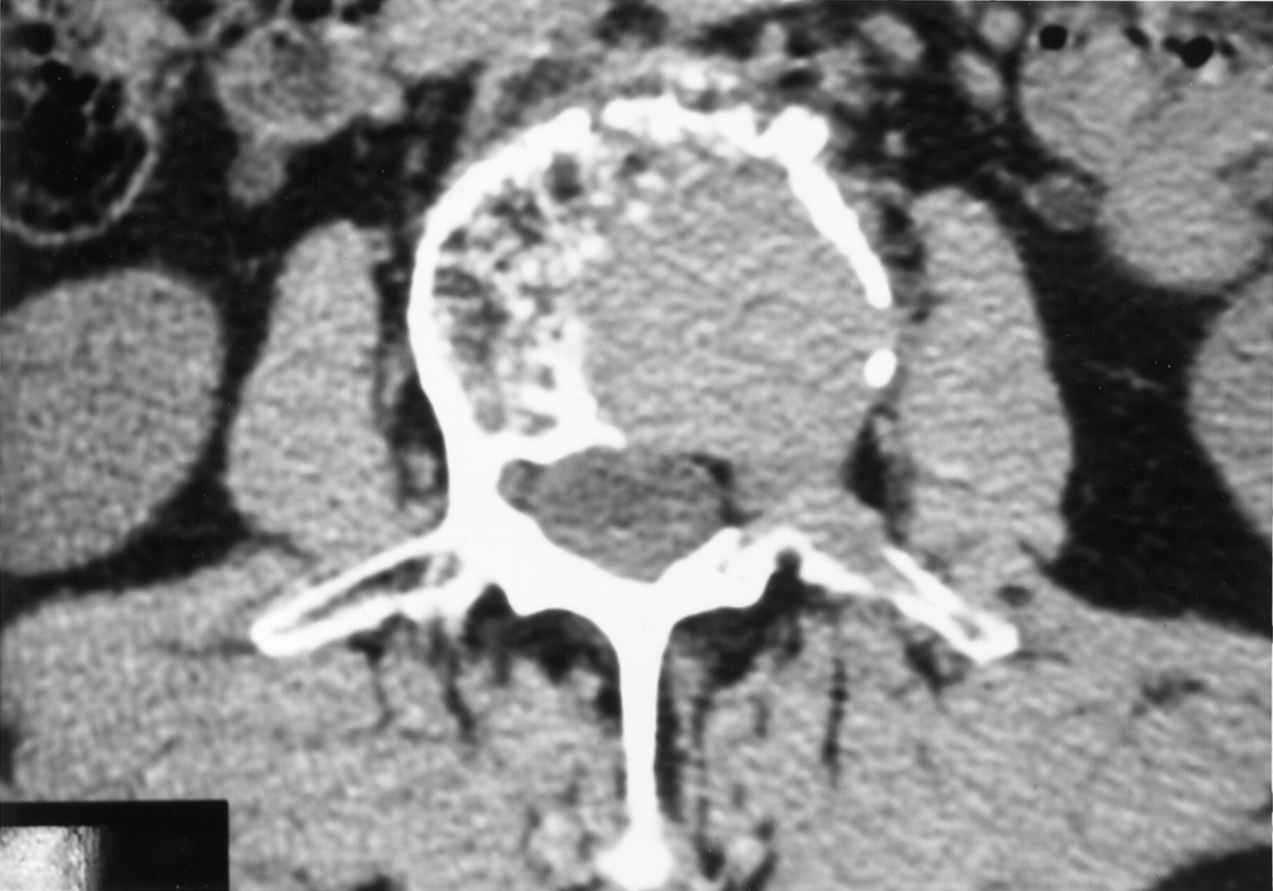
A 70-year-old man was referred to our institution for evaluation because of severe, debilitating back pain after optimal medical treatment for multiple myeloma with spine involvement. Treatment had included radiation therapy; oral, injected, and transdermal narcotics; steroid therapy; bracing; and local analgesic injections. Workup revealed an increase in his compression fracture of L2, which revealed approximately 35% loss of height (Fig 1). Despite destruction of the posterior vertebral wall and pedicle on the left side, there was no clinical or radiographic evidence of neural compromise. A trial of percutaneous vertebroplasty with intrathecal contrast medium was planned to evaluate for posterior displacement of the soft tumor mass into the spinal canal during PMMA (Parallax Medical, Mountainview, CA) injection.

Fig 1.
  • Download figure
  • Open in new tab
  • Download powerpoint
Fig 1.

CT scan at L2 level. Note destruction of posterior vertebral wall and pedicle.

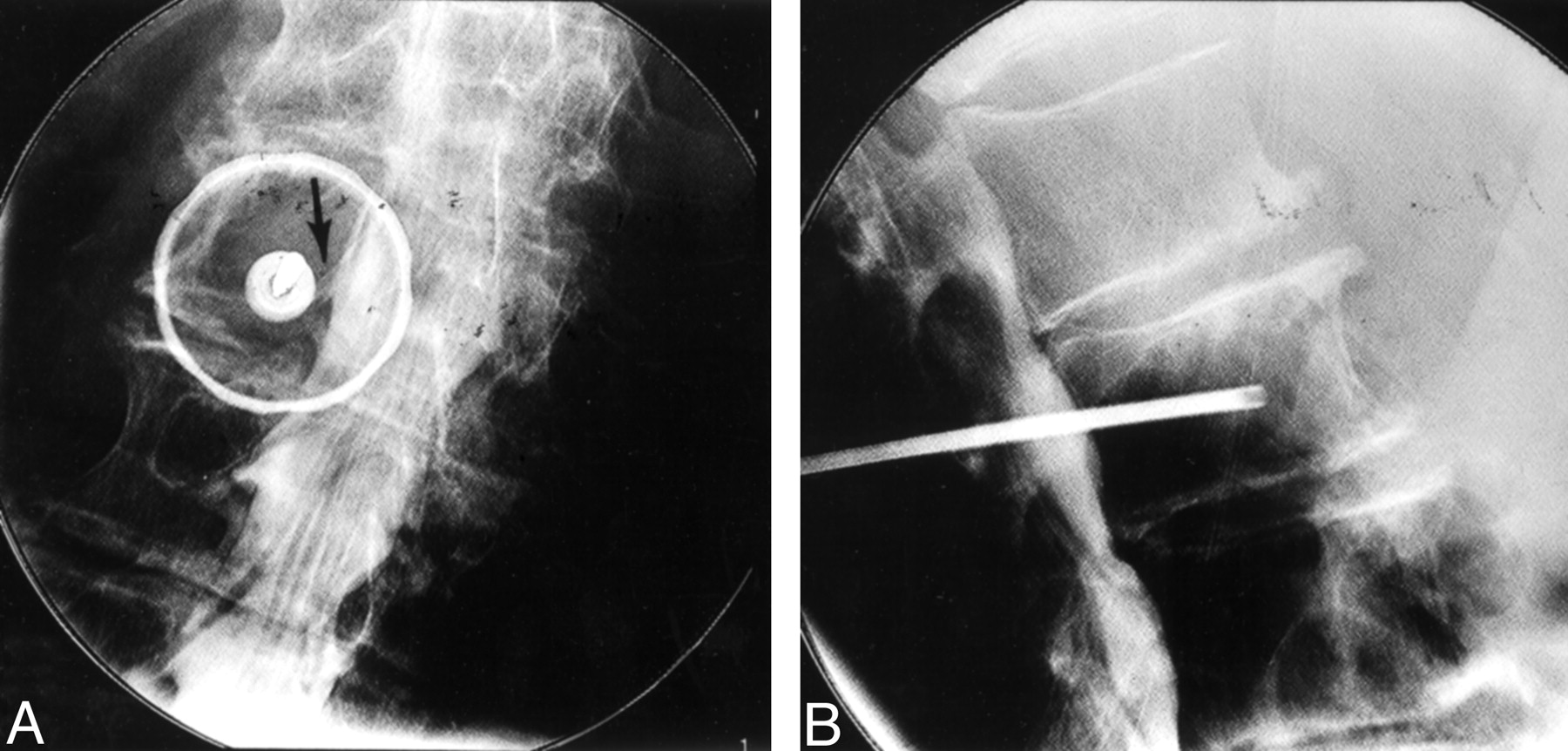
The patient was placed in prone position on the angiography table and prepared and draped in sterile fashion. An adequate plane of anesthesia was obtained by using injectable Fentanyl (Abbott Laboratories, Abbott Park, IL) and versed (Roche Laboratories, Nutley, NJ) and local injection of 1% Lidocaine (Abbott Laboratories, Chicago, IL). A 25-gauge spinal needle was placed at the L3 level, and 15 cc of Isovue M-200 (Bracco RX, Inc., Princeton, NJ) was injected via the intrathecal sac; visualization of the thecal sac and nerve roots was achieved (Fig 2). A small incision was made over the left L2 pedicle, and an 11-gauge bone trocar was advanced under fluoroscopic guidance into the L2 vertebral body. Under fluoroscopic observation, PMMA was slowly injected with a hand injector. After approximately 6 cc of PMMA was applied, the thecal sac showed signs of scalloping but no evidence of extension of PMMA to the posterior cortical margin (Fig 3). Despite lack of optimal filling of the vertebral body, we thought this evidence of thecal sac compression represented posterior displacement of the tumor mass, and no more PMMA was injected.

Fig 2.
  • Download figure
  • Open in new tab
  • Download powerpoint
Fig 2.

Fluoroscopic images of L2 after intrathecal injection of contrast material and placement of an 11-gauge trocar in the right pedicle.

A, Anteroposterior view. Contrast medium shows the proximity of the right nerve root sheath to the trocar and appropriate placement of the trocar.

B, Lateral view. Contrast medium shows minimal canal compromise by posterior tumor extension but no nerve root compromise.

Fig 3.
  • Download figure
  • Open in new tab
  • Download powerpoint
Fig 3.

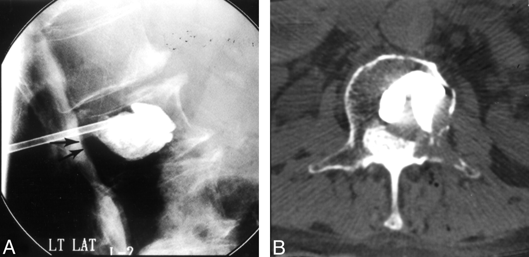
Fluoroscopic image at the L2 level after injection of PMMA (A) and CT scan at same level obtained after vertebroplasty (B).

A, Note subtle posterior displacement of thecal sac (arrows) and no extension of PMMA into the epidural venous plexus or posterior to vertebral margin.

B, Note the distance between the PMMA and posterior vertebral wall and the subsequent posterior epidural extension of the tumor that causes the scalloping visible in A.

The patient awoke from neuroleptic anesthesia without clinical evidence of neurologic compromise. Postoperative CT was performed and revealed posterior displacement of the tumor mass with encroachment on the thecal sac but no evidence of epidural extension of PMMA. The patient was discharged 2 hours after the procedure and reported notable reduction of back pain. He returned for follow-up 2 weeks later and reported a reduction of his back pain from a 10 on the visual assessment pain scale and scheduled narcotic use to a 3 with intermittent narcotic use. He had no evidence of neural compromise.

Discussion

The occurrence of neural compromise or epidural extension associated with percuatneous vertebroplasty varies and depends on the initial indications for vertebroplasty; however, in patients with spinal malignant tumors, the frequency is 10%. This compares with 2–5% of patients with vertebral angiomas and 1–3% of patients with osteoporotic lesions (1, 6–9). The increased risk is attributed to the destruction of vertebral cortex, which, according to Deramond and colleagues (2), occurs in more than 50% of patients. To date, there are few reports of spinal cord compression secondary to posterior displacement during vertebroplasty, but this likely represents a reluctance to perform the procedure in the face of posterior vertebral wall destruction. As this case demonstrates, the introduction of intrathecal contrast medium can allow for safe injection of PMMA with real-time visualization of the nerve roots and thecal sac and can demonstrate the posterior displacement of the soft-tissue tumor mass, which is not visible under conventional fluoroscopy. We have performed this procedure in six other patients with similar spinal tumors. In all six cases, the intrathecal contrast medium revealed scalloping of the thecal sac before optimal vertebral body filling, which resulted in early truncation of PMMA injection. In all of these cases, follow-up CT confirmed thecal sac encroachment that was seen at fluoroscopy. In our experience, there have been no deleterious consequences associated with intrathecal injection of contrast medium during percutaneous vertebroplasty.

References

  1. ↵
    Bascoulergue Y, Dusquesnel J, Leclercq, et al. Percutaneous injection of methylmethacrylate in the vertebral body for the treatment of various diseases: percutaneous vertebroplasty. Radiology 1988;372:169
  2. ↵
    Deramond H, Depriester C, Galibert P, Le Gars D. Percutaneous vertebroplasty with polymethylmetacrylate: technique, indications and results. Radiol Clin N Am 1998 :36:533–546
    CrossRefPubMed
  3. Galibert P, Deramond H. La vertebroplasie acrylique percutanee comme traitement des angiome vertbraux et des affection dolorigenes et fragilisantes du rachis [in French]. Chirurgie 1990;116:326–335
    PubMed
  4. ↵
    Kaemmerlen P, Thiesse P, Jonas, et al. Percutaneous injection of orthopedic cement in metastatic vertebral lesions. N Engl J Med 1989;321:121
    PubMed
  5. ↵
    Chiras J, Depriester C, Weill A, et al. Vertebroplasties percutanees: technique et indications [in French]. J Neuroradiol 1997;24:45–59
    PubMed
  6. ↵
    Cotton A, Dewatre F, Corter B, et al. Percutaneous vertebroplasty for osteolytic metastases and myelomas: effects of the percentage of lesion filling and the leakage of methylmethacrylate at clinical follow-up. Radiology 1996;200:525–530
    PubMed
  7. Dermond H, Darrason R, Gailibert P. Percutaneous vertebroplasty with acrylic cement in the treatment of aggressive spinal angiomas. Rachis 19891:143–153
  8. Gangi A, Kastler BA, Dietermann JL. Percutaneous vertebroplasty guides by a comination of CT and fluoroscopy. AJNR Am J Neuroradiol 1994;15:83–86
    Abstract/FREE Full Text
  9. ↵
    Weil A, Chiras J, Simon JM, et al. Spinal metastases: indications for and results of percutaneous injection of acrylic surgical cement. Radiology 196;199:241–247
  • Received October 3, 2001.
  • Accepted after revision July 1, 2002.
  • Copyright © American Society of Neuroradiology
View Abstract
Back to top

In this issue

American Journal of Neuroradiology: 24 (5)
American Journal of Neuroradiology
Vol. 24, Issue 5
1 May 2003
  • Table of Contents
  • Index by author
Advertisement
Print
Download PDF
Email Article

Thank you for your interest in spreading the word on American Journal of Neuroradiology.

NOTE: We only request your email address so that the person you are recommending the page to knows that you wanted them to see it, and that it is not junk mail. We do not capture any email address.

Enter multiple addresses on separate lines or separate them with commas.
Intrathecal Injection of Contrast Medium to Prevent Polymethylmethacrylate Leakage during Percutaneous Vertebroplasty
(Your Name) has sent you a message from American Journal of Neuroradiology
(Your Name) thought you would like to see the American Journal of Neuroradiology web site.
CAPTCHA
This question is for testing whether or not you are a human visitor and to prevent automated spam submissions.
Cite this article
John S. Sarzier, Avery J. Evans
Intrathecal Injection of Contrast Medium to Prevent Polymethylmethacrylate Leakage during Percutaneous Vertebroplasty
American Journal of Neuroradiology May 2003, 24 (5) 1001-1002;

Citation Manager Formats

  • BibTeX
  • Bookends
  • EasyBib
  • EndNote (tagged)
  • EndNote 8 (xml)
  • Medlars
  • Mendeley
  • Papers
  • RefWorks Tagged
  • Ref Manager
  • RIS
  • Zotero
0 Responses
Respond to this article
Share
Bookmark this article
Intrathecal Injection of Contrast Medium to Prevent Polymethylmethacrylate Leakage during Percutaneous Vertebroplasty
John S. Sarzier, Avery J. Evans
American Journal of Neuroradiology May 2003, 24 (5) 1001-1002;
del.icio.us logo Twitter logo Facebook logo Mendeley logo
  • Tweet Widget
  • Facebook Like
  • Google Plus One

Jump to section

  • Article
    • Abstract
    • Case Description and Technique
    • Discussion
    • References
  • Figures & Data
  • Info & Metrics
  • Responses
  • References
  • PDF

Related Articles

  • No related articles found.
  • PubMed
  • Google Scholar

Cited By...

  • No citing articles found.
  • Crossref
  • Google Scholar

This article has not yet been cited by articles in journals that are participating in Crossref Cited-by Linking.

More in this TOC Section

  • Bern Score Validity for SIH
  • MP2RAGE 7T in MS Lesions of the Cervical Spine
  • Resisted Inspiration for CSF-Venous Fistula
Show more Spine

Similar Articles

Advertisement

Indexed Content

  • Current Issue
  • Accepted Manuscripts
  • Article Preview
  • Past Issues
  • Editorials
  • Editors Choice
  • Fellow Journal Club
  • Letters to the Editor

Cases

  • Case Collection
  • Archive - Case of the Week
  • Archive - Case of the Month
  • Archive - Classic Case

Special Collections

  • Special Collections

Resources

  • News and Updates
  • Turn around Times
  • Submit a Manuscript
  • Author Policies
  • Manuscript Submission Guidelines
  • Evidence-Based Medicine Level Guide
  • Publishing Checklists
  • Graphical Abstract Preparation
  • Imaging Protocol Submission
  • Submit a Case
  • Become a Reviewer/Academy of Reviewers
  • Get Peer Review Credit from Publons

Multimedia

  • AJNR Podcast
  • AJNR SCANtastic
  • Video Articles

About Us

  • About AJNR
  • Editorial Board
  • Not an AJNR Subscriber? Join Now
  • Alerts
  • Feedback
  • Advertise with us
  • Librarian Resources
  • Permissions
  • Terms and Conditions

American Society of Neuroradiology

  • Not an ASNR Member? Join Now

© 2025 by the American Society of Neuroradiology All rights, including for text and data mining, AI training, and similar technologies, are reserved.
Print ISSN: 0195-6108 Online ISSN: 1936-959X

Powered by HighWire