
Abstract
BACKGROUND AND PURPOSE: We hypothesized the occurrence of characteristic hippocampal-shape alterations in young children with autistic spectrum disorder (ASD) who also exhibit deficits on neuropsychologic tests of medial temporal lobe (MTL) function.
MATERIALS AND METHODS: Coronal 3D MR images were acquired from 3- to 4-year-old children with ASD (n = 45) and age-matched children with typical development (n = 13). Children with ASD were further subclassified into those with autism disorder (AD, n = 29) or pervasive developmental disorder–not otherwise specified (PDD-NOS) (n = 16). Variations in hippocampal shape were evaluated by using large-deformation high-dimensional brain mapping.
RESULTS: Hippocampal shape measures distinguished children with ASD from those with typical development; within the ASD sample, children with AD were distinguished from those with PDD-NOS. Hippocampal-shape alterations in children with ASD were correlated with degree of mental retardation and performance deficits on tests of MTL function.
CONCLUSIONS: Children with ASD exhibited an alteration of hippocampal shape consistent with inward deformation of the subiculum. This pattern of hippocampal-shape deformations in the children with ASD was accentuated in the more severely affected subgroup of children with AD and was associated with deficits on neuropsychologic tests of MTL but not prefrontal function. Hippocampal-shape deformation in the children with ASD was observed to be similar to a pattern of hippocampal shape deformation previously reported in adults with MTL epilepsy. Although the children with ASD, and those with AD in particular, PDD-NOS are at high risk for epilepsy as they enter adolescence, the specificity and causal relationship of this pattern of hippocampal-shape deformation to the development of seizures is not yet known.
Autism spectrum disorder (ASD) is a neurodevelopmental disorder diagnosed in early childhood that has characteristic symptoms of impaired social interaction, delayed and atypical language and communication, and a restricted range of interests.1,2 Mental retardation is evident in approximately 70% of individuals with ASD and although the pattern and extent of associated mental retardation is quite variable, structures of the medial temporal lobe (MTL) have been specifically implicated.3-5 There also is a substantial risk of epilepsy in ASD; up to 38% of children with ASD develop a seizure disorder either in infancy or more typically during adolescence.6,7 Although postmortem studies of ASD have found abnormalities of neuronal size and attenuation in the MTL structures including the hippocampus,8-10 MR imaging studies have found only inconsistent evidence for gross volumetric abnormalities of the hippocampus.11-18
In the present study, we quantified the shape of the hippocampus in a sample of 3- to 4-year-old children diagnosed with ASD compared with similarly aged typically developing children. The sample of children with ASD included children with autistic disorder (AD) or pervasive developmental disorder–not otherwise specified (PDD-NOS) with or without associated mental retardation. Previous evaluation of these children, which included an expanded sample of typically developing children, found no significant hippocampal volume differences in the children with ASD, after adjustment for cerebral volume, but enlargement of the amygdala, though not of the hippocampus, that distinguished the subgroup of children with AD from children with PDD-NOS.17 However, because hippocampal dysfunction may not be reflected by gross volume abnormalities, the intent of the current study was to apply large-deformation high-dimensional brain mapping19-21 to characterize subtle shape variations of the hippocampal structure in relationship to diagnosis and symptom expression. We hypothesized that hippocampal shape alterations would be associated with a diagnosis of ASD and would be accentuated in the more severely affected children with AD and that these shape alterations would be related to the degree of mental retardation and functional impairment on neuropsychologic tests of MTL but not prefrontal function.
Materials and Methods
Participants
Two groups of children were evaluated in this study: 1) 45 children with ASD (7 girls, 38 boys; mean age, 47.9 months ± 4.2; range, 38–54 months) including 29 children with AD (3 girls, 26 boys; mean age, 46.9 months ± 4.3; range, 38–54 months) and 16 children with PDD-NOS (4 girls, 12 boys; mean age, 48.2 months ± 4.0; range, 42–54 months); and 2) 13 children with typical development (3 girls, 10 boys; mean age, 44.4 months ± 5.9; range, 36–55 months). MR imaging data from an additional 13 typically developing children studied at the National Institute of Mental Health and included in a previous volumetric study,17 were excluded because of differences in scanning parameters (ie, FOV = 24 cm, section thickness = 2 mm) that could have interfered with hippocampal-shape characterization.
Children in the ASD group received a diagnostic evaluation that included administration of the Autism Diagnostic Interview-Revised,22 a structured clinical interview with a parent, and the Autism Diagnostic Observation Schedule-Generic.23 Both instruments assess symptoms of autism as defined in the Diagnostic and Statistical Manual of Mental Disorders, 4th ed.2 In addition, clinicians made a judgment of diagnosis based on the presence/absence of ASD symptoms. On the basis of integration of clinical findings from clinical assessment and the 2 structured diagnostic tests, children with ASD were further divided into subgroups of AD and PDD-NOS on the basis of the range and severity of symptom expression.2,17 Intellectual ability and adaptive behavior were evaluated with the Mullen Scales of Early Learning and the Vineland Adaptive Behavior Scales, respectively, both well-validated normalized measures of development.24,25 Additionally, summation of performance scores from a battery of targeted neuropsychologic tasks, described in detail in Dawson et al,4,5 provided specific measures of MTL and prefrontal function for the children with ASD.
Children in the typically developing group scored within 1 SD of the normal range on the Vineland Adaptive Behavior Scales.25 Typically developing children did not meet criteria for ASD, nor did they show elevated symptoms of autism on clinical evaluation or on the Autism Diagnostic Observation Schedule-Generic.23 Moreover, there was no parental report of language, social, motor, or cognitive delay; speech therapy; emotional or psychiatric disturbances; or special services for learning problems.
For all children, a history of seizures was exclusionary for entry into the study. Additionally, a known genetic syndrome (eg, fragile X), significant motor or sensory impairment (eg, blindness, deafness), major physical abnormalities, history of serious head injury, identifiable neurologic disorder, prenatal or perinatal difficulties, metal implants such as prostheses, or taking psychoactive medications on a regular basis was exclusionary for participation in the study. Children were recruited from local parent advocacy groups, preschools, clinics, and hospitals in the greater Seattle area and the University of Washington Infant and Child Subject Pool. Written parental/guardian informed consent, approved by the University of Washington Internal Review Board, was obtained for each child participating in the study.
MR Images
Imaging studies were performed on a 1.5T Signa scanner (GE Healthcare, Milwaukee, Wis) by using a 3D spoiled gradient recalled-echo imaging sequence (TR = 33, TE = minimum, flip angle = 30°, 22-cm FOV, and 256 × 256 matrix) acquired in the coronal plane. A custom-built pediatric linear birdcage head coil was used, as described elsewhere.17 During acquisition, a 3-mm section thickness was reduced to 1.5 mm through zero-filling in the 3rd phase-encoding direction to improve resolution (ie, the effective partition thickness) without loss in signal intensity to noise ratio. Children with ASD were imaged during continuous intravenous infusion of propofol at 180–220 μg/Kg per minute.26 The typically developing children were scanned late at night while asleep; 8 typically developing children were given diphenhydramine (Benadryl, 25 mg orally) because they previously had experienced sedation when given this agent.
Preparation of a Hippocampus Template
A previously developed neuroanatomic MR template for the adult brain was adapted for use in this study.27,28 The precision of the transformations for defining the boundaries of the hippocampus has been compared with hand segmentations of the hippocampus in a subset of both adult and child scans and was found to be equivalent to the precision of an expert conducting repeat segmentations. Specifically, manual segmentation of 5 scans of 3–4 year olds from the current sample demonstrated an overlap of 74.59% (±5.68%) with automatic transformation maps and an L1 error of 0.17626 (±0.03), which is comparable with our previous assessments of precision in adult scans by using the same template.27,28
The boundaries of the left and right hippocampi in this template scan were manually outlined by using an explicit set of anatomic descriptors described elsewhere.29 The subiculum and cornu Ammonis were included as part of the hippocampus, as has been done by others.30 The white matter boundary (alveus and fimbria) between the hippocampus and caudate nucleus was not included in the measurement of the hippocampus; notably, this region was included in prior work measuring the hippocampal volume in this sample.17
Large-Deformation High-Dimensional Brain Mapping
Mapping of the template scan containing the hippocampal template to the target MR images occurred in 2 steps. In the 1st step, a coarse alignment of the template scan to each target was obtained by using landmarks placed in the template scan and in each of the target scans at external brain boundaries, at points where the anterior and posterior commissures intersected the midsagittal plane and along the surface of each hippocampus in accordance with its principal axis.31 In the 2nd step, a fluid transformation was used to achieve an exact correspondence of the template to target scans; displacements of each voxel in the template scan were constrained according to the physical properties of a viscous fluid. The continuum mechanics-based mathematic derivations that underlie large-deformation high-dimensional brain mapping are reported elsewhere.32,33
Hippocampal-Shape Quantification
All statistical analyses were performed by using SAS for Windows software (SAS System for Windows, SAS Institute, Cary, NC). To quantify both hippocampal volume and shape, we superimposed a triangulated graph on the surface of the left and right hippocampus within the template scan; these surfaces were carried along as the template scan was mapped onto the target scans. Hippocampal volumes were then estimated in each target scan by calculating the volume enclosed by the transformed hippocampal surfaces. The volumes were compared between groups by using a mixed-model repeated-measures analysis of variance (ANOVA) with diagnosis as the main effect and hemisphere as the repeated factor.
Variation in hippocampal shape was measured by using vector fields derived from the transformations of the template onto the target image; these vector fields yielded a covariance matrix that accounted for variation in the shape of the hippocampal surface within the entire study population. Eigenvalues (sorted in descending order of power) and eigenvectors were computed for this covariance to define the principal dimensions of hippocampal shape across all subjects.28,34 The first 5 eigenvectors accounted for 61.8% of the total variance in hippocampal shape, and coefficients associated with these eigenvectors were selected a priori for comparison of hippocampal shape between groups.27,28 Multivariate ANOVA (MANOVA, Wilks lambda) with diagnosis as the main effect was used to test the null hypothesis that group means of these eigen coefficients were equal across groups. Post hoc analyses included the development of logistic regression models to select the most discriminating eigenvectors. Coefficients for these eigenvectors and xbeta scores generated by the linear combination were correlated with performance measures from neuropsychologic tests of MTL and prefrontal function. For post hoc comparisons reaching significance, the Bonferroni correction was applied, dividing the alpha level by the number of eigenvectors assessed, to determine significance after correction for multiple comparisons.
Results
There was a significant difference in age (t = 2.36, df = 56, P = .022), but not sex (X2 = 0.40, df = 1, P = .53), in comparing the children with ASD and typically developing children. There was no significant difference in age (t = 0.93, df = 43, P = .36) or sex (X2 = 1.69, df = 1, P = .194) between children with AD and those with PDD-NOS. Mullen score group mean differences between children with AD and those with PDD-NOS were observed both for nonverbal intelligence quotient (IQ) (59.0 ± 16.8 and 74.7 ± 16.8, respectively; t = 2.94, df = 42, P < .005) and for verbal IQ (46.6 ± 25.08 versus 64.8 ± 20.99, respectively; t = 2.46, df = 42, P < .018). There were significant differences in MTL scores between children with AD and those with PDD-NOS (−.60 ± .92 versus 0.60 ± 1.1, respectively; t = −3.7, df = 39, P = .0006) but not significant prefrontal score differences (0.19 ±.89 versus 0.55 ± .43, respectively; t = −1.4, df = 39, P = .16).
There was no significant difference in hippocampal volumes between the children with ASD and typically developing children (ASD: left, 2752 ± 349 mm3; right, 2860 ± 341 mm3; typically developing: left, 2754 ± 297 mm3; right, 2865 ± 317 mm3) (F = 0.1, df = 1,56, P = .96). There was also no significant hemisphere effect (F = 2.1, df = 1,56, P = .15) nor a hemisphere-by-group interaction (F = 0.0, df = 1,56, P = .98). Similarly, children with AD and those with PDD-NOS had similar hippocampal volumes (AD: left, 2737 ± 338 mm3; right, 2841 ± 378 mm3; PDD-NOS: left, 2779 ± 378 mm3; right, 2894 ± 269 mm3) (F = 0.38, df = 1,43, P = .54). Comparing children with AD and those with PDD-NOS, we also found no hemisphere effect (F = 2.0, df = 1,43, P = .16) nor a hemisphere-by-diagnostic-group interaction (F = 0.0, df = 1,43, P = .94). When sex and age were additionally entered as covariates, the results were the same.
With the first 5 eigenvalues, MANOVA revealed a significant main effect of diagnostic group (ASD, typically developing) on hippocampal shape (F = 4.55, df = 5,52, P < .0016); this difference remained after adjusting for age and sex (F = 4.37, df = 5,49, P < .0023). A MANOVA comparing the children with AD and those with PDD-NOS also revealed a significant main effect of group on hippocampal shape (F = 3.09; df = 5,39; P < .019); this difference also remained after adjusting for age and sex effects (F = 2.84, df = 5,37, P < .028).
A post hoc analysis was performed to identify the individual eigenvectors that best discriminated children with ASD and typically developing children. Eigenvectors 2, 1, and 3 were selected in that order by using a logistic regression chi-square scoring procedure to discriminate children with ASD and typically developing children (X2 = 17.1, df = 3, P = .0007). With these 3 eigenvectors, a leave-one-out (jackknife) procedure correctly predicted group classification in 41 of 45 children with ASD (91.1%), but only 4 of 13 typically developing children (30.8%). For discriminating children with AD versus those with PDD-NOS, only eigenvector 2 was selected by a logistic regression chi-square scoring procedure (X2 = 12.95, df = 1, P = .0003). A leave-one-out (jackknife) procedure correctly predicted group classification in 24 of 29 children with AD (82.8%) but only 6 of 16 children with PDD-NOS (37.5%). Spearman rank ordering of the coefficients for individual eigenvectors revealed that right and left hippocampal volumes were correlated with eigenvector 1 (left: ρ = −0.33, P < .03; right: ρ = −0.31, P < .04) and eigenvector 3 (left: ρ = −0.67, P < .0001; right: ρ = −0.56; P < .0001), but not eigenvector 2 (left: ρ = 0.10, P < .46; right: ρ = 0.21, P < .11). Neither cerebral volume, age, nor sex were correlated with these eigenvalues (data not shown).
As we hypothesized, there were significant correlations across ASD subjects between the composite scores for hippocampal shape (xbeta) derived from the linear combination of coefficients for eigenvectors 1, 2, and 3 and Mullen nonverbal IQ scores (ρ = 0.43, P < .003) and Mullen verbal IQ scores (ρ = 0.39, P < .008). Post hoc analysis of the relationships between the coefficients for individual eigenvectors and Mullen IQ scores were estimated by using Spearman rho. Significant direct correlations were found between eigenvector 2 and Mullen nonverbal IQ scores (ρ = 0.36, P < .02) and Mullen verbal IQ scores (ρ = 0.42, P < .005), whereas inverse correlations were found between eigenvector 3 and Mullen nonverbal IQ scores (ρ = −0.35, P < .02) and Mullen verbal IQ scores (ρ = −0.27, P < .07). No significant correlations were found assessing eigenvector 1 and Mullen IQ scores (all ρ's <0.03). After applying the Bonferroni correction for multiple comparisons, the correlation between eigenvector 2 and Mullen verbal IQ scores remained significant at P < .05.
As further hypothesized, the children with ASD demonstrated a significant correlation between the composite scores for hippocampal shape (xbeta) and MTL performance (ρ = 0.43, P < .005). Further, post hoc analysis revealed a direct correlation (Spearman rho) between MTL composite scores and eigenvector 2 (ρ = 0.41, P < .007), as well as an inverse correlation between MTL composite scores and eigenvector 3 (ρ = −0.34, P < .03); no significant correlation was found between MTL composite scores and eigenvector 1 (ρ <0.03). After applying the Bonferroni correction for multiple comparisons, the correlation between eigenvector 2 and MTL composite scores remained significant at P < .05. As predicted, no significant correlation was found between composite scores for hippocampal shape (xbeta) and prefrontal composite scores (ρ = 0.092, P < .57), nor were there any significant correlations between individual eigenvectors and prefrontal composite scores (all ρ's <0.19).
Discussion
Using automated methods for quantifying hippocampal shape, we identified characteristic shape alterations that distinguished children with ASD from typically developing children, as shown in Fig 1, and further distinguished the more severely affected children with AD from the children with PDD-NOS, as shown in Fig 2. Group differences in hippocampal shape were observed in the absence of significant volume relationships. Additionally, group differences in hippocampal shape were captured primarily by eigenvector 2 (Fig 1E, -F), which was not correlated to hippocampal volume. Post hoc exploratory analyses suggested that hippocampal shape measures were highly sensitive, but not particularly specific, to the presence of ASD and, more specifically, to AD. The low specificity observed indicates that group differences in hippocampal shape cannot be considered diagnostic. Individual variability in hippocampal shape within the ASD group, primarily reflected by eigenvector 2, was correlated with overall level of intellectual function, as assessed by both verbal and nonverbal IQ measures and with tasks specifically tapping deficits of the MTL or MTL-ventromedial prefrontal circuit. The specificity of these observed functional relationships is supported by a lack of relationship between hippocampal shape alterations and tests of prefrontal cortex function. Overall, these observations are consistent with the proposition that assessment of neuroanatomic shapes can provide distinct and perhaps more revealing information than volume assessments alone in characterizing the neuroanatomic characteristics of neuropsychiatric disorders.19 Moreover, these findings add to the existing literature implicating developmental abnormalities of the hippocampus early during the clinical course of autism.

The left column (A and B) shows the group difference between the average hippocampal surfaces of the typically developing and ASD groups. The deformation from typically developing to ASD is painted as a flame scale (shown on the right of figure) onto the average surface of the typically developing group with inward deformation in cooler colors and outward deformation in warmer colors. The middle column (C and D) shows the group difference as reconstructed by using eigenvectors 2, 1, and 3 (in that order), selected on the basis of logistic regression analysis to best discriminate children with ASD and typically developing children. The right column (E and F) shows the group difference as reconstructed by using only eigenvector 2, the single eigenvector found to best distinguish between diagnostic groups. The similarity between the pattern of hippocampal shape deformity indicated by the empiric group difference (A and B) and the eigenvector reconstructions demonstrates how the eigenvector shape metrics represent group shape variation

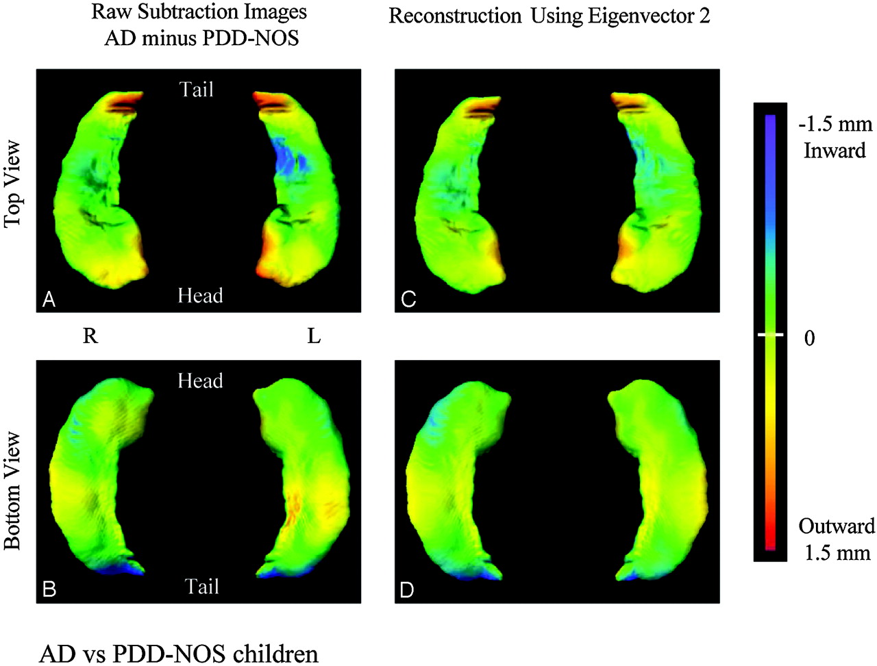
The left column (A and B) shows the group difference between the average hippocampal surfaces of the PDD-NOS group and children with AD (subgroups of the ASD group). The deformation from PDD-NOS to AD is painted as a flame scale (shown on the right of figure) onto the average surface of the PDD-NOS group with inward deformation in cooler colors and outward deformation in warmer colors. The right column (C and D) shows the group difference as reconstructed by using eigenvector 2, selected on the basis of logistic regression analysis to best discriminate children with AD from children with PDD-NOS. In comparison with Fig 1 (E and F), a similar pattern of shape deformation between children with AD and those with PDD-NOS illustrates the accentuation of characteristic ASD hippocampal-shape differences by the more severely affected children with AD.
The pattern of hippocampal shape differences that separated children with ASD from the typically developing children, and children with AD from children with PDD-NOS, was largely captured by the pattern of shape variation represented by eigenvector 2 and, to a lesser extent, by eigenvector 3. The physical representation of these dimensions of hippocampal shape variation is shown in Figures 1 and 2 and suggests an upward bending of the head and the tail of the hippocampus coupled with inward deformation of the medial aspect of the structure (ie, in the vicinity of the subiculum). Accentuation of shape differences within the ASD group was observed for the subgroup of more severely affected children with AD and can be visualized by comparing ASD–typically developing differences (reconstructed by using eigenvector 2 shown in Fig 1E, -F) to AD–PDD-NOS differences (Fig 2C, -D). The observed differences in hippocampal shape may be an indirect measure of abnormalities in the intrinsic cellular elements immediately below the structural surface. However, such alterations could also reflect changes in afferents or efferents from other structures. Alternatively, it is possible that shape changes within a particular structure may reflect physical constraints from structural alterations of adjacent brain structures, in the present case the amygdala. This consideration is of particular interest in the present study because we previously observed an enlargement of the amygdala in the children with AD, but not in those with PDD-NOS.17
ASD is a heterogeneous clinical syndrome, characterized by variable signs and symptoms, and has a variable clinical course and prognosis. As a consequence, there has been substantial interest in identifying ASD subgroups with distinct etiologies, pathophysiology, and treatment outcomes. A clinical subclassification of patients with ASD into those with AD and those with PDD-NOS was established in the psychiatric nomenclature to further this goal.2 This subclassification generally reflects a more severe clinical expression of autistic symptoms in the AD group. Shape analysis of the hippocampus revealed group differences between children with AD and those with PDD-NOS in the same direction as group differences between children with ASD and typically developing children. Thus, hippocampal shape alteration would seem to be associated with the core symptoms of autism. However, the grounds for clinical differentiation of these 2 related disorders are controversial (especially in young children), and the nature of their actual distinction remains poorly understood,35 in which case, the relationship observed in this study between hippocampal shape change and the AD subclassification could instead reflect the impact of greater MTL dysfunction and overall mental retardation.
It is also possible that group and subgroup differences in hippocampal shape observed in this study may be specific to the preschool age range evaluated and the narrow window of development represented in our sample. Brain growth is not yet complete in 3–4 year olds, and the volume of the hippocampus at this age has not yet reached typical adult size. However, because this age represents the initial developmental time point of our ongoing longitudinal study of the children with ASD, stability of hippocampal shape alterations with time are amenable to future study.
In prior work, we used large-deformation high-dimensional brain mapping to characterize hippocampal shape in adults with MTL epilepsy and also found an inward deformation of the medial aspect of the hippocampal surface (in the vicinity of the subiculum).20-21 Because a history of seizures was exclusionary for participation in the present study, the similarity observed between hippocampal shape alterations in children with ASD and adults with MTL epilepsy cannot be based on the presence of an existing seizure disorder in the children with ASD. Furthermore, any relationship between pre-existing structural abnormalities of the hippocampus and future onset of seizures in children with neurodevelopmental disorders has not been established. However, children with ASD, and in particular the children with AD or those having a lower IQ, are at high risk for seizures as they grow older.6,7 Thus, it is intriguing, though highly speculative, to consider whether the observed hippocampal shape differences may represent a risk factor for future seizure onset. Because longitudinal follow-up of the children reported in this study is ongoing and because 15%–38% of the children with ASD are expected to develop seizures during adolescence, the extent to which these hippocampal shape alterations may be related to future risk of seizure onset is testable through prospective evaluation.
Conclusions
Quantitative measures of hippocampal shape by using large-deformation high-dimensional brain mapping were found to distinguish children with ASD from typically developing children and, within the ASD sample, children with AD from those with PDD-NOS. As further predicted, hippocampal-shape alterations in the children with ASD were related to overall measures of intellectual function and performance deficits on specific tests of MTL function. There was good sensitivity for detecting characteristic shape alterations in the affected children but low specificity for diagnostic classification. The pattern of hippocampal-shape variations observed in children with ASD, and more specifically in the children with AD, was similar to that previously found in adults with MTL epilepsy. Although children with ASD are at high risk for onset of seizures as they enter adolescence, any increased vulnerability conferred by this pattern of hippocampal shape alterations remains speculative.
Acknowledgments
We gratefully acknowledge the contributions of the Diagnostic and Statistical Cores of the Autism Program Project and the parents and their children who participated in this study.
Footnotes
This research was supported by a grant from the Cure Autism Now Foundation and by a program project grant (PO1HD34565), which is part of the National Institute of Child Health and Human Development/National Institute on Deafness and Other Communication Disorders Collaborative Program of Excellence in Autism. Further support for this research came from Studies to Advance Autism Research and Treatment Center (U54 MH066399) and Conte Center (P50 MH071616) through the National Institute of Mental Health.
References
- Received April 7, 2006.
- Accepted after revision June 16, 2006.
- Copyright © American Society of Neuroradiology
In this issue


Jump to section
Related Articles
Cited By...
- The autism spectrum disorder risk gene NEXMIF alters hippocampal CA1 cellular and network dynamics
- Impaired spatial memory in adult vitamin D deficient BALB/c mice is associated with reductions in spine density, nitric oxide, and neural nitric oxide synthase in the hippocampus
- The Hippocampus and Social Impairment in Psychiatric Disorders