Advertisement

Main menu

  • Home
  • Content
    • Current Issue
    • Accepted Manuscripts
    • Article Preview
    • Past Issue Archive
    • AJNR Case Collection
    • Case of the Week Archive
    • Classic Case Archive
    • Case of the Month Archive
  • Special Collections
    • Spinal CSF Leak Articles (Jan 2020-June 2024)
    • 2024 AJNR Journal Awards
    • Most Impactful AJNR Articles
  • Multimedia
    • AJNR Podcast
    • AJNR Scantastics
    • Video Articles
  • For Authors
    • Submit a Manuscript
    • Author Policies
    • Fast publishing of Accepted Manuscripts
    • Graphical Abstract Preparation
    • Manuscript Submission Guidelines
    • Imaging Protocol Submission
    • Submit a Case for the Case Collection
  • About Us
    • About AJNR
    • Editorial Board
  • More
    • Become a Reviewer/Academy of Reviewers
    • Subscribers
    • Permissions
    • Alerts
    • Feedback
    • Advertisers
    • ASNR Home
  • Other Publications
    • ajnr

User menu

  • Alerts
  • Log in

Search

  • Advanced search
American Journal of Neuroradiology

American Journal of Neuroradiology

ASHNR American Society of Functional Neuroradiology ASHNR American Society of Pediatric Neuroradiology ASSR
  • Alerts
  • Log in

Advanced Search

  • Home
  • Content
    • Current Issue
    • Accepted Manuscripts
    • Article Preview
    • Past Issue Archive
    • AJNR Case Collection
    • Case of the Week Archive
    • Classic Case Archive
    • Case of the Month Archive
  • Special Collections
    • Spinal CSF Leak Articles (Jan 2020-June 2024)
    • 2024 AJNR Journal Awards
    • Most Impactful AJNR Articles
  • Multimedia
    • AJNR Podcast
    • AJNR Scantastics
    • Video Articles
  • For Authors
    • Submit a Manuscript
    • Author Policies
    • Fast publishing of Accepted Manuscripts
    • Graphical Abstract Preparation
    • Manuscript Submission Guidelines
    • Imaging Protocol Submission
    • Submit a Case for the Case Collection
  • About Us
    • About AJNR
    • Editorial Board
  • More
    • Become a Reviewer/Academy of Reviewers
    • Subscribers
    • Permissions
    • Alerts
    • Feedback
    • Advertisers
    • ASNR Home
  • Follow AJNR on Twitter
  • Visit AJNR on Facebook
  • Follow AJNR on Instagram
  • Join AJNR on LinkedIn
  • RSS Feeds

Welcome to the new AJNR, Updated Hall of Fame, and more. Read the full announcements.


AJNR is seeking candidates for the position of Associate Section Editor, AJNR Case Collection. Read the full announcement.


LetterLetter

Catheter Angiography, MR Angiography, and MR Perfusion in Posterior Reversible Encephalopathy Syndrome

R. Schwartz, R. Mulkern, S. Vajapeyam and D.F. Kacher
American Journal of Neuroradiology February 2009, 30 (2) e19; DOI: https://doi.org/10.3174/ajnr.A1285
R. Schwartz
  • Find this author on Google Scholar
  • Find this author on PubMed
  • Search for this author on this site
R. Mulkern
  • Find this author on Google Scholar
  • Find this author on PubMed
  • Search for this author on this site
S. Vajapeyam
  • Find this author on Google Scholar
  • Find this author on PubMed
  • Search for this author on this site
D.F. Kacher
  • Find this author on Google Scholar
  • Find this author on PubMed
  • Search for this author on this site
  • Article
  • Figures & Data
  • Info & Metrics
  • Responses
  • References
  • PDF
Loading

In their article, “Catheter Angiography, MR Angiography, and MR Perfusion in Posterior Reversible Encephalopathy Syndrome” (PRES), Drs Bartynski and Boardman1 describe their findings in hypertensive patients with PRES who were studied retrospectively with MR perfusion imaging. The authors have done a commendable job in displaying and analyzing their data and have provided interesting and thoughtful theories to support their findings.

A few words of caution are necessary, however, when interpreting perfusion data under these circumstances. The authors reported that perfusion MR imaging in PRES demonstrates reduced perfusion to the brain. However, it is important to recognize that PRES is a dynamic process, characterized by rapid and transient changes in brain hemodynamics. Bartynski and Boardman performed a retrospective analysis, and several of their patients were examined between 1 and 3 days after their hypertensive crisis. Observations made subsequent to the initiating event, especially after the patient has been treated, may not be reflective of the local processes occurring at the time when the edema was first produced, and this may result in spurious conclusions with regard to the etiology of PRES.

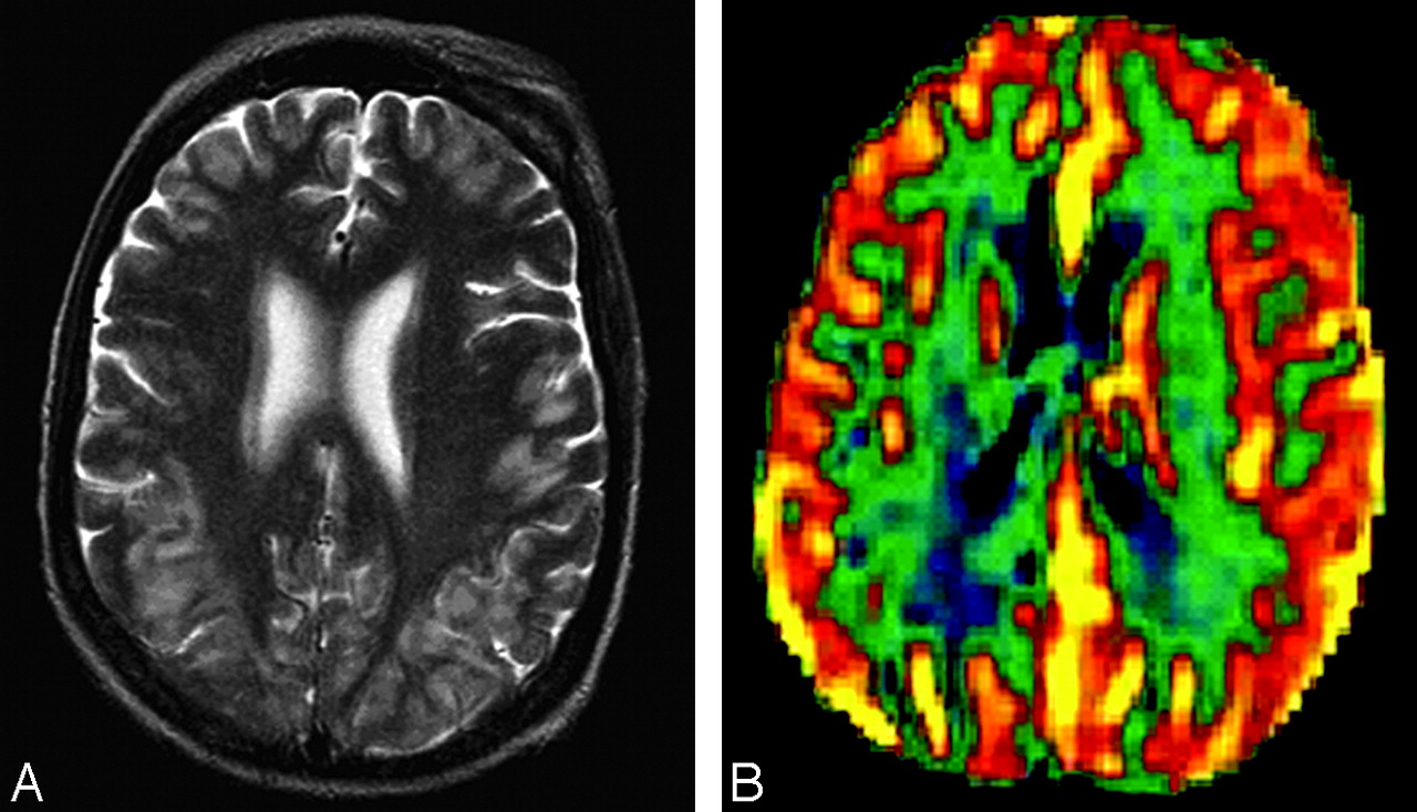
As an example, Figs 1 and 2 present 2 sets of data obtained in a patient with labile hypertension whom we studied with MR perfusion imaging; these data reflect the wide variation in intracranial perfusion that can occur with PRES in the setting of fluctuations in mean arterial blood pressures (MAP). A 41-year-old woman with a B-cell lymphoproliferative disorder, status post unrelated bone marrow transplant, was admitted with seizures and acute hypertension and was scanned within 24 hours after she received antihypertensive medication; her systemic blood pressure at the time of the first scanning was 150/90 mm Hg (MAP, 110.0 mm Hg).

While in the hospital, the patient developed renal failure and accelerated hypertension, accompanied by worsening seizures. She was scanned again within an hour of this episode, when her blood pressure was 230/130 mm Hg (MAP, 163.3 mm Hg).

We have found that brain perfusion in PRES often is markedly increased in the acute hypertensive setting, but after blood pressures are reduced, perfusion also tends to decrease; indeed, the presence of interstitial or intracellular edema would be expected to reduce local perfusion under any circumstance. Observations of perfusion data in PRES, therefore, must be placed in the appropriate temporal framework to avoid misinterpretation of the findings. A prospective study in which blood pressures are measured at the time of MR perfusion imaging would be necessary to address these issues.

Fig 1.
  • Download figure
  • Open in new tab
  • Download powerpoint
Fig 1.

A, Axial T2-weighted image. Signal-intensity abnormalities are present in the subcortical white matter of the occipital lobes. B, Regional cerebral blood volume colorized map shows mild hyperperfusion at multiple cortical sites. Reprinted from Schwartz et al.2

Fig 2.
  • Download figure
  • Open in new tab
  • Download powerpoint
Fig 2.

A, Extensive hyperintense T2 signal intensity is noted in the subcortical white matter and cortex of the occipital and parietal lobes and, to a lesser degree, in the frontal lobes. B, Regional cerebral blood volume colorized map shows markedly increased perfusion throughout the brain, with maximal perfusion shown posteriorly. Reprinted from Schwartz et al.2

References

  1. ↵
    Bartynski WS, Boardman JF. Catheter angiography, MR angiography, and MR perfusion in posterior reversible encephalopathy syndrome. AJNR Am J Neuroradiol 2008;29:447–55
    Abstract/FREE Full Text
  2. ↵
    Schwartz RB, Vandepayam S, Mulkern RV. Hypertensive encephlaopathy: perfusion MR shows increased cerebral blood flow and disturbance of autoregulation to the occipital lobes. Paper presented at: Annual Meeting of the American Society of Neuroradiology; June 7–14,2003; Washington, DC
  • Copyright © American Society of Neuroradiology
PreviousNext
Back to top

In this issue

American Journal of Neuroradiology: 30 (2)
American Journal of Neuroradiology
Vol. 30, Issue 2
February 2009
  • Table of Contents
  • Index by author
Advertisement
Print
Download PDF
Email Article

Thank you for your interest in spreading the word on American Journal of Neuroradiology.

NOTE: We only request your email address so that the person you are recommending the page to knows that you wanted them to see it, and that it is not junk mail. We do not capture any email address.

Enter multiple addresses on separate lines or separate them with commas.
Catheter Angiography, MR Angiography, and MR Perfusion in Posterior Reversible Encephalopathy Syndrome
(Your Name) has sent you a message from American Journal of Neuroradiology
(Your Name) thought you would like to see the American Journal of Neuroradiology web site.
CAPTCHA
This question is for testing whether or not you are a human visitor and to prevent automated spam submissions.
Cite this article
R. Schwartz, R. Mulkern, S. Vajapeyam, D.F. Kacher
Catheter Angiography, MR Angiography, and MR Perfusion in Posterior Reversible Encephalopathy Syndrome
American Journal of Neuroradiology Feb 2009, 30 (2) e19; DOI: 10.3174/ajnr.A1285

Citation Manager Formats

  • BibTeX
  • Bookends
  • EasyBib
  • EndNote (tagged)
  • EndNote 8 (xml)
  • Medlars
  • Mendeley
  • Papers
  • RefWorks Tagged
  • Ref Manager
  • RIS
  • Zotero
0 Responses
Respond to this article
Share
Bookmark this article
Catheter Angiography, MR Angiography, and MR Perfusion in Posterior Reversible Encephalopathy Syndrome
R. Schwartz, R. Mulkern, S. Vajapeyam, D.F. Kacher
American Journal of Neuroradiology Feb 2009, 30 (2) e19; DOI: 10.3174/ajnr.A1285
del.icio.us logo Twitter logo Facebook logo Mendeley logo
  • Tweet Widget
  • Facebook Like
  • Google Plus One
Purchase

Jump to section

  • Article
    • References
  • Figures & Data
  • Info & Metrics
  • Responses
  • References
  • PDF

Related Articles

  • No related articles found.
  • PubMed
  • Google Scholar

Cited By...

  • Posterior reversible encephalopathy syndrome (PRES) in classic Hodgkins lymphoma, complicated by anthracycline-induced cardiomyopathy
  • Crossref (15)
  • Google Scholar

This article has been cited by the following articles in journals that are participating in Crossref Cited-by Linking.

  • Arterial spin labeling MRI: Clinical applications in the brain
    Nicholas A. Telischak, John A. Detre, Greg Zaharchuk
    Journal of Magnetic Resonance Imaging 2015 41 5
  • Posterior Reversible Encephalopathy Syndrome
    Dimitre Staykov, Stefan Schwab
    Journal of Intensive Care Medicine 2012 27 1
  • Posterior Reversible Encephalopathy Syndrome
    Jeffrey B. Rykken, Alexander M. McKinney
    Seminars in Ultrasound, CT and MRI 2014 35 2
  • Asymmetric Posterior Reversible Encephalopathy Syndrome Complicating Hemodynamic Augmentation for Subarachnoid Hemorrhage-Associated Cerebral Vasospasm
    Barbara Voetsch, Nicholas Tarlov, Thanh N. Nguyen, Christina DeFusco, Glenn D. Barest, Alexander Norbash, Deborah M. Green, Joseph D. Burns
    Neurocritical Care 2011 15 3
  • Posterior Reversible Encephalopathy Syndrome and Reversible Cerebral Vasoconstriction Syndrome
    Alex Levitt, Richard Zampolin, Judah Burns, Jacqueline A. Bello, Shira E. Slasky
    Radiologic Clinics of North America 2019 57 6
  • Posterior Reversible Encephalopathy Syndrome: A Noteworthy Syndrome in End-Stage Renal Disease Patients
    Eleni Ermeidi, Olga Balafa, Giorgos Spanos, Anastasia Zikou, Maria Argyropoulou, Kostas C. Siamopoulos
    Nephron Clinical Practice 2013 123 3-4
  • Posterior reversible encephalopathy syndrome in a postpartum hemorrhagic woman without hypertension
    Chon-Fu Lio, Ying-Hua Lee, Hung-Yen Chan, Chang-Ching Yu, Nan-Jing Peng, Hung-Pin Chan
    Medicine 2017 96 16
  • Posterior reversible encephalopathy syndrome and reversible cerebral vasoconstriction syndrome associated spinal subdural hematoma
    Hanfeng Chen, Ziqi Xu, Yuan Yuan
    Medicine 2020 99 31
  • Imaging of Reversible Cerebral Vasoconstriction Syndrome and Posterior Reversible Encephalopathy Syndrome
    Bilal Battal, Mauricio Castillo
    Neuroimaging Clinics of North America 2024 34 1
  • Cerebellar variant of posterior reversible encephalopathy syndrome (PRES) after coil embolization for the hemorrhagic dissecting aneurysm
    Nobutaka Horie, Minoru Morikawa, Naoki Kitagawa, Izumi Nagata
    Acta Neurochirurgica 2011 153 5

More in this TOC Section

  • Brain AVM’s Nidus: What if We Hadn’t Understood Anything?
  • Letter to the Editor regarding “Automated Volumetric Software in Dementia: Help or Hindrance to the Neuroradiologist?”
  • Reply:
Show more Letters

Similar Articles

Advertisement

Indexed Content

  • Current Issue
  • Accepted Manuscripts
  • Article Preview
  • Past Issues
  • Editorials
  • Editors Choice
  • Fellow Journal Club
  • Letters to the Editor

Cases

  • Case Collection
  • Archive - Case of the Week
  • Archive - Case of the Month
  • Archive - Classic Case

Special Collections

  • Special Collections

Resources

  • News and Updates
  • Turn around Times
  • Submit a Manuscript
  • Author Policies
  • Manuscript Submission Guidelines
  • Evidence-Based Medicine Level Guide
  • Publishing Checklists
  • Graphical Abstract Preparation
  • Imaging Protocol Submission
  • Submit a Case
  • Become a Reviewer/Academy of Reviewers
  • Get Peer Review Credit from Publons

Multimedia

  • AJNR Podcast
  • AJNR SCANtastic
  • Video Articles

About Us

  • About AJNR
  • Editorial Board
  • Not an AJNR Subscriber? Join Now
  • Alerts
  • Feedback
  • Advertise with us
  • Librarian Resources
  • Permissions
  • Terms and Conditions

American Society of Neuroradiology

  • Not an ASNR Member? Join Now

© 2025 by the American Society of Neuroradiology All rights, including for text and data mining, AI training, and similar technologies, are reserved.
Print ISSN: 0195-6108 Online ISSN: 1936-959X

Powered by HighWire