Advertisement

Main menu

  • Home
  • Content
    • Current Issue
    • Accepted Manuscripts
    • Article Preview
    • Past Issue Archive
    • AJNR Case Collection
    • Case of the Week Archive
    • Classic Case Archive
    • Case of the Month Archive
  • Special Collections
    • Spinal CSF Leak Articles (Jan 2020-June 2024)
    • 2024 AJNR Journal Awards
    • Most Impactful AJNR Articles
  • Multimedia
    • AJNR Podcast
    • AJNR Scantastics
    • Video Articles
  • For Authors
    • Submit a Manuscript
    • Author Policies
    • Fast publishing of Accepted Manuscripts
    • Graphical Abstract Preparation
    • Manuscript Submission Guidelines
    • Imaging Protocol Submission
    • Submit a Case for the Case Collection
  • About Us
    • About AJNR
    • Editorial Board
  • More
    • Become a Reviewer/Academy of Reviewers
    • Subscribers
    • Permissions
    • Alerts
    • Feedback
    • Advertisers
    • ASNR Home
  • Other Publications
    • ajnr

User menu

  • Alerts
  • Log in
  • Log out

Search

  • Advanced search
American Journal of Neuroradiology

American Journal of Neuroradiology

ASHNR American Society of Functional Neuroradiology ASHNR American Society of Pediatric Neuroradiology ASSR
  • Alerts
  • Log in
  • Log out

Advanced Search

  • Home
  • Content
    • Current Issue
    • Accepted Manuscripts
    • Article Preview
    • Past Issue Archive
    • AJNR Case Collection
    • Case of the Week Archive
    • Classic Case Archive
    • Case of the Month Archive
  • Special Collections
    • Spinal CSF Leak Articles (Jan 2020-June 2024)
    • 2024 AJNR Journal Awards
    • Most Impactful AJNR Articles
  • Multimedia
    • AJNR Podcast
    • AJNR Scantastics
    • Video Articles
  • For Authors
    • Submit a Manuscript
    • Author Policies
    • Fast publishing of Accepted Manuscripts
    • Graphical Abstract Preparation
    • Manuscript Submission Guidelines
    • Imaging Protocol Submission
    • Submit a Case for the Case Collection
  • About Us
    • About AJNR
    • Editorial Board
  • More
    • Become a Reviewer/Academy of Reviewers
    • Subscribers
    • Permissions
    • Alerts
    • Feedback
    • Advertisers
    • ASNR Home
  • Follow AJNR on Twitter
  • Visit AJNR on Facebook
  • Follow AJNR on Instagram
  • Join AJNR on LinkedIn
  • RSS Feeds

Welcome to the new AJNR, Updated Hall of Fame, and more. Read the full announcements.


AJNR is seeking candidates for the position of Associate Section Editor, AJNR Case Collection. Read the full announcement.


LetterLETTER

Can We Perform Spinal 1H-MR Spectroscopy in Daily Clinical Practice?

L.C. Hygino da Cruz
American Journal of Neuroradiology December 2013, 34 (12) E128-E129; DOI: https://doi.org/10.3174/ajnr.A3809
L.C. Hygino da Cruz Jr
aClinicas CDPI and IRM Federal University of Rio de Janeiro Rio de Janeiro, Brazil
  • Find this author on Google Scholar
  • Find this author on PubMed
  • Search for this author on this site
  • Article
  • Figures & Data
  • Info & Metrics
  • Responses
  • References
  • PDF
Loading

I read with great interest the review article by Hock et al1 and agree with the authors that 1H-MR spectroscopy can be of great value in the assessment of a variety of spinal cord diseases. The difficulty in determining a specific diagnosis of these lesions is a challenge in their management. It is necessary to predict an appropriate treatment and avoid an unnecessary invasive intervention, such as biopsy or surgery. Thus, a reliable differential diagnosis can be obtained by using functional MR imaging techniques, which have been used for a long time to evaluate brain lesions. Technical limitations due to a low signal-to-noise ratio and the proximity to the bone and CSF interface have been described as important limitations to the use of functional MR images in the evaluation of spinal cord lesions.

We describe a case of a 79-year-old man with systemic non-Hodgkin lymphoma, with a solitary, solid, enhancing lesion in the cervical spine (Fig 1). Single-voxel 1H-MR spectroscopy was performed in a 1.5T MR imaging clinical scanner (Fig 2). Saturation bands, to reduce CSF pulsation artifacts and to suppress signals from outside the voxel, were placed surrounding the voxel. We did not use electrocardiography or respiratory triggering or gating. The voxel size used was 10 × 10 × 20 mm3. TE was 30 ms and TR was 1500 ms; the number of averages was 160, resulting in a total scanning time of 4 minutes.

Fig 1.
  • Download figure
  • Open in new tab
  • Download powerpoint
Fig 1.

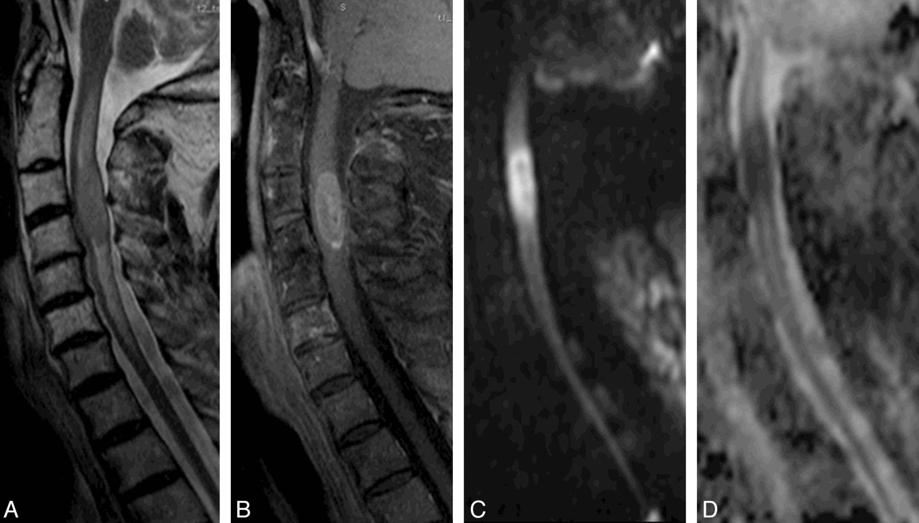
A solid spinal cord expansion lesion is demonstrated in a 79-year-old man with non-Hodgkin lymphoma. Sagittal T2-weighted image (A) shows an expansive hypointense lesion, surrounded by edema, which enhances after the intravenous contrast administration in the sagittal T1-weighted image (B). The lesion also has restricted diffusion on DWI (C), confirmed on the ADC map (D).

Fig 2.
  • Download figure
  • Open in new tab
  • Download powerpoint
Fig 2.

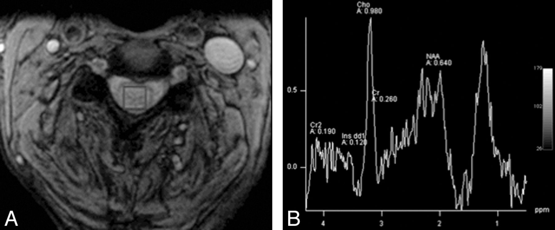
1H-MR spectroscopy was performed by placing the voxel within the lesion (A) and demonstrates characteristic findings of lymphoma: elevated lipid peaks, a high choline/creatine ratio, and a low N-acetylaspartate/creatine ratio (B).

To our knowledge and on the basis of the extended review of the literature shown by Hock et al,1 no previous publication described the MR spectroscopy changes observed in a spinal cord lymphoma. Intramedullary spinal lymphoma is a very rare neoplasm that accounts for approximately 3.3% of all CNS lymphomas, which consist of only 1% of all lymphomas in the body.2 Most are solitary lesions located in the cervical spine.

Previous reports used 1H-MR spectroscopy to evaluate brain lymphoma and basically found elevated lipid peaks, a high choline/creatine ratio, and a low N-acetylaspartate/creatine ratio.3 1H-MR spectroscopy findings of spinal lesions were similar to those described in the brain. In this particular case, we also performed diffusion-weighted MR imaging. The lesion demonstrated restricted diffusion, which has been described as related to the high cellularity and a high nuclear-cytoplasmic ratio, characteristic findings of lymphoma.3 On the basis of these findings and with the clinical history of the patient, we made a presumptive diagnosis of spinal cord lymphoma, avoiding an invasive diagnostic intervention.

We demonstrated the feasibility and the potential of applying MR spectroscopy to assess spinal cord lesions in daily clinical practice, even though some technical obstacles can be observed. We used a clinical 1.5T MR imaging scanner to obtain reliable MR spectra and in less than the average scanning time presented by Hock et al1; this scanning time, in our view, can be reliably used in clinical practice. Thus, one may conclude that it is not totally necessary to perform the examination in a 3T MR imaging scanner with a long scanning time.

In conclusion, we propose that by exploiting the higher SNR available with new 1.5T scanners with multichannel coils, MR spectra can be obtained in a 1.5T scanner with a scanning time as short as 4 minutes, without compromising quality. The clinical accuracy of this technique needs to be investigated in a broader clinical setting in patients with spinal cord lesions.

REFERENCES

  1. 1.↵
    1. Hock A,
    2. Henning A,
    3. Boesiger P,
    4. et al
    . 1H-MR spectroscopy in the human spinal cord. AJNR Am J Neuroradiol 2013;34:1682–89
    Abstract/FREE Full Text
  2. 2.↵
    1. Koeller KK,
    2. Rosenblum RS,
    3. Morrison AL
    . Neoplasms of the spinal cord and filum terminale: radiologic-pathologic correlation. Radiographics 2000;20:1721–49
    CrossRefPubMed
  3. 3.↵
    1. Haldorsen IS,
    2. Espeland A,
    3. Larsson EM
    . Central nervous system lymphoma: characteristic findings on traditional and advanced imaging. AJNR Am J Neuroradiol 2011;32:984–92
    Abstract/FREE Full Text
  • © 2013 by American Journal of Neuroradiology
PreviousNext
Back to top

In this issue

American Journal of Neuroradiology: 34 (12)
American Journal of Neuroradiology
Vol. 34, Issue 12
1 Dec 2013
  • Table of Contents
  • Index by author
  • Complete Issue (PDF)
Advertisement
Print
Download PDF
Email Article

Thank you for your interest in spreading the word on American Journal of Neuroradiology.

NOTE: We only request your email address so that the person you are recommending the page to knows that you wanted them to see it, and that it is not junk mail. We do not capture any email address.

Enter multiple addresses on separate lines or separate them with commas.
Can We Perform Spinal 1H-MR Spectroscopy in Daily Clinical Practice?
(Your Name) has sent you a message from American Journal of Neuroradiology
(Your Name) thought you would like to see the American Journal of Neuroradiology web site.
CAPTCHA
This question is for testing whether or not you are a human visitor and to prevent automated spam submissions.
Cite this article
L.C. Hygino da Cruz
Can We Perform Spinal 1H-MR Spectroscopy in Daily Clinical Practice?
American Journal of Neuroradiology Dec 2013, 34 (12) E128-E129; DOI: 10.3174/ajnr.A3809

Citation Manager Formats

  • BibTeX
  • Bookends
  • EasyBib
  • EndNote (tagged)
  • EndNote 8 (xml)
  • Medlars
  • Mendeley
  • Papers
  • RefWorks Tagged
  • Ref Manager
  • RIS
  • Zotero
0 Responses
Respond to this article
Share
Bookmark this article
Can We Perform Spinal 1H-MR Spectroscopy in Daily Clinical Practice?
L.C. Hygino da Cruz
American Journal of Neuroradiology Dec 2013, 34 (12) E128-E129; DOI: 10.3174/ajnr.A3809
del.icio.us logo Twitter logo Facebook logo Mendeley logo
  • Tweet Widget
  • Facebook Like
  • Google Plus One
Purchase

Jump to section

  • Article
    • REFERENCES
  • Figures & Data
  • Info & Metrics
  • Responses
  • References
  • PDF

Related Articles

  • Reply:
  • PubMed
  • Google Scholar

Cited By...

  • No citing articles found.
  • Crossref (1)
  • Google Scholar

This article has been cited by the following articles in journals that are participating in Crossref Cited-by Linking.

  • Functional Imaging in Oncology
    Mauricio Castillo, Majda M. Thurnher
    2014

More in this TOC Section

  • Brain AVM’s Nidus: What if We Hadn’t Understood Anything?
  • Letter to the Editor regarding “Automated Volumetric Software in Dementia: Help or Hindrance to the Neuroradiologist?”
  • Reply:
Show more Letters

Similar Articles

Advertisement

Indexed Content

  • Current Issue
  • Accepted Manuscripts
  • Article Preview
  • Past Issues
  • Editorials
  • Editors Choice
  • Fellow Journal Club
  • Letters to the Editor

Cases

  • Case Collection
  • Archive - Case of the Week
  • Archive - Case of the Month
  • Archive - Classic Case

Special Collections

  • Special Collections

Resources

  • News and Updates
  • Turn around Times
  • Submit a Manuscript
  • Author Policies
  • Manuscript Submission Guidelines
  • Evidence-Based Medicine Level Guide
  • Publishing Checklists
  • Graphical Abstract Preparation
  • Imaging Protocol Submission
  • Submit a Case
  • Become a Reviewer/Academy of Reviewers
  • Get Peer Review Credit from Publons

Multimedia

  • AJNR Podcast
  • AJNR SCANtastic
  • Video Articles

About Us

  • About AJNR
  • Editorial Board
  • Not an AJNR Subscriber? Join Now
  • Alerts
  • Feedback
  • Advertise with us
  • Librarian Resources
  • Permissions
  • Terms and Conditions

American Society of Neuroradiology

  • Not an ASNR Member? Join Now

© 2025 by the American Society of Neuroradiology All rights, including for text and data mining, AI training, and similar technologies, are reserved.
Print ISSN: 0195-6108 Online ISSN: 1936-959X

Powered by HighWire