
Article Figures & Data
Figures
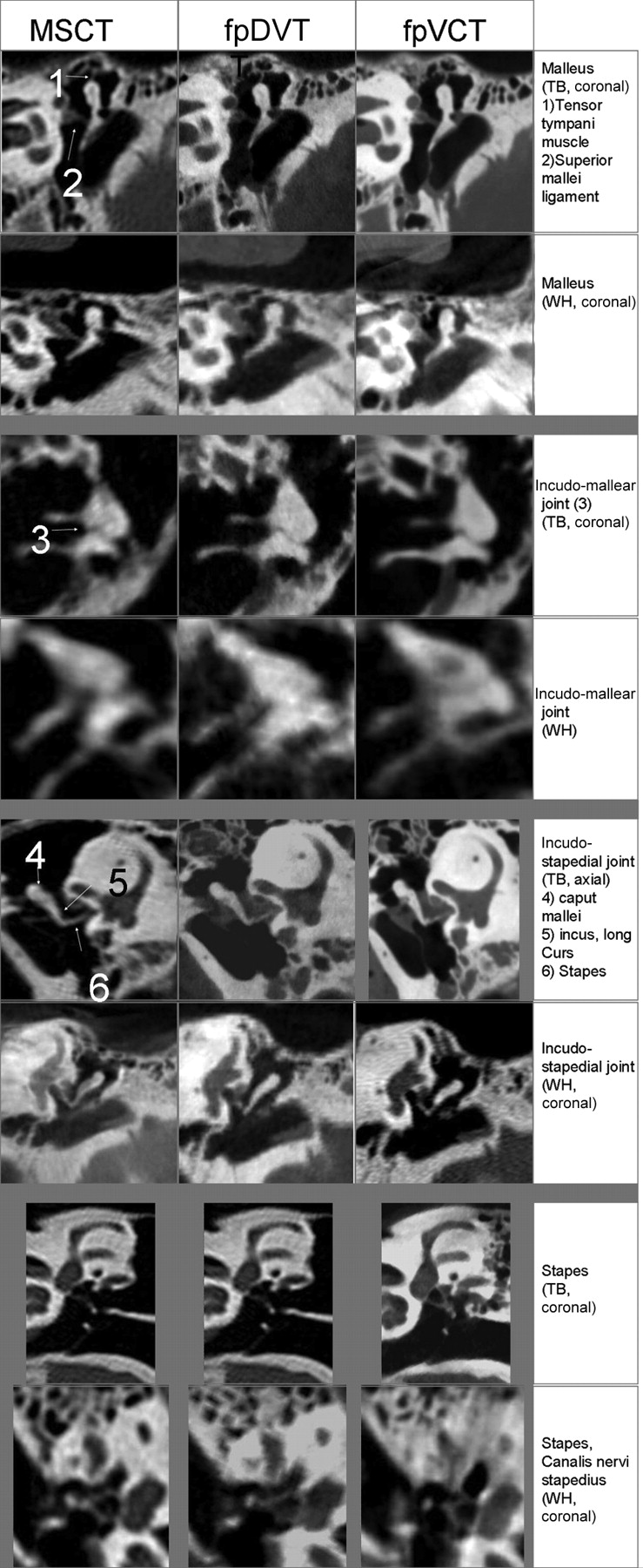
- Fig 1.
Images of the incus, ambomalleolar joint, incostapedial joint, and the stapes in explanted temporal bone (TB) and whole-head specimens (WH) obtained by 3 different scanners.
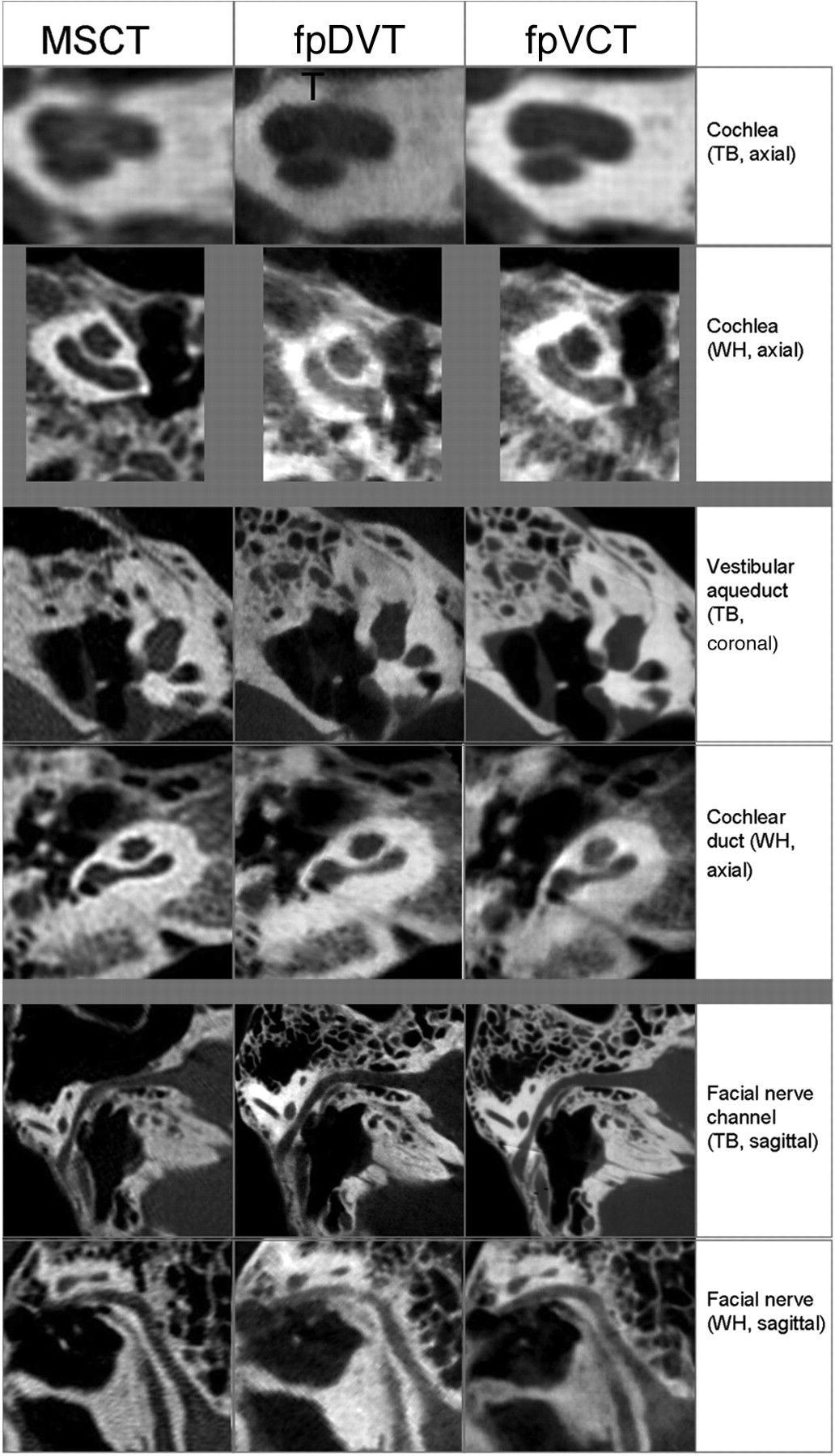
- Fig 2.
Images of the cochlea, vestibular duct, cochlear duct, and facial nerve in explanted temporal bone (TB) and whole-head specimens (WH).
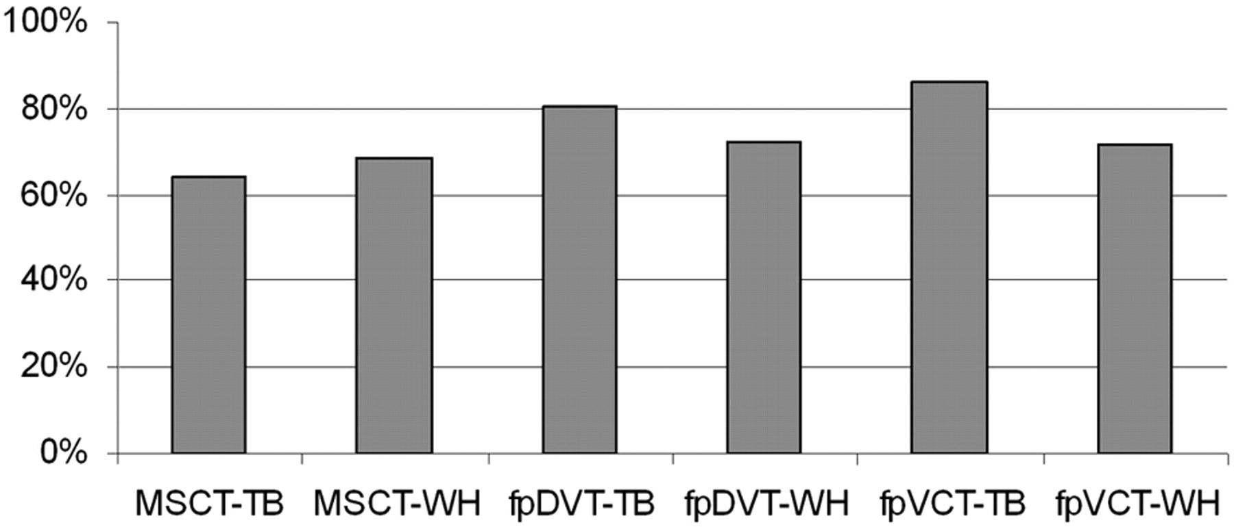
- Fig 3.
Three different examiners rated image quality with regard to delimitation of 38 different anatomic substructures as viewed in explanted temporal bones (TB) and whole-head (WH) specimens, each specimen being scanned with an MSCT scanner, an fpDVT scanner, and an fpVCT scanner.


In this issue


Jump to section
Related Articles
Cited By...
- Comparison of a Photon-Counting-Detector CT with an Energy-Integrating-Detector CT for Temporal Bone Imaging: A Cadaveric Study
- Flat Panel Angiography in the Cross-Sectional Imaging of the Temporal Bone: Assessment of Image Quality and Radiation Dose Compared with a 64-Section Multisection CT Scanner