
Article Figures & Data
Figures
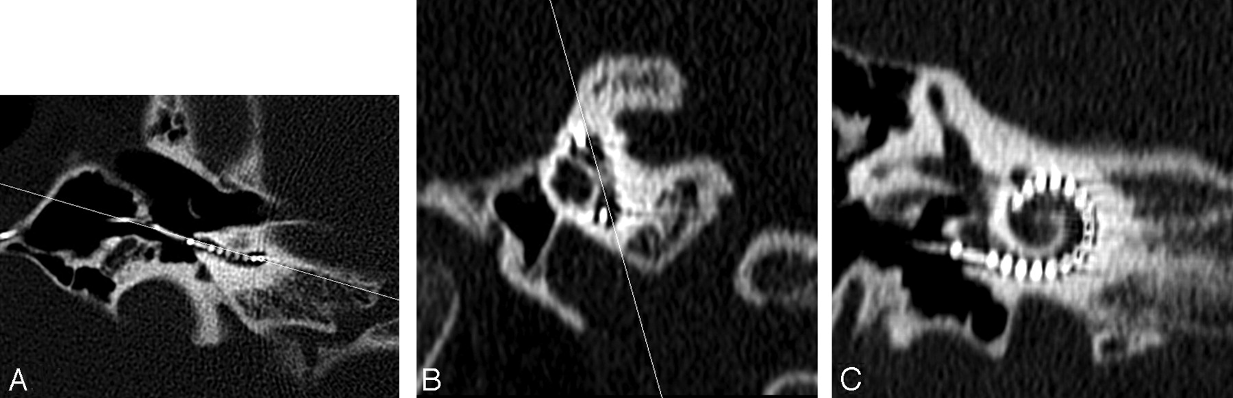
- Fig 1.
Axial CT image (A) through the inferior segment of the basal turn in a postimplant cochlea with a line shown angled to the basal turn. An oblique sagittal reformat (B) is obtained perpendicular to the line through the basal turn in (A). A line joining the midsuperior and -inferior segments of the basal turn in (B) is used to obtain the double oblique coronal reformat of the basal turn shown in (C).
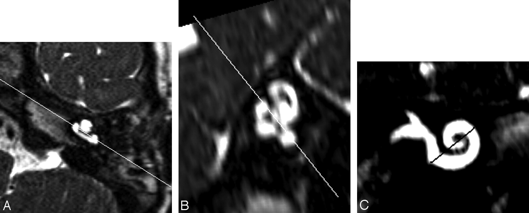
- Fig 2.
Axial 3D-DRIVE MR image (A) through the inferior segment of the basal turn in a preimplant cochlea with a line shown angled to the basal turn. An oblique sagittal reformat (B) is obtained perpendicular to the line through the basal turn in (A). A line joining the mid superior and inferior segments of the basal turn in (B) is used to obtain the double oblique coronal reformat of the basal turn shown in (C).
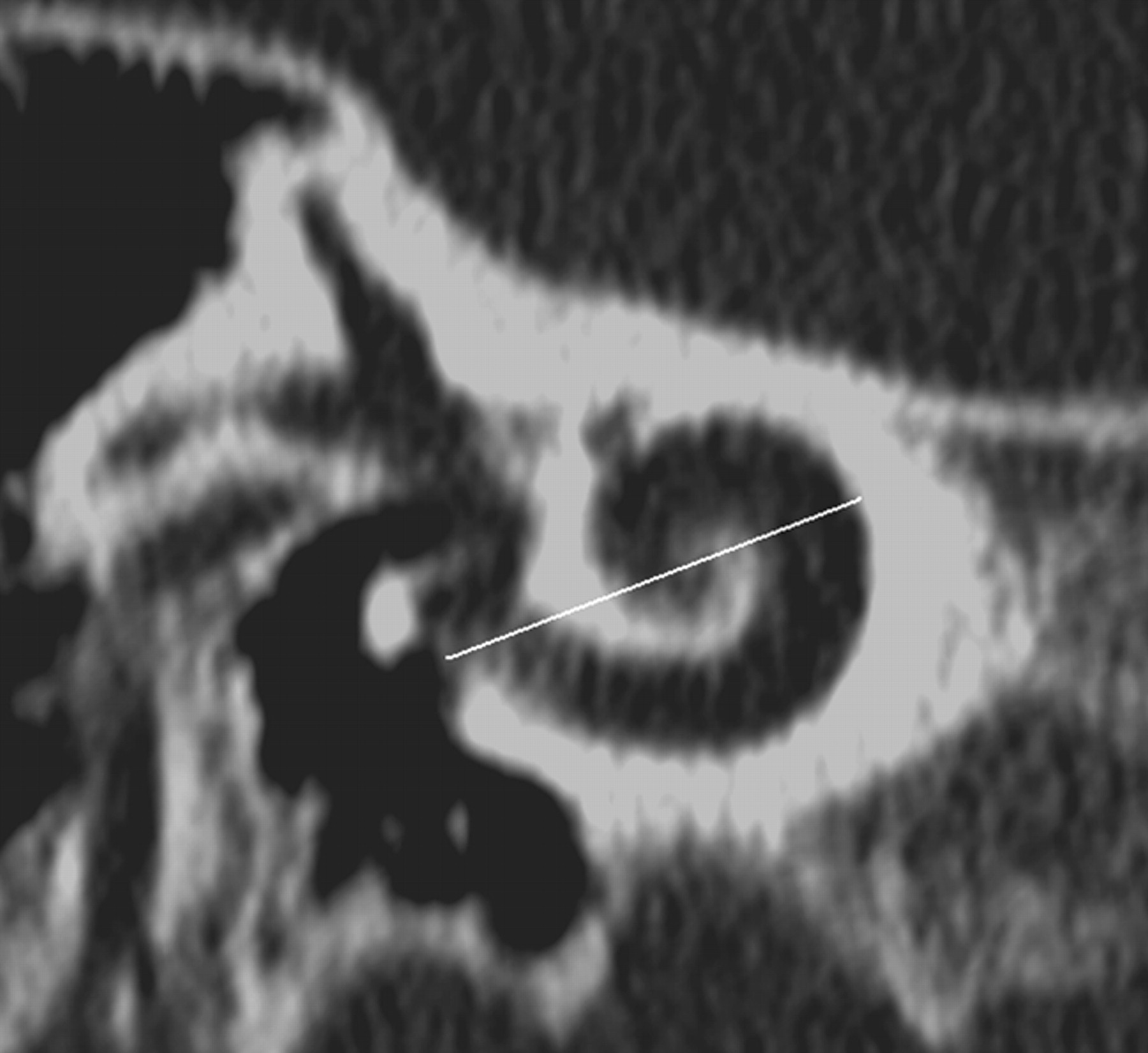
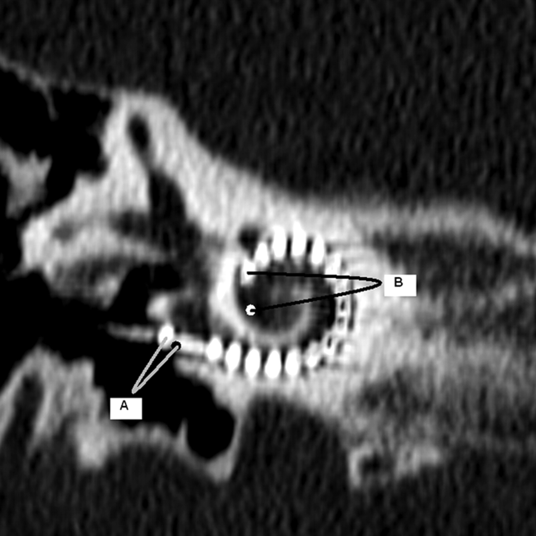
- Fig 3.
CD measurement is illustrated on a double oblique 1-mm thick reformatted CT through the basal turn of the cochlea. CD is measured from the midround window through the midmodiolar axis (confirmed by scrolling through adjacent images in this case) to the opposite wall of the basal turn. The midround window was identified on the reformatted MR imaging section by referring to an adjacent, more posterior image where the midinferior point of the round window “keyhole” could be extrapolated. The line indicating the CD measurement for MR imaging is shown in Fig 2C.
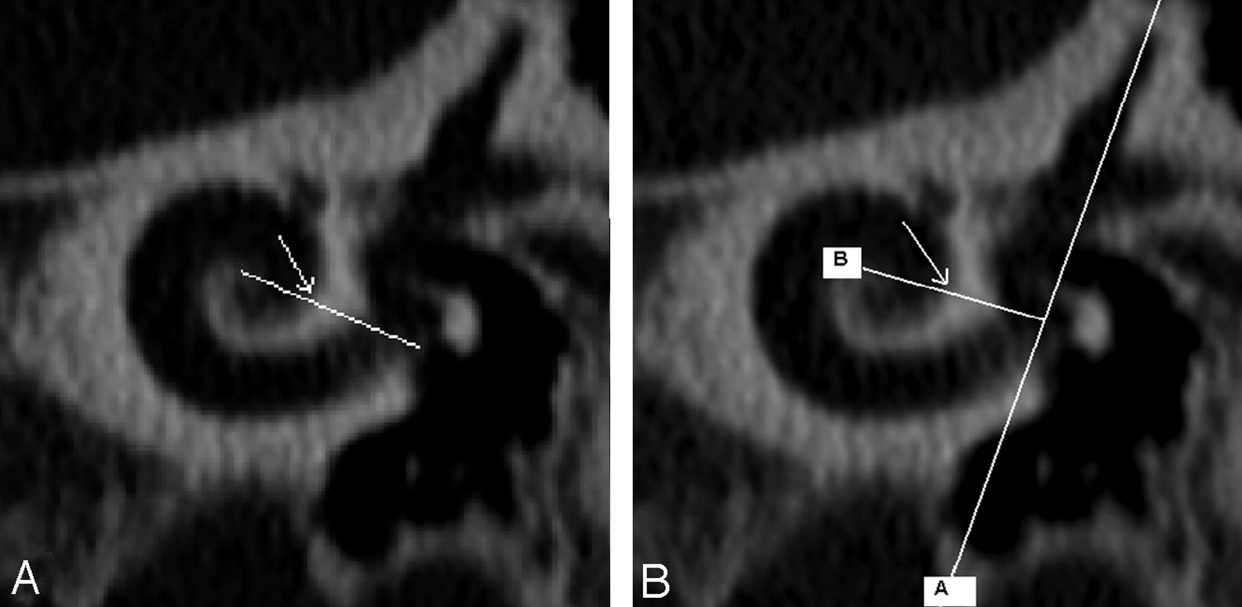
- Fig 4.
The distal reference (360° point) was defined in 2 ways according to Xu et al and Marsh et al, depending on where projected imaginary lines crossed the cochlear lumen [arrows in (A) and (B)]. For the Xu et al measurement (A), the imaginary line extended from the center of the cochlea to the midround window. For the Marsh et al measurement (B), the imaginary line extended from the center of the cochlea to a perpendicular vertical line through the superior semicircular canal and the vestibule.
- Fig 5.
The CALC 360 was measured by use of the distance from the reference electrode to the wire at the midround window (A, black mark indicates midround window) and the distance from the distal-most contact to the 360° point (B, white mark indicates the 360° point as per Xu et al and as seen in Fig 4A).The distance between the reference electrode and the distal-most contact was 19 mm; thus, it was possible to calculate the length of the array extending through 1 complete turn to the 360° point (CALC 360).


Tables
- Table 1:
Intraobserver reproducibility (mean difference [SD] and Pearson linear correlation coefficient) for CT and MR imaging cochlear dimensions measurements
Intraobserver Reproducibility (Observation 1-Observation 2) Mean Difference (SD) (mm) Intraobserver Reproducibility (Pearson R) CT (observer A/observer B) −0.03 (0.21)/0.06 (0.33) 0.85/0.55 MRI (observer A/observer B) −0.01 (0.23)/−0.04 (0.34) 0.87/0.67 - Table 2:
Interobserver reproducibility (mean difference [SD] and Pearson linear correlation coefficient) for CT and MR imaging cochlear dimensions measurements
Interobserver Reproducibility (Observer A-Observer B) Mean Difference (SD) (mm) Interobserver Reproducibility (Pearson R) CT −0.19 (0.28) 0.68 MRI −0.16 (0.26) 0.83 - Table 3:
Comparison of CT and MR imaging cochlear dimension measurements (mean difference [SD] and Pearson linear correlation coefficient)
Mean (SD) Difference (MR Imaging−CT) Pearson r A 0.044 (0.19) mm 0.8 B 0.023 (0.33) mm 0.4 A+B 0.07 (0.19) mm 0.75 - Table 4:
Comparison of EST 360 vs CALC 360 for Xu et al9 and Marsh et al17 methods (mean difference [SD], range, and Pearson linear correlation coefficient)
Xu et al9 Marsh et al17 Mean difference (SD) (and range): CALC 360 − EST 360 Pearson r Mean difference (SD) (and range): CALC 360 − EST 360 Pearson r CT A −0.79 (0.84) mm (−0.54–2.4) 0.67 −1.49 (1.04) mm (−1.6–2.9) 0.62 B −1.36 (0.90) mm (−0.43–2.9) 0.49 −2.05 (1.13) mm (−1.6–3.7) 0.47 A+B −1.06 (0.84) mm (−0.80–2.2) 0.61 −1.75 (1.09) mm (−1.6–3.2) 0.53 MR Imaging A −0.76 (0.81) mm (−0.70–2.3) 0.75 −1.45 (1.15) mm (−1.9–3.1) 0.57 B −1.30 (1.05) mm (−0.70–3.4) 0.52 −1.98 (1.29) mm (−1.9–3.7) 0.43 A+B −1.0 (0.87) mm (−0.70–2.9) 0.67 −1.72 (1.17) mm (−1.9–3.4) 0.52 Note:—EST 360 indicates an estimate of the insertion depth to the 360° point based on the cochlear distance measured; CALC 360, calculation of the length of the array extending through 1 complete turn to the 360° point on postoperative CT.


