Advertisement

Main menu

  • Home
  • Content
    • Current Issue
    • Accepted Manuscripts
    • Article Preview
    • Past Issue Archive
    • AJNR Case Collection
    • Case of the Week Archive
    • Classic Case Archive
    • Case of the Month Archive
  • Special Collections
    • Spinal CSF Leak Articles (Jan 2020-June 2024)
    • 2024 AJNR Journal Awards
    • Most Impactful AJNR Articles
  • Multimedia
    • AJNR Podcast
    • AJNR Scantastics
    • Video Articles
  • For Authors
    • Submit a Manuscript
    • Author Policies
    • Fast publishing of Accepted Manuscripts
    • Graphical Abstract Preparation
    • Manuscript Submission Guidelines
    • Imaging Protocol Submission
    • Submit a Case for the Case Collection
  • About Us
    • About AJNR
    • Editorial Board
  • More
    • Become a Reviewer/Academy of Reviewers
    • Subscribers
    • Permissions
    • Alerts
    • Feedback
    • Advertisers
    • ASNR Home
  • Other Publications
    • ajnr

User menu

  • Alerts
  • Log in

Search

  • Advanced search
American Journal of Neuroradiology

American Journal of Neuroradiology

ASHNR American Society of Functional Neuroradiology ASHNR American Society of Pediatric Neuroradiology ASSR
  • Alerts
  • Log in

Advanced Search

  • Home
  • Content
    • Current Issue
    • Accepted Manuscripts
    • Article Preview
    • Past Issue Archive
    • AJNR Case Collection
    • Case of the Week Archive
    • Classic Case Archive
    • Case of the Month Archive
  • Special Collections
    • Spinal CSF Leak Articles (Jan 2020-June 2024)
    • 2024 AJNR Journal Awards
    • Most Impactful AJNR Articles
  • Multimedia
    • AJNR Podcast
    • AJNR Scantastics
    • Video Articles
  • For Authors
    • Submit a Manuscript
    • Author Policies
    • Fast publishing of Accepted Manuscripts
    • Graphical Abstract Preparation
    • Manuscript Submission Guidelines
    • Imaging Protocol Submission
    • Submit a Case for the Case Collection
  • About Us
    • About AJNR
    • Editorial Board
  • More
    • Become a Reviewer/Academy of Reviewers
    • Subscribers
    • Permissions
    • Alerts
    • Feedback
    • Advertisers
    • ASNR Home
  • Follow AJNR on Twitter
  • Visit AJNR on Facebook
  • Follow AJNR on Instagram
  • Join AJNR on LinkedIn
  • RSS Feeds

Welcome to the new AJNR, Updated Hall of Fame, and more. Read the full announcements.


AJNR is seeking candidates for the position of Associate Section Editor, AJNR Case Collection. Read the full announcement.


Review ArticleHEAD & NECK
Open Access

“Finding a Voice”: Imaging Features after Phonosurgical Procedures for Vocal Fold Paralysis

B.A. Vachha, D.T. Ginat, P. Mallur, M. Cunnane and G. Moonis
American Journal of Neuroradiology September 2016, 37 (9) 1574-1580; DOI: https://doi.org/10.3174/ajnr.A4781
B.A. Vachha
aFrom the Department of Radiology (B.A.V.), Memorial Sloan Kettering Cancer Center, New York, New York
bDepartment of Radiology (B.A.V.), Massachusetts General Hospital, Harvard Medical School, Boston, Massachusetts
  • Find this author on Google Scholar
  • Find this author on PubMed
  • Search for this author on this site
  • ORCID record for B.A. Vachha
D.T. Ginat
cDepartment of Radiology (D.T.G.), University of Chicago, Chicago, Illinois
  • Find this author on Google Scholar
  • Find this author on PubMed
  • Search for this author on this site
  • ORCID record for D.T. Ginat
P. Mallur
dDepartment of Otology and Laryngology (P.M.), Beth Israel Deaconess Medical Center, Harvard Medical School, Boston, Massachusetts
  • Find this author on Google Scholar
  • Find this author on PubMed
  • Search for this author on this site
  • ORCID record for P. Mallur
M. Cunnane
eDepartment of Radiology (M.C.), Massachusetts Eye and Ear Infirmary, Harvard Medical School, Boston, Massachusetts
  • Find this author on Google Scholar
  • Find this author on PubMed
  • Search for this author on this site
  • ORCID record for M. Cunnane
G. Moonis
fDepartment of Radiology (G.M.), Columbia University Medical Center, New York, New York.
  • Find this author on Google Scholar
  • Find this author on PubMed
  • Search for this author on this site
  • ORCID record for G. Moonis
  • Article
  • Figures & Data
  • Info & Metrics
  • Responses
  • References
  • PDF
Loading

Article Figures & Data

Figures

  • Tables
  • Fig 1.
    • Download figure
    • Open in new tab
    • Download powerpoint
    Fig 1.

    Schematic of the main categories of procedures for treating vocal cord paralysis: injection laryngoplasty (A), medialization laryngoplasty (B), and arytenoid adduction (C).
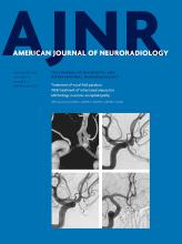
  • Fig 2.
    • Download figure
    • Open in new tab
    • Download powerpoint
    Fig 2.

    Hyaluronic acid gel. Axial (A) and coronal (B) contrast-enhanced CT at the level of the vocal folds demonstrates fluid attenuation material within the left vocal fold (black arrow).
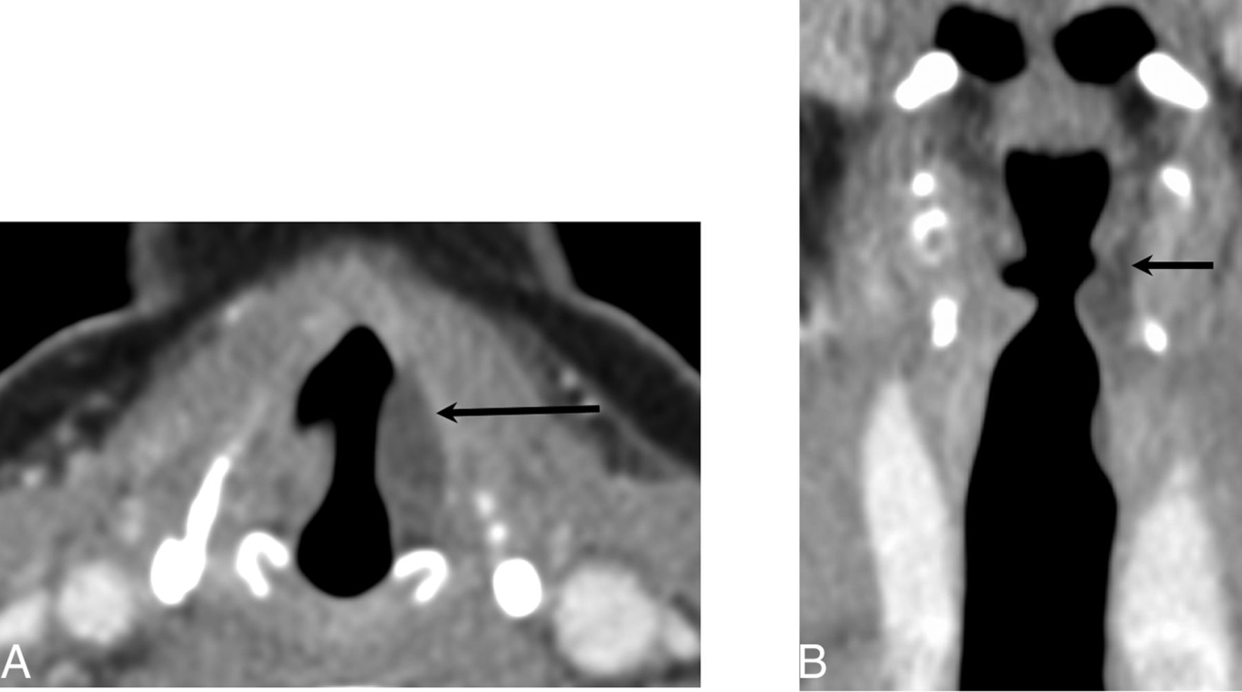
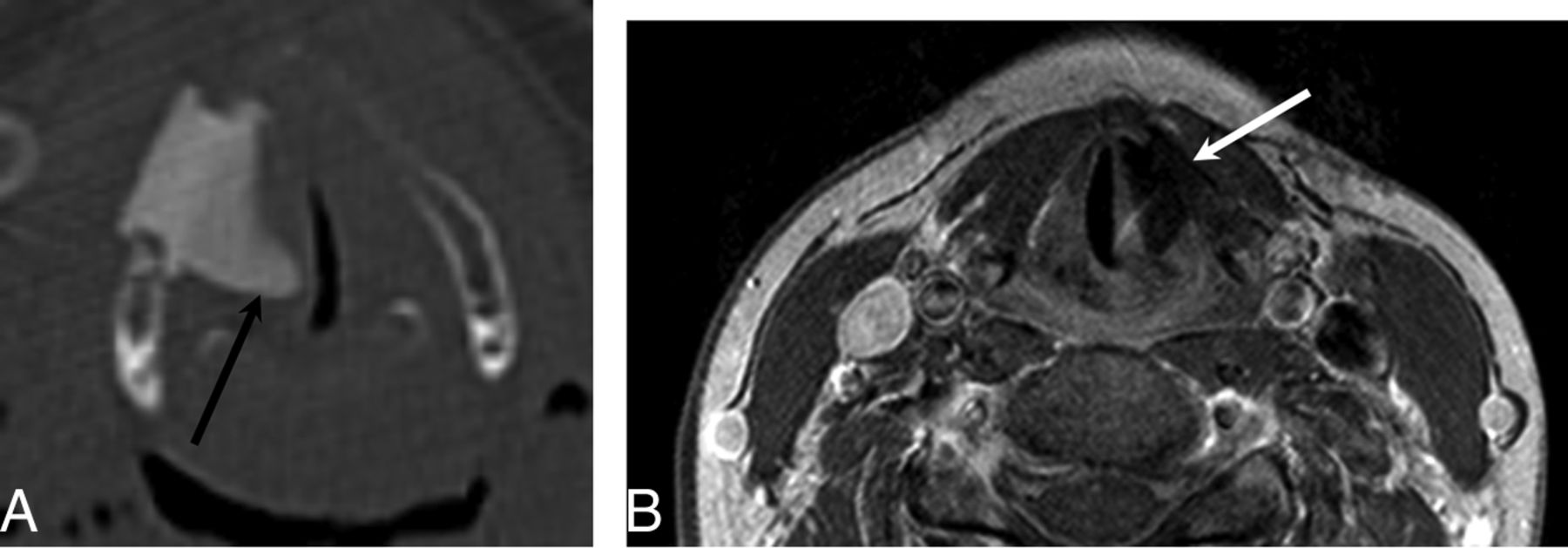
  • Fig 3.
    • Download figure
    • Open in new tab
    • Download powerpoint
    Fig 3.

    Micronized collagen (Cymetra) approximately 11 months after injection. A, Unenhanced T1-weighted image at the level of the true vocal folds demonstrates T1 hyperintensity within the right vocal fold (arrow; due to volume averaging, portions of the false cord are also partially seen). B, T2-weighted image demonstrates a hyperintense region in the true vocal fold on the right corresponding to the site of injection (arrow).
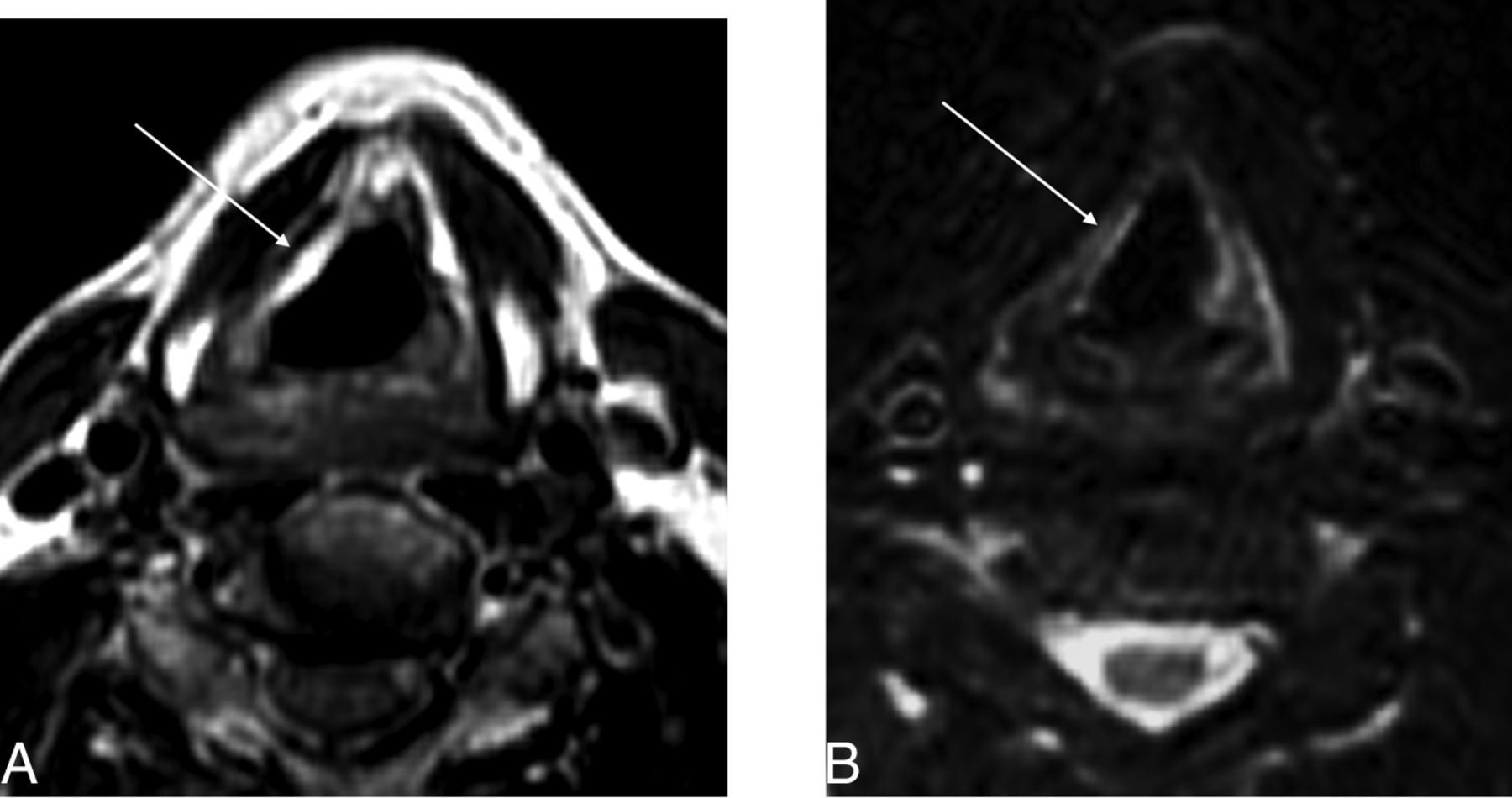
  • Fig 4.
    • Download figure
    • Open in new tab
    • Download powerpoint
    Fig 4.

    Calcium hydroxylapatite. A, Axial contrast-enhanced CT with a bone algorithm demonstrates hyperattenuating material in the region of the left vocal fold (black arrow). B, Axial positron-emission tomography demonstrates hypermetabolism at the site of the injected material, which can be a false-positive finding for malignant disease (white arrow).

  • Fig 5.
    • Download figure
    • Open in new tab
    • Download powerpoint
    Fig 5.

    Autologous fat. A, Coronal CT at the level of the vocal folds demonstrates transglottic carcinoma on the right (white dotted line). B, Coronal CT posttreatment shows low-attenuation material within the right vocal fold status post fat augmentation (arrow).

  • Fig 6.
    • Download figure
    • Open in new tab
    • Download powerpoint
    Fig 6.

    Teflon. A, Axial CT demonstrates hyperattenuating material in the bilateral vocal folds (arrows) consistent with Teflon injection. B, Axial CT in a different patient status post Teflon injection into the right vocal fold demonstrates a slightly nodular contour (arrowheads) consistent with granuloma formation, a common complication of Teflon.

  • Fig 7.
    • Download figure
    • Open in new tab
    • Download powerpoint
    Fig 7.

    Montgomery Implant. A, Axial CT demonstrates a Montgomery Implant (black arrow) and appropriate medialization of the right vocal cord. Note the triangular configuration of the Montgomery Implant. B, Axial contrast-enhanced T1-weighted MR imaging in a different patient demonstrates triangular hypointensity adjacent to the left vocal fold, representing the Montgomery Implant (white arrow), with appropriate medialization of left vocal fold.
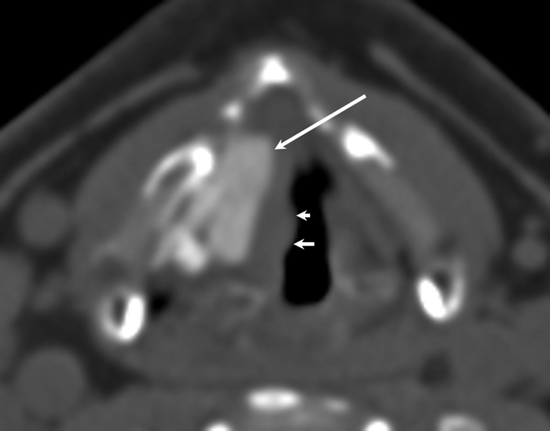
  • Fig 8.
    • Download figure
    • Open in new tab
    • Download powerpoint
    Fig 8.

    PTFE. Axial CT demonstrates hyperattenuating PTFE in the region of the right vocal fold with appropriate medialization of the right vocal fold. Note the slight nodular contour of the medial margin of the vocal fold (smaller arrows) indicating mild granuloma formation in this patient.

  • Fig 9.
    • Download figure
    • Open in new tab
    • Download powerpoint
    Fig 9.

    Malpositioned Montgomery Implant. Axial CT demonstrates an externally rotated Montgomery Implant, which is not flush against the thyroid window (arrow).
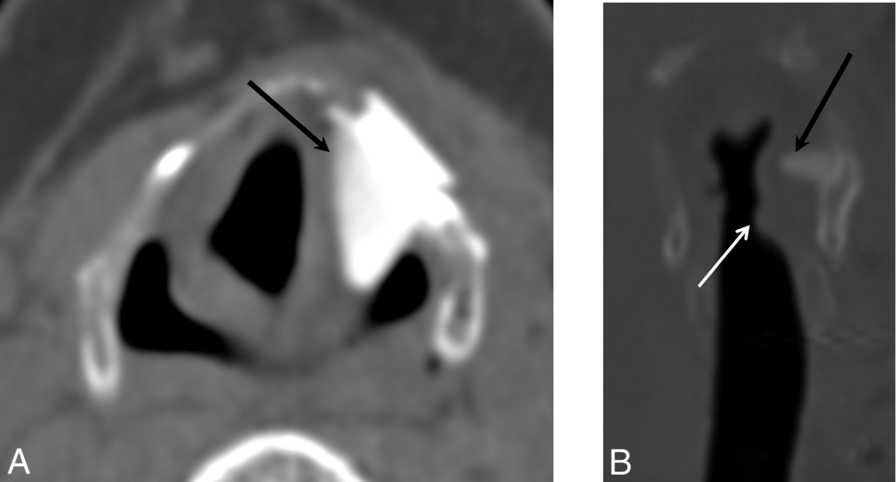
  • Fig 10.
    • Download figure
    • Open in new tab
    • Download powerpoint
    Fig 10.

    Malpositioned Montgomery Implant. Axial (A) and coronal (B) images demonstrate a Montgomery Implant that is displaced superiorly into the aryepiglottic fold and false cord (black arrow). White arrow indicates the appropriate level of the implant.
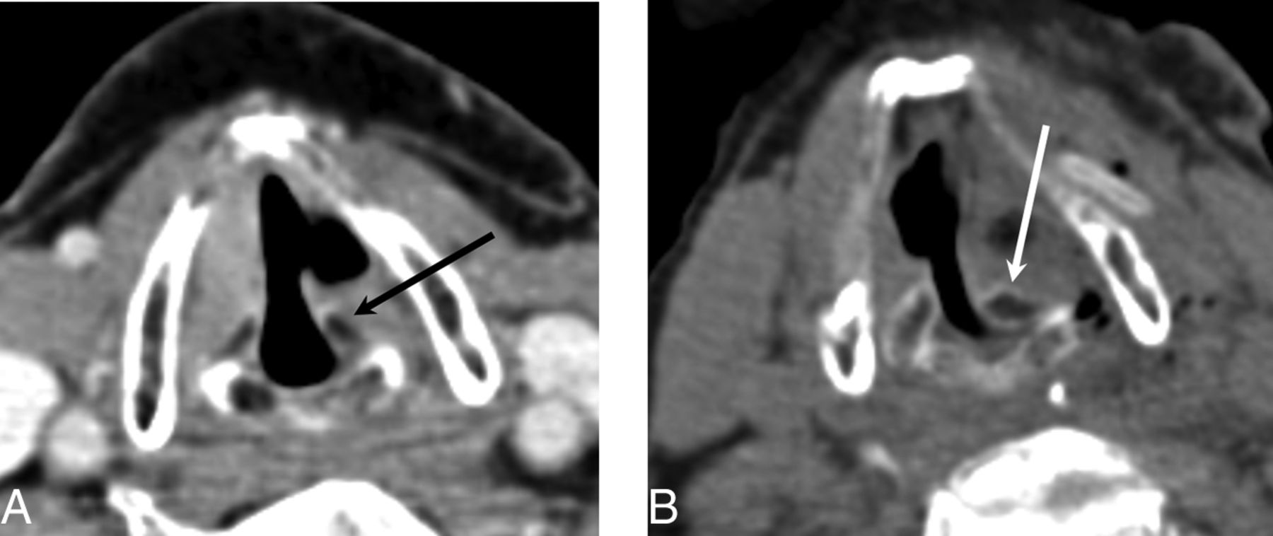
  • Fig 11.
    • Download figure
    • Open in new tab
    • Download powerpoint
    Fig 11.

    Arytenoid adduction. Preoperative axial CT image (A) and postoperative axial CT image (B) demonstrate the medially rotated (adducted) left arytenoid cartilage (white arrow) compared with the preoperative position (black arrow). Note that adduction turns the arytenoid in toward the vocal cord to ensure that no air leaks through the posterior glottic gap. This result helps patients achieve a more robust voice quality after the operation.

Tables

  • Figures
    • View popup
    Table 1:

    Overview of vocal cord procedures

    ProceduresIndicationsTechniquesImaging Findings
    Injection laryngoplastyTemporary correction of glottic incompetence due to unilateral vocal fold paralysis and long-term correction of mild-moderate glottic insufficiencyPercutaneous, transnasal, or peroral injection of filler material into the vocal cord or paraglottic spaceVaries depending on agent injected; also refer to the text and Table 2
    Medialization laryngoplastyPermanent correction; should be reserved for cases of vocal fold paralysis in which recovery of motion is definitively not expected (time >6 months from onset, surgical recurrent nerve sacrifice, or malignant invasion)Insertion of an implant into the vocal fold through a window in the thyroid, which results in displacement of the paralyzed vocal fold to a more medial positionThe implants typically used appear as hyperattenuating on CT and low signal on T1- and T2-weighted sequences, with a triangular shape on axial images; a defect in the adjacent thyroid cartilage may be visible
    Arytenoid adduction (adduction arytenopexy)To enhance posterior glottal closure in patients with paralytic dysphonia by reproducing lateral cricoarytenoid muscle function; can be performed in conjunction with medialization laryngoplastyThe inner perichondrium of the thyroid cartilage is opened and the muscular process of the arytenoid is identified and sutured to the thyroid or cricoid cartilageMedially rotated (adducted) arytenoid cartilage and narrowing of the posterior glottic gap
    • View popup
    Table 2:

    Description of selected agents used for injection laryngoplasty

    AgentsLongevityImaging Findings
    Hyaluronic acid gels (Restylane, Hylaform)TemporaryNearly fluid attenuation on CT; T1 hypointense and T2 hyperintense, similar to fluid on MRI; may display peripheral enhancement initially
    Micronized cadaveric dermis (Cymetra)TemporaryT1 and T2 hyperintense within the first year of injection
    Bovine collagen preparations (Zyplast)TemporaryGenerally fluid attenuation on CT; T1 hypointense and T2 hyperintense, similar to fluid on MRI; may display peripheral enhancement initially
    Calcium hydroxylapatite (Radiesse Voice)Long-lastingHigh attenuation on CT (280–700 HU); hypermetabolic on 18FDG-PET
    Autologous fatLong-lastingAttenuation and signal characteristics of adipose tissue
    TeflonPermanentGlobular high attenuation on CT (200–400 HU range)
PreviousNext
Back to top

In this issue

American Journal of Neuroradiology: 37 (9)
American Journal of Neuroradiology
Vol. 37, Issue 9
1 Sep 2016
  • Table of Contents
  • Index by author
  • Complete Issue (PDF)
Advertisement
Print
Download PDF
Email Article

Thank you for your interest in spreading the word on American Journal of Neuroradiology.

NOTE: We only request your email address so that the person you are recommending the page to knows that you wanted them to see it, and that it is not junk mail. We do not capture any email address.

Enter multiple addresses on separate lines or separate them with commas.
“Finding a Voice”: Imaging Features after Phonosurgical Procedures for Vocal Fold Paralysis
(Your Name) has sent you a message from American Journal of Neuroradiology
(Your Name) thought you would like to see the American Journal of Neuroradiology web site.
CAPTCHA
This question is for testing whether or not you are a human visitor and to prevent automated spam submissions.
Cite this article
B.A. Vachha, D.T. Ginat, P. Mallur, M. Cunnane, G. Moonis
“Finding a Voice”: Imaging Features after Phonosurgical Procedures for Vocal Fold Paralysis
American Journal of Neuroradiology Sep 2016, 37 (9) 1574-1580; DOI: 10.3174/ajnr.A4781

Citation Manager Formats

  • BibTeX
  • Bookends
  • EasyBib
  • EndNote (tagged)
  • EndNote 8 (xml)
  • Medlars
  • Mendeley
  • Papers
  • RefWorks Tagged
  • Ref Manager
  • RIS
  • Zotero
0 Responses
Respond to this article
Share
Bookmark this article
“Finding a Voice”: Imaging Features after Phonosurgical Procedures for Vocal Fold Paralysis
B.A. Vachha, D.T. Ginat, P. Mallur, M. Cunnane, G. Moonis
American Journal of Neuroradiology Sep 2016, 37 (9) 1574-1580; DOI: 10.3174/ajnr.A4781
del.icio.us logo Twitter logo Facebook logo Mendeley logo
  • Tweet Widget
  • Facebook Like
  • Google Plus One
Purchase

Jump to section

  • Article
    • Abstract
    • ABBREVIATIONS:
    • Conclusions
    • Footnotes
    • References
  • Figures & Data
  • Info & Metrics
  • Responses
  • References
  • PDF

Related Articles

  • No related articles found.
  • PubMed
  • Google Scholar

Cited By...

  • No citing articles found.
  • Crossref (18)
  • Google Scholar

This article has been cited by the following articles in journals that are participating in Crossref Cited-by Linking.

  • Injectable Silk Protein Microparticle-based Fillers: A Novel Material for Potential Use in Glottic Insufficiency
    Joseph E. Brown, Christopher P. Gulka, Jodie E.M. Giordano, Maria P. Montero, Anh Hoang, Thomas L. Carroll
    Journal of Voice 2019 33 5
  • Laryngeal framework surgery: current strategies
    Markus M. Hess, Susanne Fleischer
    Current Opinion in Otolaryngology & Head & Neck Surgery 2016 24 6
  • Surgical effects of type-I thyroplasty and fat injection laryngoplasty on voice recovery
    Akihito Hasukawa, Ryuichi Mochizuki, Hiramori Sakamoto, Akira Shibano, Tadashi Kitahara
    Auris Nasus Larynx 2021 48 2
  • Development of three‐dimensional laryngostroboscopy for office‐based laryngeal diagnostics and phonosurgical therapy
    Philipp P. Caffier, Tadeus Nawka, Ahmed Ibrahim‐Nasr, Björn Thomas, Holger Müller, Seo‐Rin Ko, Wen Song, Manfred Gross, Sebastian Weikert
    The Laryngoscope 2018 128 12
  • Imaging of Treated Thyroid and Parathyroid Disease
    Kalen Riley, Yoshimi Anzai
    Neuroimaging Clinics of North America 2022 32 1
  • Comparison of arytenoid vertical height discrepancy in normal versus patients with vocal cord palsy
    Eugene H. Wong, Murray Smith, Richard Tjahjono, Danielle B. Stone, Niranjan Sritharan, Carsten E. Palme, Mark C. Smith, Faruque Riffat
    American Journal of Otolaryngology 2020 41 1
  • Phonosurgery for Adult Unilateral Vocal Fold Paralysis
    Nupur Kapoor Nerurkar, Gauri Kapre Vaidya, Farha Naaz Kazi
    Current Otorhinolaryngology Reports 2021 9 2
  • Postoperative Pharynx and Larynx
    Jason Hostetter, Sandrine Yazbek
    Neuroimaging Clinics of North America 2022 32 1
  • Surgical Implants in the Head and Neck: Normal and Abnormal Imaging Appearances
    Daniel Ginat
    Seminars in Roentgenology 2023 58 3
  • Type I Thyroplasty or Fat Injection Laryngoplasty Versus Arytenoid Adduction: Effects of Surgery on Voice Recovery in Patients With Unilateral Vocal Fold Paralysis
    Akihito Hasukawa, Ryuichi Mochizuki, Hiramori Sakamoto, Akira Shibano, Tadashi Kitahara
    Ear, Nose & Throat Journal 2023

More in this TOC Section

  • Chondrosarcoma vs Synovial Chondromatosis: Imaging
  • NI-RADS for HEAD&NECK Cancer Recurrence
  • WHO Classification Update: Nasal&Skull Base Tumors
Show more HEAD & NECK

Similar Articles

Advertisement

Indexed Content

  • Current Issue
  • Accepted Manuscripts
  • Article Preview
  • Past Issues
  • Editorials
  • Editors Choice
  • Fellow Journal Club
  • Letters to the Editor

Cases

  • Case Collection
  • Archive - Case of the Week
  • Archive - Case of the Month
  • Archive - Classic Case

Special Collections

  • Special Collections

Resources

  • News and Updates
  • Turn around Times
  • Submit a Manuscript
  • Author Policies
  • Manuscript Submission Guidelines
  • Evidence-Based Medicine Level Guide
  • Publishing Checklists
  • Graphical Abstract Preparation
  • Imaging Protocol Submission
  • Submit a Case
  • Become a Reviewer/Academy of Reviewers
  • Get Peer Review Credit from Publons

Multimedia

  • AJNR Podcast
  • AJNR SCANtastic
  • Video Articles

About Us

  • About AJNR
  • Editorial Board
  • Not an AJNR Subscriber? Join Now
  • Alerts
  • Feedback
  • Advertise with us
  • Librarian Resources
  • Permissions
  • Terms and Conditions

American Society of Neuroradiology

  • Not an ASNR Member? Join Now

© 2025 by the American Society of Neuroradiology All rights, including for text and data mining, AI training, and similar technologies, are reserved.
Print ISSN: 0195-6108 Online ISSN: 1936-959X

Powered by HighWire