
Article Figures & Data
Figures
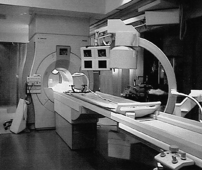
Fig 1. Superconducting short-bore system. The solution proposed by Phillips, sited in this example at the University of Minnesota, includes a standard short-bore superconducting 1.5 T MR imager adjacent to a surgical work-space that includes C-arm fluoroscopy. A custom table can slide from the surgical work-space into the magnet for intermittent imaging at appropriate times during the surgical intervention. The C-arm fluoroscopic unit can also assist with angiographic procedures. (Photo courtesy of Charles L. Truwit, MD, University of Minnesota)
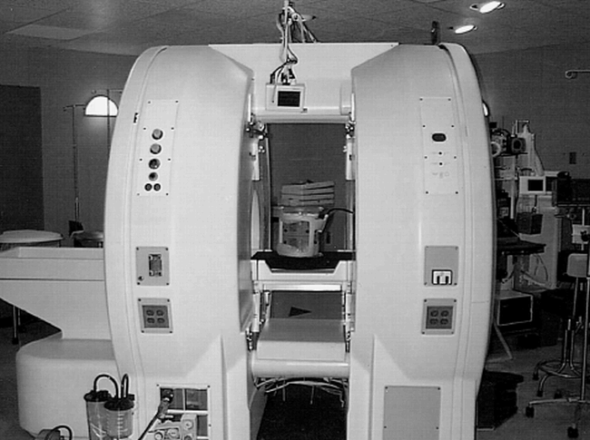
Fig 2. “Double donut” design is shown. General Electric Medical Systems Signa SP scanner consists of two very short superconducting cylindrical magnets separated by a 54-cm space to allow physician or nurse access. The space between the two magnet halves allows patient access from the top or sides, typically used with the surgeon and assistant on opposite sides of the patient's head. An optically linked frameless stereotactic tracking system is integrated into the top support piece. The scanner is designed for an operating-room environment, with electrical outlets and anesthesia gases integrated into the magnet covers. This system has a large fringe field, and all surgical instruments and accessory equipment in proximity to the system must be MR-safe or MR-compatible. (Photo courtesy of John Schenck, MD, PhD, and Robert W. Newman, MS, General Electric Medical Systems.)
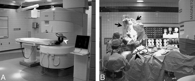
Fig 3. Biplanar system is shown.
A, Siemens Magnetom Open Viva system located in the operating room complex at University Hospitals of Cleveland. With head in imaging position, side access is possible, but the upper magnet pole blocks direct vertical access. Head fixation during surgery is provided with a prototype coil developed by the Heidelberg Neurosurgical Group (arrowhead). Concentric markings on the floor signify 20-mT (200-gauss), 0.5-mT (5- gauss), and 0.15-mT (1.5-gauss) field lines. Anesthesia gas columns, telephone, computer connections, and surgical lighting and ventilation meet same requirements as adjacent standard surgical suites.
B, As a variation on the “neighboring work-space” concept, the head end of the table can be smoothly rotated 120° from the magnet bore on a fixed base for surgical procedures, placing the surgical field (arrowhead) beyond the 5-gauss line. In this rotated operating position, the table can be tilted or raised to allow standard surgical positioning of the patient, and access for the surgeons and anesthesiologist is identical to a standard surgical suite. Because the surgical field is at less than 5-gauss field strength, standard surgical instruments, a standard Zeiss neurosurgical microscope (arrows), as well as standard ultrasonic suction-aspirator, electrocautery, and cortical stimulation equipment can be used. Retractors, curettes, and other equipment used in the magnet bore during image acquisition for monitoring or guidance must be MR-compatible.
Fig 4. Continuous MR imaging guidance mode. Images from continuous series obtained at 7 seconds/image with Fast Imaging with Steady-state Precession (FISP) sequence (18/7/4/90°, TR/TE/number of signal averages/ flip angle) obtained during guidance of needle insertion in a 68-year-old man with C1–2 vertebral and prevertebral mass. Previous attempt at surgical transoral biopsy had been unsuccessful.
A, An image obtained early during insertion shows needle tip passing through the left parotid space (arrow). A poorly defined mass can be seen in the prevertebral space (arrowheads).
B, High vascular conspicuity resulting from 2D Fourier transform technique allows ready visualization of flow-related enhancement within the internal carotid (arrowhead) and vertebral (curved arrow) arteries. The needle tip (straight arrow) can be interactively directed to avoid these major vascular structures. The internal jugular vein is only visible during portions of the respiratory cycle. Although visible on the right, the vein was obstructed on the left side of this patient.
C, The needle (arrow) is interactively redirected more anteriorly once safely beyond the internal carotid artery (ICA) to allow deployment of the central stylet of the 18-gauge core-cutting needle.
D, After extending the central stylet of the side-notch cutting needle, the notch location is shown as an area of thinning of the distal needle tip (between arrows). Histologic findings revealed chronic osteomyelitis and cellulitis and the offending organism was successfully isolated.
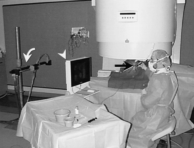
Fig 5. C-arm system for percutaneous intervention is shown. MR suite set up for radiologic intervention has three-camera video sensor array (curved arrow) that detects the location and orientation of a hand-held probe (black arrow). The system automatically acquires continuous MR images based on the probe position, and automatically updates display of four images on shielded LCD monitor adjacent to the scanner (arrowhead). A computer mouse on the LCD console and foot pedals (not shown) allow the scanner to be operated by the radiologist throughout the procedure
- fig 7.
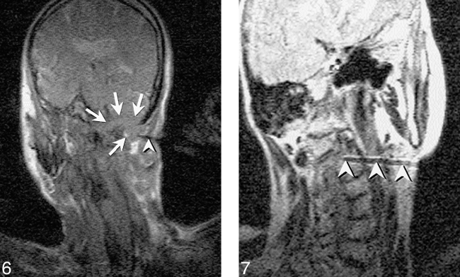
Fig 6. FISP image shows a skull base mass in a 71-year-old man. A single frame from the continuous series of FISP images (18/7/1/90°) obtained at approximately 1.7 s/image shows needle (arrowhead) directed into skull base mass (arrows) that destroys the occipital condyle and extends upward into the posterior fossa. This retromastoid approach benefits from the oblique scan planes achieved with MR enabling imaging of the entire craniad and anteriorly angled needle. As typically performed, this scan plane was alternated with the orthogonal view along the path of the needle. Pathologic diagnosis was plasmacytoma.
MR imaging reveals C2 vertebral body lesion in a 77-year-old woman who reported neck pain. Capability to guide biopsy with oblique imaging plane also allows needle to be angled around internal carotid and vertebral artery for biopsy of high cervical spinal lesions. Turbo spin-echo (SE) T2-weighted image (2000/105/3/17 [TR/TE/NSA/ETL], 51-s scan time) obtained prior to sampling with 18-gauge Menghini-type needle. Frequency encoding must be performed perpendicular to the needle shaft with Turbo SE or SE images to maximize needle visibility. Histologic diagnosis was plasmacytoma, and the patient subsequently developed multiple myeloma.
- fig 7.
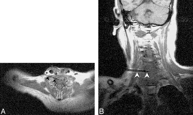
Fig 8. New C6–7 lesion underlying resection site is revealed in a 65-year-old man with history of laryngectomy. Needle placement was performed using FISP guidance (2 s/image), with turbo SE imaging to confirm final placement prior to tissue sampling. Turbo SE T1-weighted axial (A) and oblique coronal (B) images (680/24/3 [TR/TE/NSA], 106 s scanning time for 3 images) used to confirm 18-gauge Menghini cutting needle placement. The needle (arrowheads) can be noted in disk space away from spinal canal or vertebral arteries. Needle was placed through anterior scalene muscle to avoid phrenic nerve and brachial plexus, and was kept anterior to the adjacent vertebral artery. Final diagnosis was fungal osteomyelitis
- fig 7.
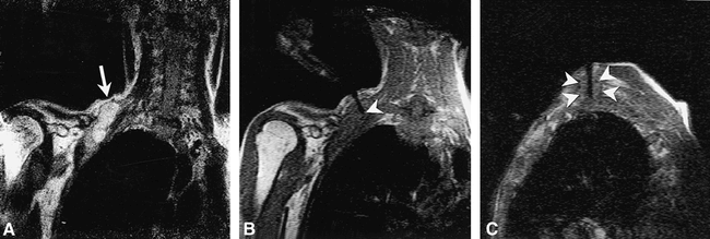
Fig 9. Right supraclavicular fullness is shown in a 52-year-old man who previously underwent left lung resection for carcinoma. The surgeon's physical examination revealed generalized fullness, but no discrete mass.
A, Turbo SE T2-weighted image (2000/105/2 [TR/TE/NSA], 59-s scanning time) reveals focal mass displacing the brachial plexus (arrow).
B, Single frame from continuous series of coronal FISP images (18/7/2/90°[TR/TE/NSA/flip angle], 3.5 s/image) shows 18-gauge side-notch cutting needle (arrowhead) inserted into the dominant mass.
C, Oblique parasagittal FISP image, orthogonal to and with same parameters as B, reveals needle centered within mass (arrowheads) with central stylet extended. The continuous imaging sequence allowed the mass to be separated from adjacent vessels as well as from the adjacent portion of the brachial plexus. Pathologic diagnosis from the biopsy was poorly differentiated large-cell carcinoma.
- fig 7.
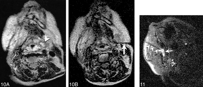
Fig 10. Imaging of an 80-year-old woman after radiation therapy for squamous cell carcinoma of the tongue is shown.
A, T2-weighted Turbo SE image shows small high-signal intensity mass (arrowhead) adjacent to pyriform sinus that was difficult to discern on T1-weighted images. Endoscopic examination of this region was performed twice, yielding no abnormal findings.
B, T2-weighted Turbo spin-echo image (200/105/2/17 [TR/TE/NSA/ETL]) obtained with 59-s scanning time shows needle (arrow) with tip positioned within solid anterior portion of lesion. Cytologic analysis revealed squamous cell carcinoma.
Reproduced with permission from reference 27.
Fig 11. MR-guided sclerotherapy performed in a 31-year-old man with a large, low-flow vascular malformation in the right masticator space. A single frame from a continuous series of FISP images (18/7/1/90°[TR/TE/NSA/flip angle], 1.8-s scanning time) obtained during treatment session shows needle placed from a paramaxillary approach into the deep right masticator space (arrowheads). Collection of high signal around needle tip represents a mixture of sclerosing and contrast agents (arrow). The needle was repositioned interactively until the entire targeted portion was injected. Follow-up images revealed thrombosis and fibrosis of the malformation, and symptoms of bleeding on dental manipulation and inability to open mouth completely resolved after treatment.
- fig 7.
Fig 12. Intraoperative images obtained during tumor resection in a 32-year-old woman are shown. Turbo SE T2-weighted images (2845/102/2 [TR/TE/NSA]; 3-min, 20-s scanning time) were obtained intermittently to monitor resection of a nonenhancing glioma.
A, Frontal pole is immediately adjacent to the dura prior to resection but after craniotomy and dural incision (arrow).
B, During tumor removal, the frontal pole and adjacent tissues shift posteriorly, creating an approximate 5-mm gap between the inner surface of the dura and the adjacent cortex (arrow).
C, At a later stage during the debulking process, there is further frontal-pole shift (arrow) as intracranial volume decreases with tumor resection. Resection was continued after obtaining this image (not shown). All images were obtained on the 0.2 T surgical suite-based C-arm system depicted in figure 3.
- fig 7.
Fig 13. Imaging of a 49-year-old woman with recurrent skull-base cyst after prior transmaxillary drainage is shown.
A and B, Turbo SE T2-weighted axial images (4083/105/5/17 [TR/TE/NSA/ETL], 3-min, 29-s scanning time) show large, multiloculated cyst replacing the petrous apex and body and pterygoid process of the sphenoid bone on the left (arrowheads). The bony Eustachian tube is obstructed with a secondary mastoid effusion.
C, FISP image (17.8/8.1/4/90° [TR/TE/NSA/flip angle], 9 s/image) obtained during drainage procedure shows gadolinium-filled catheter extending through Caldwell Luc incision (arrow), and pterygopalatine fossa resection site (arrowhead) into skull base cyst. Several of the loculations within the cyst are already drained, with residual fluid layering posteriorly (asterisk). This frame was one of a continuous series of images obtained during this portion of the procedure.
D and E, Turbo SE T2-weighted images, with same parameters as A and B, show air filling the multiple loculations of the lesion, with a small posterior air-fluid level within the petrous apex portion of the abnormality. Mastoid fluid remains unchanged. Biopsy of cyst wall again revealed normal respiratory epithelium.
- fig 7.
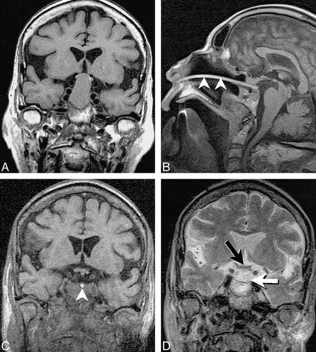
Fig 14. Imaging of a 76 year-old woman with pituitary macroadenoma resulting in visual symptoms is shown.
A, Preoperative coronal T1-weighted (680/14/3) image depicts large macroadenoma resulting in marked elevation and compression of the optic chiasm.
B, Intraoperative sagittal T1-weighted SE image (432/26/3 [TR/TE/NSA], 202 × 256 matrix, 20-cm FOV) shows localizer probe, which can be seen as a bright line (arrowheads) extending through the nasal cavity into the sella extending to the posterior aspect of the resection bed.
C, Intraoperative coronal 3D T1-weighted FLASH image (120/12/1/60° [TR/TE/NSA/flip angle], 256 × 256 matrix, 20-cm FOV, 8-min, 15-s scanning time) shows complete decompression of the optic chiasm with a CSF-filled suprasellar cistern. The localizer probe can be seen as a bright circle within the sella (arrowhead). Blood and secretions pooling within the sella make it difficult to detect residual tumor on unenhanced T1-weighted images.
D, Coronal turbo SE T2-weighted image (3500/102/1/7 [TR/TE/NSA/ETL], 210 × 256 matrix, 20-cm FOV, 5-min, 2-s scanning time) obtained later in the procedure again reveals decompression of the optic chiasm (black arrow) and relaxation of the diaphragma sellae (white arrow). On T2-weighted images, fluid or blood pooling within the sella can more easily be distinguished from residual tumor. Contrast-enhanced images are of less help than might be predicted owing to pooling of contrast-enhanced blood within the sella if any oozing occurs before or during image acquisition.
Reproduced with permission from reference 58.
- fig 7.
Fig 15. Imaging of a 40-year-old woman with sphenoid wing meningioma is shown.
A, Intraoperative turbo SE T2-weighted oblique image (2598/105/5/17 [TR/TE/NSA/ETL], 2-min, 40-s scanning time) reveals sphenoid wing mass encasing the bifurcation of the middle cerebral artery (MCA) (arrowhead).
B and C, Coronal (B) and axial (C) contrast-enhanced FLASH T1-weighted images (110/9/5/70° [TR/TE/NSA/flip angle], 1-min, 48-s scanning time) reveals meningioma after craniotomy and surgical exposure. The margins of the lesion and location of the optic chiasm, ICA, and primary MCA branches were determined interactively using gadolinium-filled markers.
D and E, Coronal (D) and axial (E) images obtained after resection, with same parameters as A, show gross debulking of the tumor with a thin rim of enhancing tissue intentionally left along the cavernous sinus and encased arteries (arrowheads).


