
Article Figures & Data
Figures
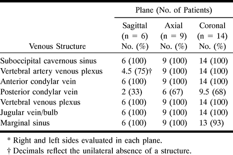
- fig 1.
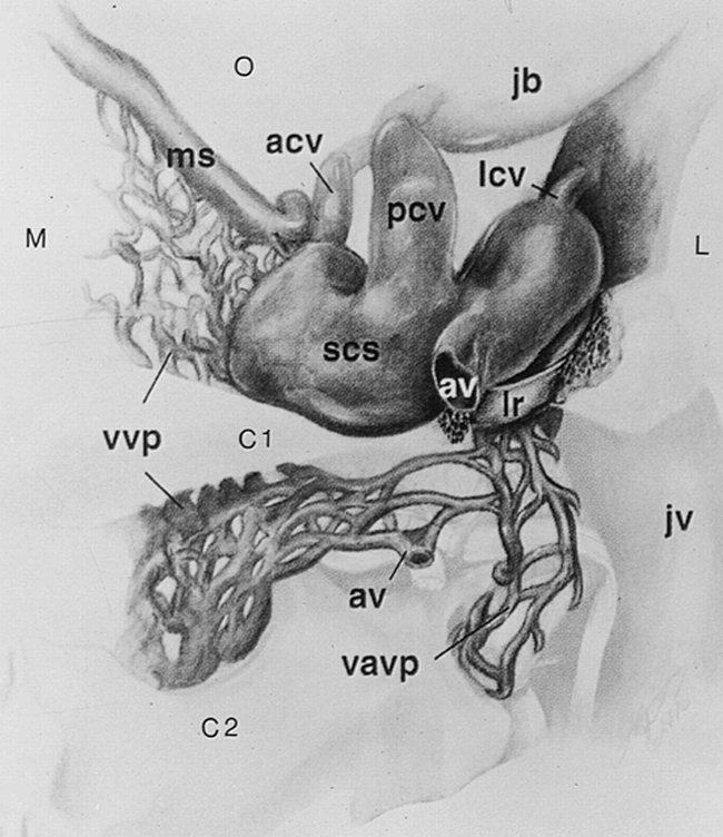
Posterior view of the right suboccipital cavernous sinus and its venous communications at the skull base. The vertebral artery has been removed. The left suboccipital cavernous sinus would form a mirror image, with direct connection of the marginal sinus and vertebral venous plexus across the midline. M indicates medial; L, lateral; C1, atlas; C2, axis; O, occipital region; acv, anterior condylar vein; av, anastomotic vein; jb, jugular bulb; jv, jugular vein; lcv, lateral condylar vein; lr, lateral ring; ms, marginal sinus; pcv, posterior condylar vein; scs, suboccipital cavernous sinus; vavp, vertebral artery venous plexus; vvp, vertebral venous plexus. Adapted from (1) with permission
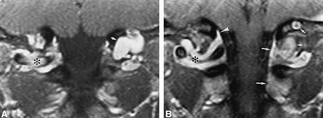
- fig 2.
45-year-old man referred for follow-up of a presumed schwannoma posterior to the left occipital condyle, shown to be the left posterior condylar vein.
A and B, Contrast-enhanced fat-suppressed T1-weighted (640/14/3) coronal images (A posterior to B) show the suboccipital cavernous sinus (asterisk), marginal sinus (arrowhead), posterior condylar vein (short arrows), and vertebral venous plexus (long arrows). The right posterior condylar vein is diminutive.
- fig 3.
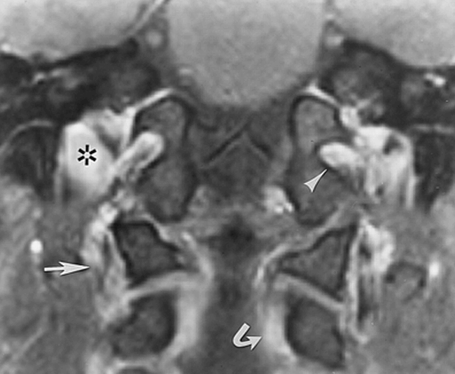
43-year-old man. Contrast-enhanced fat-suppressed T1-weighted (950/12/2) coronal image in a plane anterior to figure 2 shows the anterior condylar vein (arrowhead) and the jugular vein (asterisk) at the jugular tubercle, the vertebral venous plexus (curved arrow), and the vertebral artery venous plexus (straight arrow)
- fig 4.
68-year-old woman.
A and B, Contrast-enhanced fat-suppressed T1-weighted (630/20/2) axial images (superior to inferior) show the anterior condylar vein (large straight arrow), suboccipital cavernous sinus (curved arrow), vertebral venous plexus (arrowhead), vertebral artery venous plexus (small straight arrows), and anastomotic vein (wavy arrow).
- fig 5.
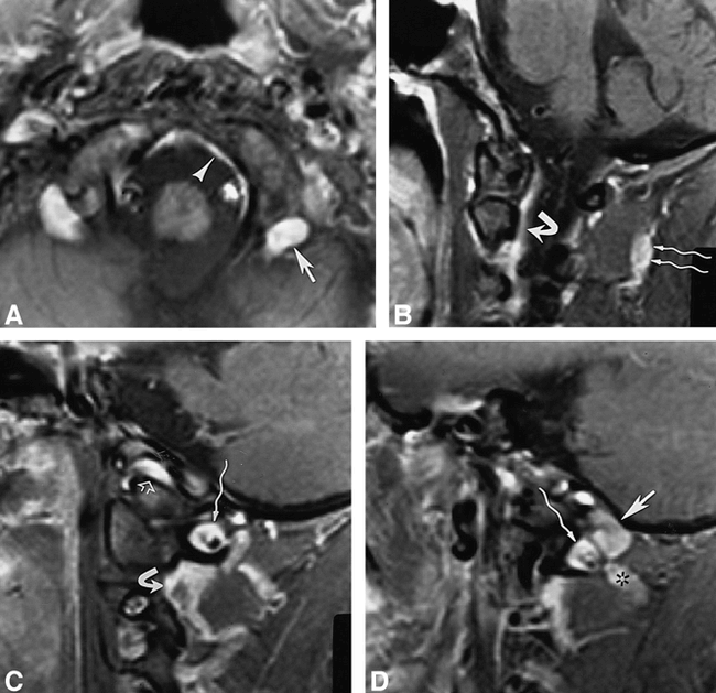
34-year-old woman.
A–D, Contrast-enhanced fat-suppressed T1-weighted (820/12/2) axial image (in a plane between fig 4A and B) (A) and T1-weighted (700/12/2) sagittal images (medial to lateral) (B–D) show the posterior condylar vein (straight arrow, A and D) at the posterior aspect of the occipital condyle, marginal sinus (arrowhead, A), suboccipital cavernous sinus (wavy arrow, C and D), vertebral venous plexus (curved arrow, B and C), suboccipital venous plexus (wavy arrows, B), anterior condylar vein (open arrow, C), and anastomotic vein (asterisk, D).


Tables
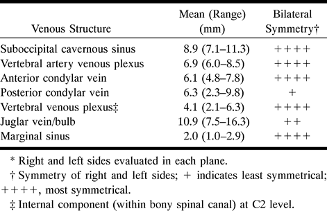
 Table 1:
Normal suboccipital venous structures seen in 14 patients using contrast-enhanced T1-weighted fat-suppressed sequences*
- Table 2:
Summary of measured diameters of normal suboccipital venous structures in 14 patients using contrast-enhanced T1-weighted fat-suppressed MR sequences*
In this issue


Jump to section
Related Articles
Cited By...
- Cerebral venous anatomy: implications for the neurointerventionalist
- Cerebral venous anatomy: implications for the neurointerventionalist
- Venous structures at the craniocervical junction: anatomical variations evaluated by multidetector row CT
- Vertebral Artery Dissection with a Normal-Appearing Lumen at Multisection CT Angiography: The Importance of Identifying Wall Hematoma