
Article Figures & Data
Figures
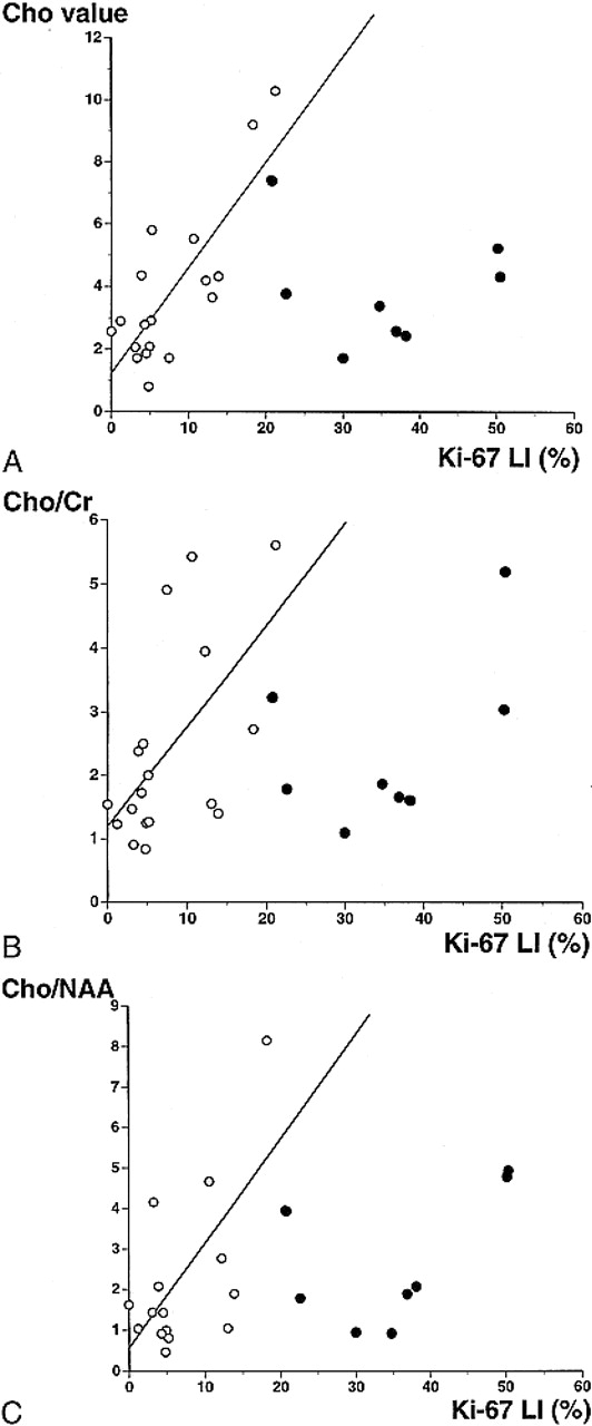
- fig 1.
A–C, Relationships between the Ki-67 labeling index and the Cho value (A), the Cho/Cr ratio (B), and the Cho/NAA ratio (C). Open circles indicate that the spectroscopic voxel is homogeneous on MR images with absent, faint, or homogeneous enhancement by contrast material. Closed circles indicate that the spectroscopic voxel is heterogeneous on MR images before or after contrast administration (cases 17–24, Table). There is a strong linear relationship between the Ki-67 labeling index and the Cho value, indicated by open circles (y = 1.24 + 0.34 * x, r = .81, P < .0001). There is a weak correlation between the Ki-67 labeling index and the Cho/Cr or Cho/NAA ratio, indicated by open circles (y = 1.22 + 0.15 * x, r = .58, P < .02 and y = 0.65 + 0.23 * x, r = .60, P < .02, respectively). Extreme data for the Cho/NAA ratio from cases 6, 13, and 25 are not shown in the graph.
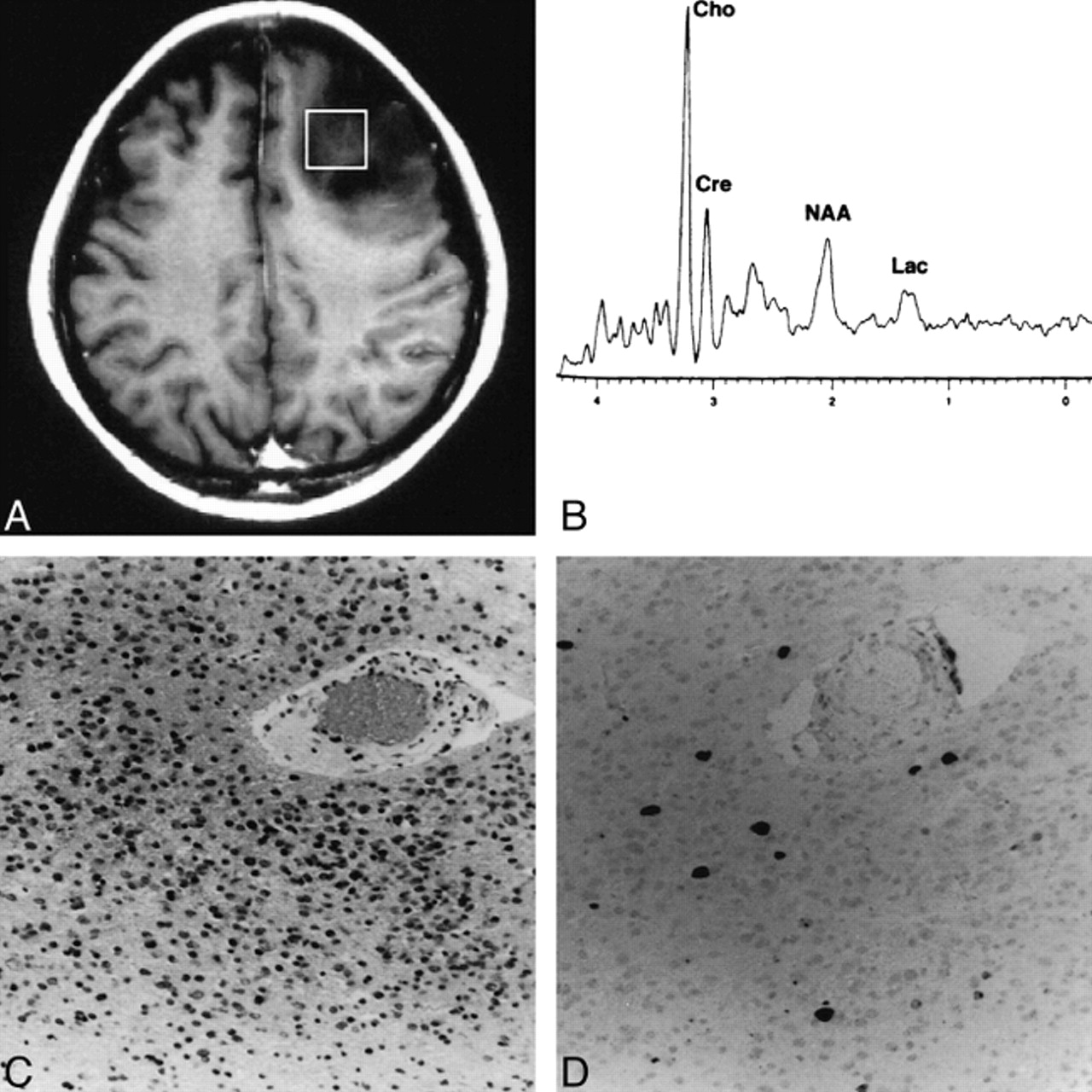
- fig 2.
Case 11: A typical case in which the spectroscopic voxel is homogeneous without contrast enhancement.
A, Contrast-enhanced T1-weighted MR image shows the spectroscopic voxel (square) filled with unenhanced homogeneous tumor.
B, Proton spectrum obtained from the region indicated in A shows a Cho value of 2.06.
C, Photomicrograph of the resected tumor indicates anaplastic astrocytoma (grade 3) (hematoxylin-eosin, original magnification ×100).
D, Photomicrograph of Ki-67 staining of the same tumor specimen shows a Ki-67 labeling index of 3.10% (original magnification ×100).
- fig 3.
A and B, T1-weighted MR images of case 8 (A) and case 25 (B) in which the spectroscopic voxel is homogeneous and only faintly enhanced. The voxels for MR spectroscopy are indicated by squares. These contrast-enhanced images were obtained after spectroscopic examinations, and the location of the voxels was superimposed afterward. Note that in case 25, the tumor outside the voxel is heterogeneous
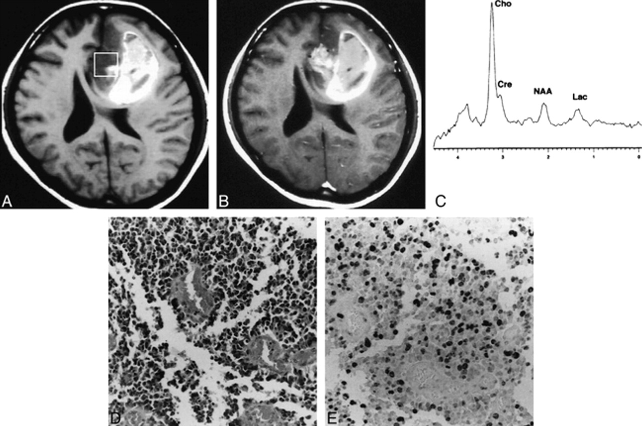
- fig 4.
Case 18: A typical case in which the spectroscopic voxel is heterogeneous on MR images before and after contrast agent administration.
A, T1-weighted MR image shows the spectroscopic voxel (square) filled with heterogeneous tumor, including intratumoral hemorrhage appearing as a hyperintense area.
B, Contrast-enhanced T1-weighted MR image shows heterogeneous enhancement within the voxel.
C, Proton spectrum obtained from the region indicated in A shows a Cho value of 5.21.
D, Photomicrograph of the resected tumor indicates glioblastoma multiforme (grade 4) (hematoxylin-eosin, original magnification ×100).
E, Photomicrograph of Ki-67 staining of the same tumor specimen shows a Ki-67 labeling index of 50.20% (original magnification ×100).
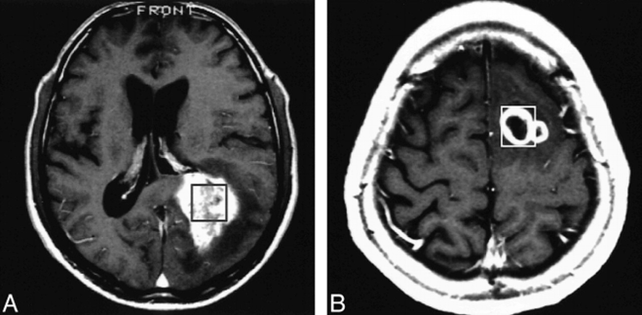
- fig 5.
A and B, Contrast-enhanced T1-weighted MR images of case 19 (A) and case 22 (B) in which the spectroscopic voxel appearance is heterogeneous. The voxels for MR spectroscopy are indicated by squares. These contrast-enhanced images were obtained after spectroscopic examination, and the location of the voxels was superimposed afterward


Tables

Findings in 26 patients with glioma

In this issue


Jump to section
Related Articles
Cited By...
- Impact of dietary intake on brain choline levels: A 3 Tesla magnetic resonance spectroscopy study
- CT Imaging Correlates of Genomic Expression for Oral Cavity Squamous Cell Carcinoma
- Multimodal Elucidation of Choline Metabolism in a Murine Glioma Model Using Magnetic Resonance Spectroscopy and 11C-Choline Positron Emission Tomography
- Magnetic resonance spectroscopy of the brain
- Multimodality Assessment of Brain Tumors and Tumor Recurrence
- Diagnostic performance of spectroscopic and perfusion MRI for distinction of brain tumors
- Noninvasive Magnetic Resonance Spectroscopic Imaging Biomarkers to Predict the Clinical Grade of Pediatric Brain Tumors
- NMR Spectroscopy and Pediatric Brain Tumors
- Tuning in on Tumor Activity with Proton MR Spectroscopy