Advertisement

Main menu

  • Home
  • Content
    • Current Issue
    • Accepted Manuscripts
    • Article Preview
    • Past Issue Archive
    • AJNR Case Collection
    • Case of the Week Archive
    • Classic Case Archive
    • Case of the Month Archive
  • Special Collections
    • Spinal CSF Leak Articles (Jan 2020-June 2024)
    • 2024 AJNR Journal Awards
    • Most Impactful AJNR Articles
  • Multimedia
    • AJNR Podcast
    • AJNR Scantastics
    • Video Articles
  • For Authors
    • Submit a Manuscript
    • Author Policies
    • Fast publishing of Accepted Manuscripts
    • Graphical Abstract Preparation
    • Manuscript Submission Guidelines
    • Imaging Protocol Submission
    • Submit a Case for the Case Collection
  • About Us
    • About AJNR
    • Editorial Board
  • More
    • Become a Reviewer/Academy of Reviewers
    • Subscribers
    • Permissions
    • Alerts
    • Feedback
    • Advertisers
    • ASNR Home
  • Other Publications
    • ajnr

User menu

  • Alerts
  • Log in

Search

  • Advanced search
American Journal of Neuroradiology

American Journal of Neuroradiology

ASHNR American Society of Functional Neuroradiology ASHNR American Society of Pediatric Neuroradiology ASSR
  • Alerts
  • Log in

Advanced Search

  • Home
  • Content
    • Current Issue
    • Accepted Manuscripts
    • Article Preview
    • Past Issue Archive
    • AJNR Case Collection
    • Case of the Week Archive
    • Classic Case Archive
    • Case of the Month Archive
  • Special Collections
    • Spinal CSF Leak Articles (Jan 2020-June 2024)
    • 2024 AJNR Journal Awards
    • Most Impactful AJNR Articles
  • Multimedia
    • AJNR Podcast
    • AJNR Scantastics
    • Video Articles
  • For Authors
    • Submit a Manuscript
    • Author Policies
    • Fast publishing of Accepted Manuscripts
    • Graphical Abstract Preparation
    • Manuscript Submission Guidelines
    • Imaging Protocol Submission
    • Submit a Case for the Case Collection
  • About Us
    • About AJNR
    • Editorial Board
  • More
    • Become a Reviewer/Academy of Reviewers
    • Subscribers
    • Permissions
    • Alerts
    • Feedback
    • Advertisers
    • ASNR Home
  • Follow AJNR on Twitter
  • Visit AJNR on Facebook
  • Follow AJNR on Instagram
  • Join AJNR on LinkedIn
  • RSS Feeds

Welcome to the new AJNR, Updated Hall of Fame, and more. Read the full announcements.


AJNR is seeking candidates for the position of Associate Section Editor, AJNR Case Collection. Read the full announcement.


Getting new auth cookie, if you see this message a lot, tell someone!
Research ArticleBRAIN

Diffusion Measurements in Intracranial Hematomas: Implications for MR Imaging of Acute Stroke

Scott W. Atlas, Philip DuBois, Michael B. Singer and Dongfeng Lu
American Journal of Neuroradiology August 2000, 21 (7) 1190-1194;
Scott W. Atlas
aFrom the Department of Radiology (S.W.A.), Stanford University Medical Center, Stanford, CA, and the Department of Radiology (S.W.A., P.D., M.B.S., D.L.), Mount Sinai Medical Center, New York, NY.
  • Find this author on Google Scholar
  • Find this author on PubMed
  • Search for this author on this site
Philip DuBois
aFrom the Department of Radiology (S.W.A.), Stanford University Medical Center, Stanford, CA, and the Department of Radiology (S.W.A., P.D., M.B.S., D.L.), Mount Sinai Medical Center, New York, NY.
  • Find this author on Google Scholar
  • Find this author on PubMed
  • Search for this author on this site
Michael B. Singer
aFrom the Department of Radiology (S.W.A.), Stanford University Medical Center, Stanford, CA, and the Department of Radiology (S.W.A., P.D., M.B.S., D.L.), Mount Sinai Medical Center, New York, NY.
  • Find this author on Google Scholar
  • Find this author on PubMed
  • Search for this author on this site
Dongfeng Lu
aFrom the Department of Radiology (S.W.A.), Stanford University Medical Center, Stanford, CA, and the Department of Radiology (S.W.A., P.D., M.B.S., D.L.), Mount Sinai Medical Center, New York, NY.
  • Find this author on Google Scholar
  • Find this author on PubMed
  • Search for this author on this site
  • Article
  • Figures & Data
  • Info & Metrics
  • Responses
  • References
  • PDF
Loading

Article Figures & Data

Figures

  • Tables
  • fig 1.
    • Download figure
    • Open in new tab
    • Download powerpoint
    fig 1.

    Average ADC value versus putative biophysical state in intracranial hematomas

  • fig 2.
    • Download figure
    • Open in new tab
    • Download powerpoint
    fig 2.

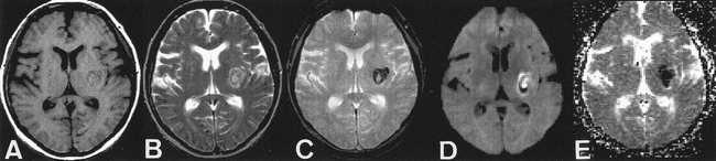
    ADC map in case of hyperacute intracranial hematoma.

    A, T1-weighted images (600/11/1). Left subinsular hematoma is isointense to brain.

    B, T2-weighted images (3600/90/1). Left subinsular hematoma is hyperintense to brain, with thin, peripheral rim of marked hypointensity.

    C, T2*-weighted gradient-echo images (500/30/1). Left subinsular hematoma is more prominent.

    D, Diffusion-weighted images show central high intensity.

    E, Calculated ADC maps show marked hypointensity indicating restricted diffusion.

Tables

  • Figures
  • TABLE 1:
    • View popup
    • Download powerpoint
    TABLE 1:

    Average ADC in intracranial hematomas

  • TABLE 2:
    • View popup
    • Download powerpoint
    TABLE 2:

    Statistical comparisons between ADC of stages of intracranial hematomas

PreviousNext
Back to top

In this issue

American Journal of Neuroradiology
Vol. 21, Issue 7
1 Aug 2000
  • Table of Contents
  • Index by author
Advertisement
Print
Download PDF
Email Article

Thank you for your interest in spreading the word on American Journal of Neuroradiology.

NOTE: We only request your email address so that the person you are recommending the page to knows that you wanted them to see it, and that it is not junk mail. We do not capture any email address.

Enter multiple addresses on separate lines or separate them with commas.
Diffusion Measurements in Intracranial Hematomas: Implications for MR Imaging of Acute Stroke
(Your Name) has sent you a message from American Journal of Neuroradiology
(Your Name) thought you would like to see the American Journal of Neuroradiology web site.
CAPTCHA
This question is for testing whether or not you are a human visitor and to prevent automated spam submissions.
Cite this article
Scott W. Atlas, Philip DuBois, Michael B. Singer, Dongfeng Lu
Diffusion Measurements in Intracranial Hematomas: Implications for MR Imaging of Acute Stroke
American Journal of Neuroradiology Aug 2000, 21 (7) 1190-1194;

Citation Manager Formats

  • BibTeX
  • Bookends
  • EasyBib
  • EndNote (tagged)
  • EndNote 8 (xml)
  • Medlars
  • Mendeley
  • Papers
  • RefWorks Tagged
  • Ref Manager
  • RIS
  • Zotero
0 Responses
Respond to this article
Share
Bookmark this article
Diffusion Measurements in Intracranial Hematomas: Implications for MR Imaging of Acute Stroke
Scott W. Atlas, Philip DuBois, Michael B. Singer, Dongfeng Lu
American Journal of Neuroradiology Aug 2000, 21 (7) 1190-1194;
del.icio.us logo Twitter logo Facebook logo Mendeley logo
  • Tweet Widget
  • Facebook Like
  • Google Plus One

Jump to section

  • Article
    • Abstract
    • Methods
    • Results
    • Discussion
    • Conclusion
    • Footnotes
    • References
  • Figures & Data
  • Info & Metrics
  • Responses
  • References
  • PDF

Related Articles

  • No related articles found.
  • PubMed
  • Google Scholar

Cited By...

  • Centrally Reduced Diffusion Sign for Differentiation between Treatment-Related Lesions and Glioma Progression: A Validation Study
  • Blood Pressure Reduction, Decreased Diffusion on MRI, and Outcomes After Intracerebral Hemorrhage
  • Diffusion Findings in Blood Clot: The Last Word?
  • Restricted Diffusion within Ring Enhancement Is Not Pathognomonic for Brain Abscess
  • Computing Diffusion Rates in T2-dark Hematomas and Areas of Low T2 Signal
  • Crossref
  • Google Scholar

This article has not yet been cited by articles in journals that are participating in Crossref Cited-by Linking.

More in this TOC Section

  • SWI or T2*: Which MRI Sequence to Use in the Detection of Cerebral Microbleeds? The Karolinska Imaging Dementia Study
  • Progression of Microstructural Damage in Spinocerebellar Ataxia Type 2: A Longitudinal DTI Study
  • Usefulness of Quantitative Susceptibility Mapping for the Diagnosis of Parkinson Disease
Show more Brain

Similar Articles

Advertisement

Indexed Content

  • Current Issue
  • Accepted Manuscripts
  • Article Preview
  • Past Issues
  • Editorials
  • Editors Choice
  • Fellow Journal Club
  • Letters to the Editor

Cases

  • Case Collection
  • Archive - Case of the Week
  • Archive - Case of the Month
  • Archive - Classic Case

Special Collections

  • Special Collections

Resources

  • News and Updates
  • Turn around Times
  • Submit a Manuscript
  • Author Policies
  • Manuscript Submission Guidelines
  • Evidence-Based Medicine Level Guide
  • Publishing Checklists
  • Graphical Abstract Preparation
  • Imaging Protocol Submission
  • Submit a Case
  • Become a Reviewer/Academy of Reviewers
  • Get Peer Review Credit from Publons

Multimedia

  • AJNR Podcast
  • AJNR SCANtastic
  • Video Articles

About Us

  • About AJNR
  • Editorial Board
  • Not an AJNR Subscriber? Join Now
  • Alerts
  • Feedback
  • Advertise with us
  • Librarian Resources
  • Permissions
  • Terms and Conditions

American Society of Neuroradiology

  • Not an ASNR Member? Join Now

© 2025 by the American Society of Neuroradiology All rights, including for text and data mining, AI training, and similar technologies, are reserved.
Print ISSN: 0195-6108 Online ISSN: 1936-959X

Powered by HighWire