
Article Figures & Data
Figures
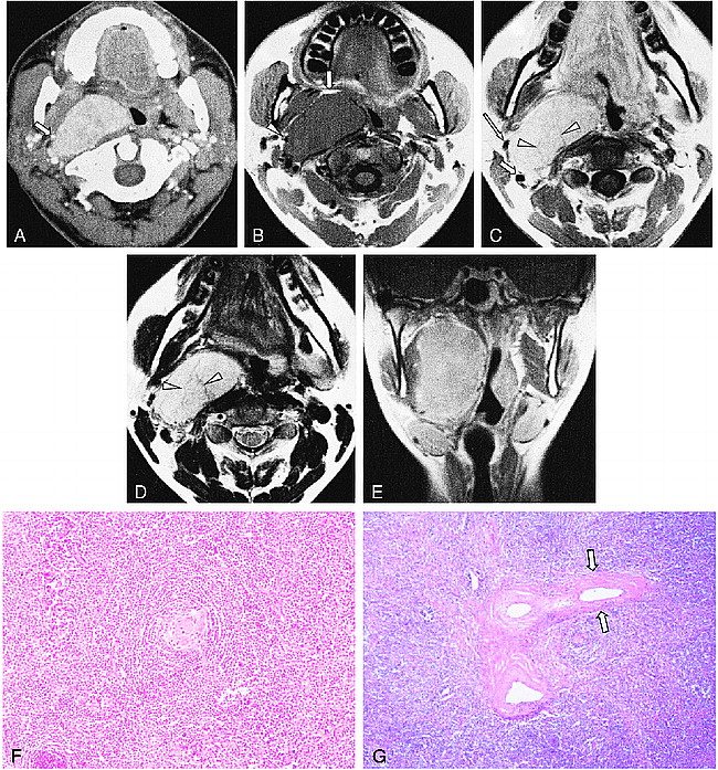
- fig 1.
Images from the case of a 34-year-old woman with an enlarging mass on the right side of her neck.
A, Contrast-enhanced axial CT scan shows a well-marginated, intensely enhancing mass in the right retropharyngeal space. Note the styloid process, which is anterolaterally displaced by the mass (arrow).
B, T1-weighed (450/12) axial MR image shows a well-marginated mass isointense to the muscle in the right retropharyngeal space. The mass displaces the parapharyngeal fat anteriorly (arrow), pharyngeal mucosal space medially, styloid process anterolaterally (arrowhead), and internal carotid artery laterally.
C, Contrast-enhanced T1-weighed (450/12) axial MR image (obtained at a slightly lower level than that shown in panel B) reveals strong enhancement of the lesion. The displaced right internal carotid artery (short arrow) and external carotid artery (long arrow) are separated. Note the linear hypointense signal within the mass (arrowheads).
D, T2-weighted (3700/99) axial MR image (obtained at the same level as that shown in panel C) shows the lesion hyperintense to the muscle. The linear hypointense signal seen in panel C is also seen as a hypointense signal (arrowheads).
E, Contrast-enhanced T1-weighed (513/12) coronal MR image shows the mass occupying the entire right retropharyngeal and parapharyngeal space from below the skull base to the hyoid bone level.
F, Photomicrograph (original magnification, ×100; hematoxylin and eosin stain) of the right retropharyngeal lymph node shows a follicle with an onion skin–appearing, prominent mantle and a penetrating hyalinizing vessel into the germinal center, consistent with hyaline-vascular-type Castleman's disease.
G, Low-power photomicrograph (original magnification, ×40; hematoxylin and eosin stain) of the right retropharyngeal lymph node shows perivascular lamellar fibrosis (arrows), which corresponds to the linear hypointense signals in an arborizing pattern on the MR images.


