Advertisement

Main menu

  • Home
  • Content
    • Current Issue
    • Accepted Manuscripts
    • Article Preview
    • Past Issue Archive
    • Video Articles
    • AJNR Case Collection
    • Case of the Week Archive
    • Case of the Month Archive
    • Classic Case Archive
  • Special Collections
    • AJNR Awards
    • Low-Field MRI
    • Alzheimer Disease
    • ASNR Foundation Special Collection
    • Photon-Counting CT
    • View All
  • Multimedia
    • AJNR Podcasts
    • AJNR SCANtastic
    • Trainee Corner
    • MRI Safety Corner
    • Imaging Protocols
  • For Authors
    • Submit a Manuscript
    • Submit a Video Article
    • Submit an eLetter to the Editor/Response
    • Manuscript Submission Guidelines
    • Statistical Tips
    • Fast Publishing of Accepted Manuscripts
    • Graphical Abstract Preparation
    • Imaging Protocol Submission
    • Author Policies
  • About Us
    • About AJNR
    • Editorial Board
    • Editorial Board Alumni
  • More
    • Become a Reviewer/Academy of Reviewers
    • Subscribers
    • Permissions
    • Alerts
    • Feedback
    • Advertisers
    • ASNR Home

User menu

  • Alerts
  • Log in

Search

  • Advanced search
American Journal of Neuroradiology

American Journal of Neuroradiology

ASHNR American Society of Functional Neuroradiology ASHNR American Society of Pediatric Neuroradiology ASSR
  • Alerts
  • Log in

Advanced Search

  • Home
  • Content
    • Current Issue
    • Accepted Manuscripts
    • Article Preview
    • Past Issue Archive
    • Video Articles
    • AJNR Case Collection
    • Case of the Week Archive
    • Case of the Month Archive
    • Classic Case Archive
  • Special Collections
    • AJNR Awards
    • Low-Field MRI
    • Alzheimer Disease
    • ASNR Foundation Special Collection
    • Photon-Counting CT
    • View All
  • Multimedia
    • AJNR Podcasts
    • AJNR SCANtastic
    • Trainee Corner
    • MRI Safety Corner
    • Imaging Protocols
  • For Authors
    • Submit a Manuscript
    • Submit a Video Article
    • Submit an eLetter to the Editor/Response
    • Manuscript Submission Guidelines
    • Statistical Tips
    • Fast Publishing of Accepted Manuscripts
    • Graphical Abstract Preparation
    • Imaging Protocol Submission
    • Author Policies
  • About Us
    • About AJNR
    • Editorial Board
    • Editorial Board Alumni
  • More
    • Become a Reviewer/Academy of Reviewers
    • Subscribers
    • Permissions
    • Alerts
    • Feedback
    • Advertisers
    • ASNR Home
  • Follow AJNR on Twitter
  • Visit AJNR on Facebook
  • Follow AJNR on Instagram
  • Join AJNR on LinkedIn
  • RSS Feeds

AJNR Awards, New Junior Editors, and more. Read the latest AJNR updates

Research ArticleHead and Neck Imaging

Intrathyroidal Lymphoepithelial (Branchial) Cyst: Sonographic Features of a Rare Lesion

Anil T. Ahuja, Alexander R. Chang, Edward To, Peter Pang, Alex S.C. Ching, Ann D. King and Con Metreweli
American Journal of Neuroradiology August 2000, 21 (7) 1340-1343;
Anil T. Ahuja
aFrom the Departments of Diagnostic Radiology and Organ Imaging (A.T.A., A.S.C.C., A.D.K., C.M.), Anatomical and Cellular Pathology (A.R.C.), and Surgery (E.T., P.P.), Chinese University of Hong Kong, Shatin, N.T., Hong Kong.
  • Find this author on Google Scholar
  • Find this author on PubMed
  • Search for this author on this site
Alexander R. Chang
aFrom the Departments of Diagnostic Radiology and Organ Imaging (A.T.A., A.S.C.C., A.D.K., C.M.), Anatomical and Cellular Pathology (A.R.C.), and Surgery (E.T., P.P.), Chinese University of Hong Kong, Shatin, N.T., Hong Kong.
  • Find this author on Google Scholar
  • Find this author on PubMed
  • Search for this author on this site
Edward To
aFrom the Departments of Diagnostic Radiology and Organ Imaging (A.T.A., A.S.C.C., A.D.K., C.M.), Anatomical and Cellular Pathology (A.R.C.), and Surgery (E.T., P.P.), Chinese University of Hong Kong, Shatin, N.T., Hong Kong.
  • Find this author on Google Scholar
  • Find this author on PubMed
  • Search for this author on this site
Peter Pang
aFrom the Departments of Diagnostic Radiology and Organ Imaging (A.T.A., A.S.C.C., A.D.K., C.M.), Anatomical and Cellular Pathology (A.R.C.), and Surgery (E.T., P.P.), Chinese University of Hong Kong, Shatin, N.T., Hong Kong.
  • Find this author on Google Scholar
  • Find this author on PubMed
  • Search for this author on this site
Alex S.C. Ching
aFrom the Departments of Diagnostic Radiology and Organ Imaging (A.T.A., A.S.C.C., A.D.K., C.M.), Anatomical and Cellular Pathology (A.R.C.), and Surgery (E.T., P.P.), Chinese University of Hong Kong, Shatin, N.T., Hong Kong.
  • Find this author on Google Scholar
  • Find this author on PubMed
  • Search for this author on this site
Ann D. King
aFrom the Departments of Diagnostic Radiology and Organ Imaging (A.T.A., A.S.C.C., A.D.K., C.M.), Anatomical and Cellular Pathology (A.R.C.), and Surgery (E.T., P.P.), Chinese University of Hong Kong, Shatin, N.T., Hong Kong.
  • Find this author on Google Scholar
  • Find this author on PubMed
  • Search for this author on this site
Con Metreweli
aFrom the Departments of Diagnostic Radiology and Organ Imaging (A.T.A., A.S.C.C., A.D.K., C.M.), Anatomical and Cellular Pathology (A.R.C.), and Surgery (E.T., P.P.), Chinese University of Hong Kong, Shatin, N.T., Hong Kong.
  • Find this author on Google Scholar
  • Find this author on PubMed
  • Search for this author on this site
  • Article
  • Figures & Data
  • Info & Metrics
  • Responses
  • References
  • PDF
Loading

Article Figures & Data

Figures

  • fig 1.
    • Download figure
    • Open in new tab
    • Download powerpoint
    fig 1.

    Transverse sonogram of thyroid showing multiloculated nodule (black arrows). Note homogeneous internal architecture and presence of comet tail artifact within (small white arrows) created by cholesterol crystals. This represents the pseudosolid appearance of a congenital neck cyst. Transducer pressure on nodule produces shifting of contents, suggesting its cystic nature. Aspiration yielded viscous yellowish puslike fluid. Open arrow identifies internal jugular vein, and large white arrow marks common carotid artery.fig 2. Clump of mature squamous cells (hematoxylin and eosin stain; original magnification, ×200).fig 3. T2-weighted (2500/100 [TR/TE]; number of acquisitions, two) axial-view image obtained through thyroid shows well-defined, markedly hyperintense cystic nodule (arrows) in right lobe of thyroid. There are no other specific features to suggest that it may be of congenital origin
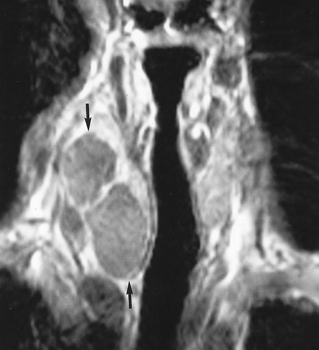
  • fig 4.
    • Download figure
    • Open in new tab
    • Download powerpoint
    fig 4.

    T1-weighted (500/20; number of acquisitions, one) coronal-view image shows multiloculated, slightly hyperintense nodule (arrows) in right lobe of the thyroid
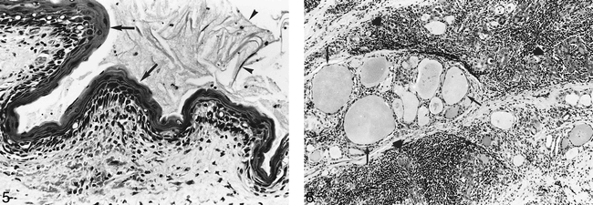
  • fig 5.
    • Download figure
    • Open in new tab
    • Download powerpoint
    fig 5.

    Higher-power view shows squamous epithelium (arrows) and desquamating keratin (arrowheads). Lymphocytes are seen in the underlying connective tissue (hematoxylin and eosin stain; original magnification, ×250).fig 6. Thyroid follicles of variable size (arrows) can be seen in this view. Some are compressed by diffuse lymphocyte infiltrate (large arrows) (hematoxylin and eosin stain; original magnification, ×100)

PreviousNext
Back to top

In this issue

American Journal of Neuroradiology
Vol. 21, Issue 7
1 Aug 2000
  • Table of Contents
  • Index by author
Advertisement
Print
Download PDF
Email Article

Thank you for your interest in spreading the word on American Journal of Neuroradiology.

NOTE: We only request your email address so that the person you are recommending the page to knows that you wanted them to see it, and that it is not junk mail. We do not capture any email address.

Enter multiple addresses on separate lines or separate them with commas.
Intrathyroidal Lymphoepithelial (Branchial) Cyst: Sonographic Features of a Rare Lesion
(Your Name) has sent you a message from American Journal of Neuroradiology
(Your Name) thought you would like to see the American Journal of Neuroradiology web site.
CAPTCHA
This question is for testing whether or not you are a human visitor and to prevent automated spam submissions.
Cite this article
Anil T. Ahuja, Alexander R. Chang, Edward To, Peter Pang, Alex S.C. Ching, Ann D. King, Con Metreweli
Intrathyroidal Lymphoepithelial (Branchial) Cyst: Sonographic Features of a Rare Lesion
American Journal of Neuroradiology Aug 2000, 21 (7) 1340-1343;

Citation Manager Formats

  • BibTeX
  • Bookends
  • EasyBib
  • EndNote (tagged)
  • EndNote 8 (xml)
  • Medlars
  • Mendeley
  • Papers
  • RefWorks Tagged
  • Ref Manager
  • RIS
  • Zotero
0 Responses
Respond to this article
Share
Bookmark this article
Intrathyroidal Lymphoepithelial (Branchial) Cyst: Sonographic Features of a Rare Lesion
Anil T. Ahuja, Alexander R. Chang, Edward To, Peter Pang, Alex S.C. Ching, Ann D. King, Con Metreweli
American Journal of Neuroradiology Aug 2000, 21 (7) 1340-1343;
del.icio.us logo Twitter logo Facebook logo Mendeley logo
  • Tweet Widget
  • Facebook Like
  • Google Plus One

Jump to section

  • Article
    • Abstract
    • Case Report
    • Discussion
    • Conclusion
    • Footnotes
    • References
  • Figures & Data
  • Info & Metrics
  • Responses
  • References
  • PDF

Related Articles

  • No related articles found.
  • PubMed
  • Google Scholar

Cited By...

  • No citing articles found.
  • Crossref
  • Google Scholar

This article has not yet been cited by articles in journals that are participating in Crossref Cited-by Linking.

More in this TOC Section

  • Hydrops Herniation into the Semicircular Canals
  • DIVr of NPC Predicted Outcome
  • MRI of the Parasellar Ligaments
Show more HEAD AND NECK IMAGING

Similar Articles

Advertisement

Indexed Content

  • Current Issue
  • Accepted Manuscripts
  • Article Preview
  • Past Issues
  • Editorials
  • Editor's Choice
  • Fellows' Journal Club
  • Letters to the Editor
  • Video Articles

Cases

  • Case Collection
  • Archive - Case of the Week
  • Archive - Case of the Month
  • Archive - Classic Case

More from AJNR

  • Trainee Corner
  • Imaging Protocols
  • MRI Safety Corner
  • Book Reviews

Multimedia

  • AJNR Podcasts
  • AJNR Scantastics

Resources

  • Turnaround Time
  • Submit a Manuscript
  • Submit a Video Article
  • Submit an eLetter to the Editor/Response
  • Manuscript Submission Guidelines
  • Statistical Tips
  • Fast Publishing of Accepted Manuscripts
  • Graphical Abstract Preparation
  • Imaging Protocol Submission
  • Evidence-Based Medicine Level Guide
  • Publishing Checklists
  • Author Policies
  • Become a Reviewer/Academy of Reviewers
  • News and Updates

About Us

  • About AJNR
  • Editorial Board
  • Editorial Board Alumni
  • Alerts
  • Permissions
  • Not an AJNR Subscriber? Join Now
  • Advertise with Us
  • Librarian Resources
  • Feedback
  • Terms and Conditions
  • AJNR Editorial Board Alumni

American Society of Neuroradiology

  • Not an ASNR Member? Join Now

© 2025 by the American Society of Neuroradiology All rights, including for text and data mining, AI training, and similar technologies, are reserved.
Print ISSN: 0195-6108 Online ISSN: 1936-959X

Powered by HighWire