Advertisement

Main menu

  • Home
  • Content
    • Current Issue
    • Accepted Manuscripts
    • Article Preview
    • Past Issue Archive
    • AJNR Case Collection
    • Case of the Week Archive
    • Classic Case Archive
    • Case of the Month Archive
  • Special Collections
    • Spinal CSF Leak Articles (Jan 2020-June 2024)
    • 2024 AJNR Journal Awards
    • Most Impactful AJNR Articles
  • Multimedia
    • AJNR Podcast
    • AJNR Scantastics
    • Video Articles
  • For Authors
    • Submit a Manuscript
    • Author Policies
    • Fast publishing of Accepted Manuscripts
    • Graphical Abstract Preparation
    • Manuscript Submission Guidelines
    • Imaging Protocol Submission
    • Submit a Case for the Case Collection
  • About Us
    • About AJNR
    • Editorial Board
  • More
    • Become a Reviewer/Academy of Reviewers
    • Subscribers
    • Permissions
    • Alerts
    • Feedback
    • Advertisers
    • ASNR Home
  • Other Publications
    • ajnr

User menu

  • Alerts
  • Log in

Search

  • Advanced search
American Journal of Neuroradiology

American Journal of Neuroradiology

ASHNR American Society of Functional Neuroradiology ASHNR American Society of Pediatric Neuroradiology ASSR
  • Alerts
  • Log in

Advanced Search

  • Home
  • Content
    • Current Issue
    • Accepted Manuscripts
    • Article Preview
    • Past Issue Archive
    • AJNR Case Collection
    • Case of the Week Archive
    • Classic Case Archive
    • Case of the Month Archive
  • Special Collections
    • Spinal CSF Leak Articles (Jan 2020-June 2024)
    • 2024 AJNR Journal Awards
    • Most Impactful AJNR Articles
  • Multimedia
    • AJNR Podcast
    • AJNR Scantastics
    • Video Articles
  • For Authors
    • Submit a Manuscript
    • Author Policies
    • Fast publishing of Accepted Manuscripts
    • Graphical Abstract Preparation
    • Manuscript Submission Guidelines
    • Imaging Protocol Submission
    • Submit a Case for the Case Collection
  • About Us
    • About AJNR
    • Editorial Board
  • More
    • Become a Reviewer/Academy of Reviewers
    • Subscribers
    • Permissions
    • Alerts
    • Feedback
    • Advertisers
    • ASNR Home
  • Follow AJNR on Twitter
  • Visit AJNR on Facebook
  • Follow AJNR on Instagram
  • Join AJNR on LinkedIn
  • RSS Feeds

Welcome to the new AJNR, Updated Hall of Fame, and more. Read the full announcements.


AJNR is seeking candidates for the position of Associate Section Editor, AJNR Case Collection. Read the full announcement.


Research ArticleBRAIN

Measurement of Cerebral Hemodynamics with Perfusion-weighted MR Imaging: Comparison with Pre- and Post-acetazolamide 133Xe-SPECT in Occlusive Carotid Disease

Keiichi Kikuchi, Kenya Murase, Hitoshi Miki, Takanori Kikuchi, Yoshifumi Sugawara, Teruhito Mochizuki, Junpei Ikezoe and Shiro Ohue
American Journal of Neuroradiology February 2001, 22 (2) 248-254;
Keiichi Kikuchi
  • Find this author on Google Scholar
  • Find this author on PubMed
  • Search for this author on this site
Kenya Murase
  • Find this author on Google Scholar
  • Find this author on PubMed
  • Search for this author on this site
Hitoshi Miki
  • Find this author on Google Scholar
  • Find this author on PubMed
  • Search for this author on this site
Takanori Kikuchi
  • Find this author on Google Scholar
  • Find this author on PubMed
  • Search for this author on this site
Yoshifumi Sugawara
  • Find this author on Google Scholar
  • Find this author on PubMed
  • Search for this author on this site
Teruhito Mochizuki
  • Find this author on Google Scholar
  • Find this author on PubMed
  • Search for this author on this site
Junpei Ikezoe
  • Find this author on Google Scholar
  • Find this author on PubMed
  • Search for this author on this site
Shiro Ohue
  • Find this author on Google Scholar
  • Find this author on PubMed
  • Search for this author on this site
  • Article
  • Figures & Data
  • Info & Metrics
  • Responses
  • References
  • PDF
Loading

Article Figures & Data

Figures

  • Tables
  • fig 1.
    • Download figure
    • Open in new tab
    • Download powerpoint
    fig 1.

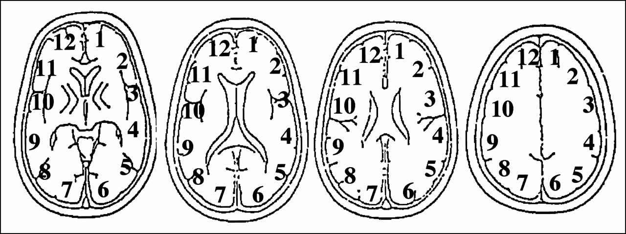
    ROIs used for the analysis of cerebral perfusion. ROIs were placed on six regions in the brain parenchyma of each hemisphere. The ROIs were obtained from four slices on 133Xe-SPECT scans and from two to four slices from PWI images. ROI 1 corresponded to the left ACA territory, ROI 2 to the left ABZ territory, ROIs 3 and 4 to the left MCA territory, ROI 5 to the left PBZ territory, and ROI 6 to the left PCA territory. ROIs 7 through 12 corresponded to the same territories in the right hemisphere. The ROI setting was consistent between 133Xe-SPECT and PWI. Data from 10 regions were obtained for each patient

  • fig 2.
    • Download figure
    • Open in new tab
    • Download powerpoint
    fig 2.

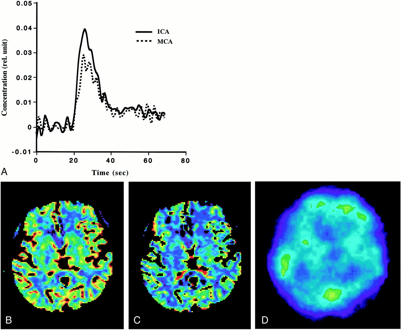
    Example of PWI and 133Xe-SPECT studies in patient 7.

    A, Comparison of the AIFs obtained from the ICA and MCA. Note that they vary slightly and that the AIF from the ICA is somewhat better than that from the MCA.

    B, rCBF image generated from PWI (rCBF-PWI).

    C, rCBV image generated from PWI (rCBV-PWI).

    D, 133Xe-SPECT scan. Note that images generated from PWI were superior to 133Xe-SPECT scan in spatial resolution, and that rCBF and rCBV values in the deep brain structure can be evaluated on rCBF-PWI and rCBV-PWI images.

  • fig 3.
    • Download figure
    • Open in new tab
    • Download powerpoint
    fig 3.

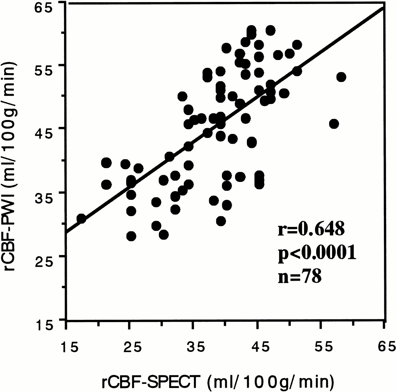
    Relationship between the rCBF values obtained from 133Xe-SPECT (rCBF-SPECT) and those obtained PWI (rCBF-PWI). A significant correlation was found between them (r = .648, P < .0001, n = 78)
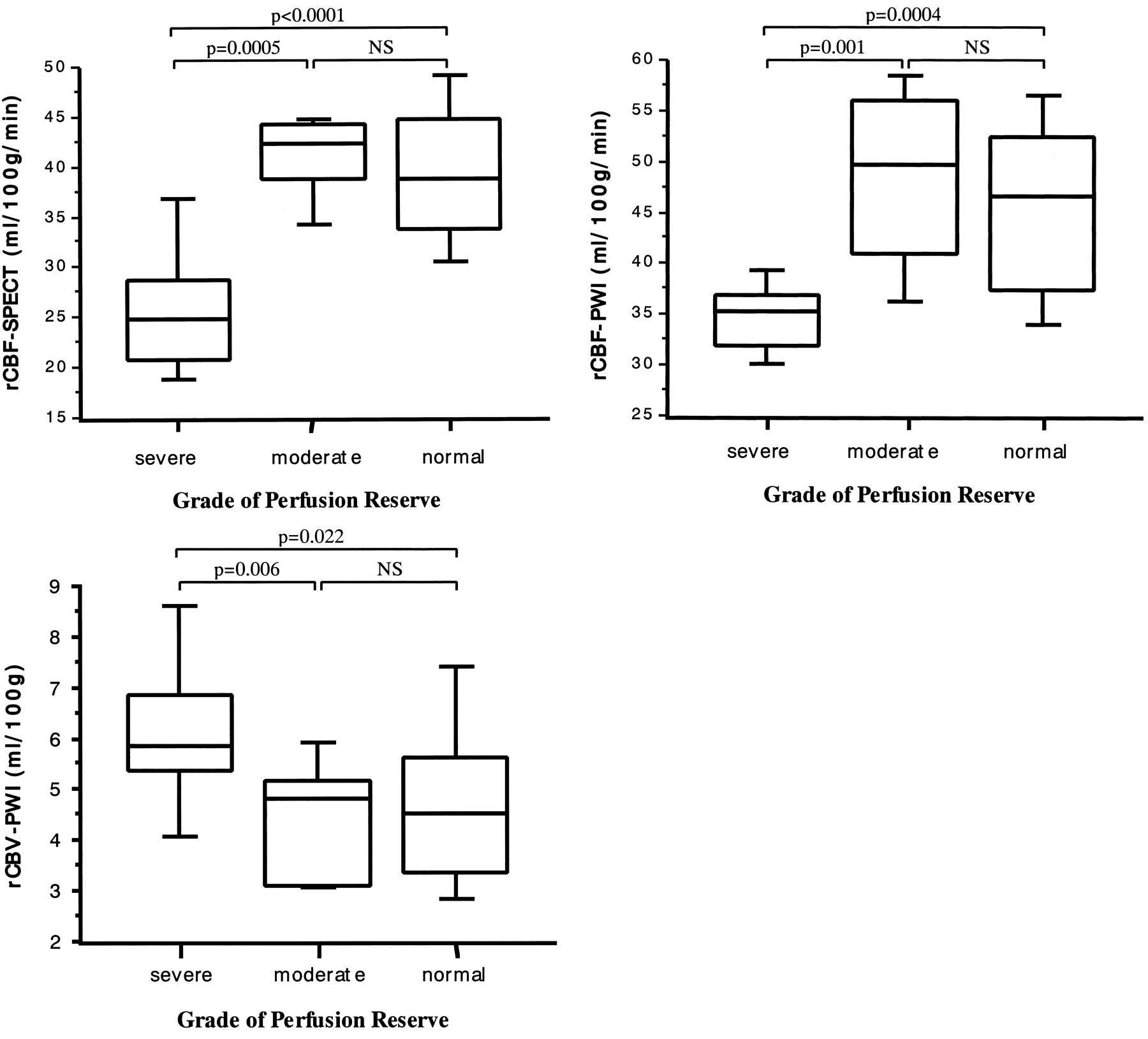
  • fig 4.
    • Download figure
    • Open in new tab
    • Download powerpoint
    fig 4.

    A–C, Box-and-whisker plot of rCBF-SPECT (A), rCBF-PWI (B), and rCBV-PWI (C) in the regions with severely decreased (% increase ≤ 0%), moderately decreased (0 < % increase ≤ 15%), and normal (15% < % increase) perfusion reserve. % increase was defined as [(post-rCBF) − (pre-rCBF)]/(pre-rCBF) × 100, where pre-rCBF and post-rCBF denote the CBF values before and after acetazolamide CBF as measured on 133Xe-SPECT scans, respectively. Boxes represent 25% to 70% range with bisecting lines showing medium values; horizontal lines represent 10% to 90% range; NS indicates not significant. Note that the regions with severely decreased perfusion reserve show significantly lower rCBF values on 133Xe-SPECT (A) and rCBF-PWI (B) studies than do those with moderately decreased or normal perfusion reserve. They also show significantly higher rCBV values on rCBV-PWI study (C) than do those with moderately decreased or normal perfusion reserve.

Tables

  • Figures
  • Table1

    Data for eight patients with occlusive carotid disease

PreviousNext
Back to top

In this issue

American Journal of Neuroradiology
Vol. 22, Issue 2
1 Feb 2001
  • Table of Contents
  • Index by author
Advertisement
Print
Download PDF
Email Article

Thank you for your interest in spreading the word on American Journal of Neuroradiology.

NOTE: We only request your email address so that the person you are recommending the page to knows that you wanted them to see it, and that it is not junk mail. We do not capture any email address.

Enter multiple addresses on separate lines or separate them with commas.
Measurement of Cerebral Hemodynamics with Perfusion-weighted MR Imaging: Comparison with Pre- and Post-acetazolamide 133Xe-SPECT in Occlusive Carotid Disease
(Your Name) has sent you a message from American Journal of Neuroradiology
(Your Name) thought you would like to see the American Journal of Neuroradiology web site.
CAPTCHA
This question is for testing whether or not you are a human visitor and to prevent automated spam submissions.
Cite this article
Keiichi Kikuchi, Kenya Murase, Hitoshi Miki, Takanori Kikuchi, Yoshifumi Sugawara, Teruhito Mochizuki, Junpei Ikezoe, Shiro Ohue
Measurement of Cerebral Hemodynamics with Perfusion-weighted MR Imaging: Comparison with Pre- and Post-acetazolamide 133Xe-SPECT in Occlusive Carotid Disease
American Journal of Neuroradiology Feb 2001, 22 (2) 248-254;

Citation Manager Formats

  • BibTeX
  • Bookends
  • EasyBib
  • EndNote (tagged)
  • EndNote 8 (xml)
  • Medlars
  • Mendeley
  • Papers
  • RefWorks Tagged
  • Ref Manager
  • RIS
  • Zotero
0 Responses
Respond to this article
Share
Bookmark this article
Measurement of Cerebral Hemodynamics with Perfusion-weighted MR Imaging: Comparison with Pre- and Post-acetazolamide 133Xe-SPECT in Occlusive Carotid Disease
Keiichi Kikuchi, Kenya Murase, Hitoshi Miki, Takanori Kikuchi, Yoshifumi Sugawara, Teruhito Mochizuki, Junpei Ikezoe, Shiro Ohue
American Journal of Neuroradiology Feb 2001, 22 (2) 248-254;
del.icio.us logo Twitter logo Facebook logo Mendeley logo
  • Tweet Widget
  • Facebook Like
  • Google Plus One

Jump to section

  • Article
    • Abstract
    • Methods
    • Results
    • Discussion
    • Conclusion
    • Footnotes
    • References
  • Figures & Data
  • Info & Metrics
  • Responses
  • References
  • PDF

Related Articles

  • No related articles found.
  • PubMed
  • Google Scholar

Cited By...

  • Computed tomography perfusion parameters predictive of symptomatic intracranial hemorrhage after mechanical thrombectomy in patients with cerebral large vessel occlusion
  • Progressive changes in cerebral perfusion after carotid stenting: a dynamic susceptibility contrast perfusion weighted imaging study
  • Elevated Cerebral Blood Volume Contributes to Increased FLAIR Signal in the Cerebral Sulci of Propofol-Sedated Children
  • Blood Pressure and Vascular Dysfunction Underlie Elevated Cerebral Blood Flow in Systemic Lupus Erythematosus
  • Vasodilatory Capacity of the Cerebral Vasculature in Patients with Carotid Artery Stenosis
  • Reporting standards for angioplasty and stent-assisted angioplasty for intracranial atherosclerosis
  • Elevated Cerebral Blood Flow and Volume in Systemic Lupus Measured by Dynamic Susceptibility Contrast Magnetic Resonance Imaging
  • Reporting Standards for Angioplasty and Stent-Assisted Angioplasty for Intracranial Atherosclerosis
  • Simple Assessment of Cerebral Hemodynamics Using Single-Slab 3D Time-of-Flight MR Angiography in Patients with Cervical Internal Carotid Artery Steno-Occlusive Diseases: Comparison with Quantitative Perfusion Single-Photon Emission CT
  • Quantitative Assessment of Cerebral Hemodynamics Using Perfusion-Weighted MRI in Patients With Major Cerebral Artery Occlusive Disease: Comparison With Positron Emission Tomography
  • Cerebral Hemodynamic Evaluation Using Perfusion-Weighted Magnetic Resonance Imaging: Comparison With Positron Emission Tomography Values in Chronic Occlusive Carotid Disease
  • Quantitative Measurement of Regional Cerebrovascular Reactivity to Acetazolamide Using 123I-N-Isopropyl-p-Iodoamphetamine Autoradiography with SPECT: Validation Study Using H215O with PET
  • Usefulness of Brain SPECT to Evaluate Brain Tolerance and Hemodynamic Changes During Temporary Balloon Occlusion Test and After Permanent Carotid Occlusion
  • Crossref
  • Google Scholar

This article has not yet been cited by articles in journals that are participating in Crossref Cited-by Linking.

More in this TOC Section

  • Progression of Microstructural Damage in Spinocerebellar Ataxia Type 2: A Longitudinal DTI Study
  • Usefulness of Quantitative Susceptibility Mapping for the Diagnosis of Parkinson Disease
  • Evaluating the Effects of White Matter Multiple Sclerosis Lesions on the Volume Estimation of 6 Brain Tissue Segmentation Methods
Show more Brain

Similar Articles

Advertisement

Indexed Content

  • Current Issue
  • Accepted Manuscripts
  • Article Preview
  • Past Issues
  • Editorials
  • Editors Choice
  • Fellow Journal Club
  • Letters to the Editor

Cases

  • Case Collection
  • Archive - Case of the Week
  • Archive - Case of the Month
  • Archive - Classic Case

Special Collections

  • Special Collections

Resources

  • News and Updates
  • Turn around Times
  • Submit a Manuscript
  • Author Policies
  • Manuscript Submission Guidelines
  • Evidence-Based Medicine Level Guide
  • Publishing Checklists
  • Graphical Abstract Preparation
  • Imaging Protocol Submission
  • Submit a Case
  • Become a Reviewer/Academy of Reviewers
  • Get Peer Review Credit from Publons

Multimedia

  • AJNR Podcast
  • AJNR SCANtastic
  • Video Articles

About Us

  • About AJNR
  • Editorial Board
  • Not an AJNR Subscriber? Join Now
  • Alerts
  • Feedback
  • Advertise with us
  • Librarian Resources
  • Permissions
  • Terms and Conditions

American Society of Neuroradiology

  • Not an ASNR Member? Join Now

© 2025 by the American Society of Neuroradiology All rights, including for text and data mining, AI training, and similar technologies, are reserved.
Print ISSN: 0195-6108 Online ISSN: 1936-959X

Powered by HighWire