
Article Figures & Data
Figures
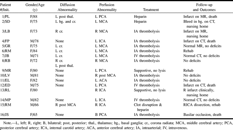
- fig 1.
Images from the case of patient 1, an 88-year-old woman.
A, Isotropic trace diffusion image (5900/159/2 [TR/TE/excitations]) shows normal signal (left). Time-to-peak perfusion map (2199/82/35) (right) shows hypoperfusion throughout the left posterior cerebral artery territory. Although these images were obtained within a therapeutic window, no symptoms referable to this distribution were encountered until hours later. The patient was treated “conservatively” in an intensive care unit and received heparin. The results were evident at the 48-hour follow-up examination.
B, Repeat isotropic trace diffusion image shows the progression to infarct rather than ischemia in the left posterior cerebral artery territory (left). A T2-weighted (3500/96/1) image (right) reveals not only infarct but also hemorrhage, with mass effect presumably from the combination of infarct and heparin
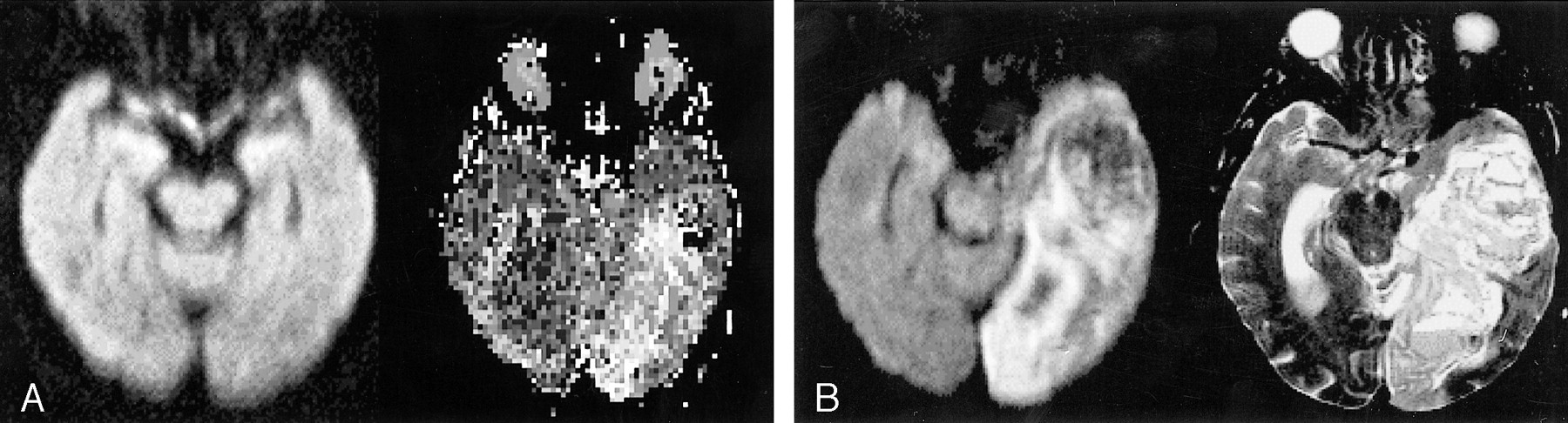
- fig 2.
Images from the case of patient 3, a 71-year-old man.
A, Three sections of isotropic trace diffusion images (5900/159/2) show abnormal increased signal in a contiguous 2-cm region of the left corona radiata, without any associated signal change in cortical regions.
B, Three time-to-peak perfusion maps (2199/82/35), obtained immediately after the diffusion images and at the same section positions, show slight hyperperfusion in the distribution of the abnormal diffusion signal shown in A. In addition, they show the otherwise unseen hypoperfusion throughout the left middle cerebral artery territory.
C, Representative sections from follow-up CT of the head, obtained 5 hours after the IV administration of thrombolytic therapy, show no infarct and no hemorrhage. The patient had no residual symptoms at the 48-hour follow-up examination
- fig 3.
Images from the case of patient 5, a 75-year-old woman.
A, Three sections of isotropic trace diffusion images (5900/159/2) show abnormal hyperintensities limited to the left corona radiata and deep frontal white matter.
B, Three time-to-peak perfusion maps (2199/82/35), obtained immediately after the diffusion images and at the same section positions, show marked hypoperfusion in the territory of the posterior division of the left middle cerebral artery.
C, Angiographic frames obtained before (left) and after (right) the intraarterial administration of thrombolytic therapy. Note the wedge of absent posterior vessels (to the right of the left image) compared with the addition of three large branches after therapy (white arrows).
D, Isotropic trace diffusion images, obtained at the 24-hour follow-up examination, reveal only scattered subcortical white matter hyperintensities (thin white arrows), without evidence for cortical infarct.
E, Follow-up perfusion maps obtained at the same levels show normal symmetrical time-to-peak effects


Tables

Clinical patient summary
In this issue


Jump to section
Related Articles
Cited By...
- Emerging impact of CTA/perfusion CT on acute stroke thrombolysis in a community hospital
- Management of Stroke in Infants and Children: A Scientific Statement From a Special Writing Group of the American Heart Association Stroke Council and the Council on Cardiovascular Disease in the Young
- Neuroimaging applications of multislice CT perfusion
- Guidelines and Recommendations for Perfusion Imaging in Cerebral Ischemia: A Scientific Statement for Healthcare Professionals by the Writing Group on Perfusion Imaging, From the Council on Cardiovascular Radiology of the American Heart Association
- Quantification of Perfusion Using Bolus Tracking Magnetic Resonance Imaging in Stroke: Assumptions, Limitations, and Potential Implications for Clinical Use