Advertisement

Main menu

  • Home
  • Content
    • Current Issue
    • Accepted Manuscripts
    • Article Preview
    • Past Issue Archive
    • AJNR Case Collection
    • Case of the Week Archive
    • Classic Case Archive
    • Case of the Month Archive
  • Special Collections
    • Spinal CSF Leak Articles (Jan 2020-June 2024)
    • 2024 AJNR Journal Awards
    • Most Impactful AJNR Articles
  • Multimedia
    • AJNR Podcast
    • AJNR Scantastics
    • Video Articles
  • For Authors
    • Submit a Manuscript
    • Author Policies
    • Fast publishing of Accepted Manuscripts
    • Graphical Abstract Preparation
    • Manuscript Submission Guidelines
    • Imaging Protocol Submission
    • Submit a Case for the Case Collection
  • About Us
    • About AJNR
    • Editorial Board
  • More
    • Become a Reviewer/Academy of Reviewers
    • Subscribers
    • Permissions
    • Alerts
    • Feedback
    • Advertisers
    • ASNR Home
  • Other Publications
    • ajnr

User menu

  • Alerts
  • Log in

Search

  • Advanced search
American Journal of Neuroradiology

American Journal of Neuroradiology

ASHNR American Society of Functional Neuroradiology ASHNR American Society of Pediatric Neuroradiology ASSR
  • Alerts
  • Log in

Advanced Search

  • Home
  • Content
    • Current Issue
    • Accepted Manuscripts
    • Article Preview
    • Past Issue Archive
    • AJNR Case Collection
    • Case of the Week Archive
    • Classic Case Archive
    • Case of the Month Archive
  • Special Collections
    • Spinal CSF Leak Articles (Jan 2020-June 2024)
    • 2024 AJNR Journal Awards
    • Most Impactful AJNR Articles
  • Multimedia
    • AJNR Podcast
    • AJNR Scantastics
    • Video Articles
  • For Authors
    • Submit a Manuscript
    • Author Policies
    • Fast publishing of Accepted Manuscripts
    • Graphical Abstract Preparation
    • Manuscript Submission Guidelines
    • Imaging Protocol Submission
    • Submit a Case for the Case Collection
  • About Us
    • About AJNR
    • Editorial Board
  • More
    • Become a Reviewer/Academy of Reviewers
    • Subscribers
    • Permissions
    • Alerts
    • Feedback
    • Advertisers
    • ASNR Home
  • Follow AJNR on Twitter
  • Visit AJNR on Facebook
  • Follow AJNR on Instagram
  • Join AJNR on LinkedIn
  • RSS Feeds

Welcome to the new AJNR, Updated Hall of Fame, and more. Read the full announcements.


AJNR is seeking candidates for the position of Associate Section Editor, AJNR Case Collection. Read the full announcement.


Research ArticleBRAIN

Abnormal Magnetization Transfer Ratios in Normal-appearing White Matter on Conventional MR Images of Patients with Occlusive Cerebrovascular Disease

Hirotsugu Kado, Hirohiko Kimura, Tatsuro Tsuchida, Yoshiharu Yonekura, Tomoo Tokime, Yasuhiko Tokuriki and Harumi Itoh
American Journal of Neuroradiology May 2001, 22 (5) 922-927;
Hirotsugu Kado
  • Find this author on Google Scholar
  • Find this author on PubMed
  • Search for this author on this site
Hirohiko Kimura
  • Find this author on Google Scholar
  • Find this author on PubMed
  • Search for this author on this site
Tatsuro Tsuchida
  • Find this author on Google Scholar
  • Find this author on PubMed
  • Search for this author on this site
Yoshiharu Yonekura
  • Find this author on Google Scholar
  • Find this author on PubMed
  • Search for this author on this site
Tomoo Tokime
  • Find this author on Google Scholar
  • Find this author on PubMed
  • Search for this author on this site
Yasuhiko Tokuriki
  • Find this author on Google Scholar
  • Find this author on PubMed
  • Search for this author on this site
Harumi Itoh
  • Find this author on Google Scholar
  • Find this author on PubMed
  • Search for this author on this site
  • Article
  • Figures & Data
  • Info & Metrics
  • Responses
  • References
  • PDF
Loading

Article Figures & Data

Figures

  • Tables
  • fig 1.
    • Download figure
    • Open in new tab
    • Download powerpoint
    fig 1.

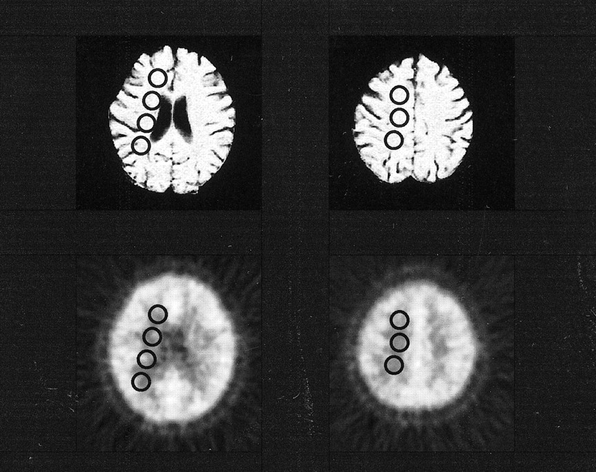
    The location of selected ROIs. Four ROIs were selected in the lesional deep white matter and three in the lesional centrum semiovale. Although this figure shows that ROIs were placed only on MTR maps and CBF images, actually the same ROIs were selected on T2-weighted images and other PET scans (CMRO2 and OEF images). PET scans were registered to the MR images of each subject. Circular ROIs were placed in right and left symmetrical locations, so that the AI could be calculated

  • fig 2.
    • Download figure
    • Open in new tab
    • Download powerpoint
    fig 2.

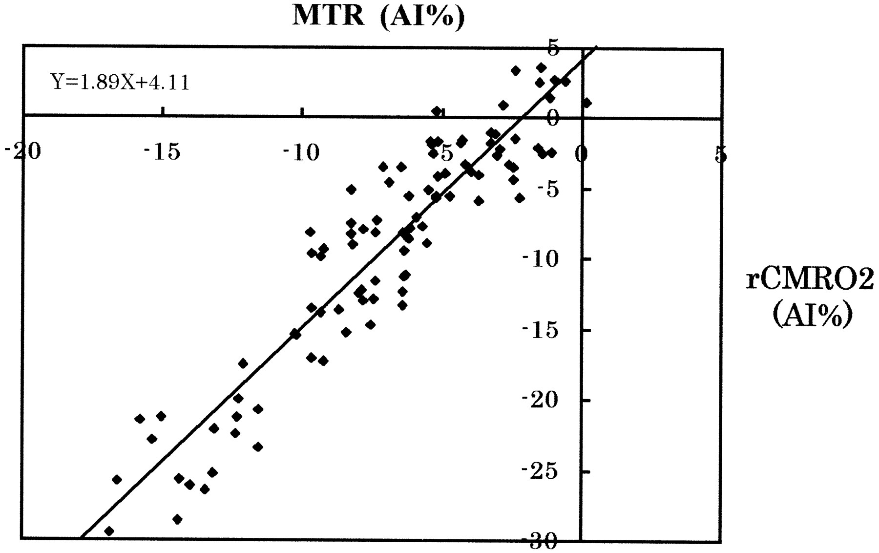
    2D plot of MTR and rCMRO2 values with an AI from groups 0, 1, and 2. The correlation coefficient was .85 (P = .001)

Tables

  • Figures
  • TABLE 1:
    • View popup
    • Download powerpoint
    TABLE 1:

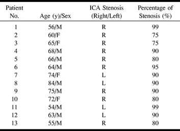
    Demographic and clinical data for 13 patient with unilateral severe stenosis of the internal carotid artery (ICA)

  • TABLE 2:
    • View popup
    • Download powerpoint
    TABLE 2:

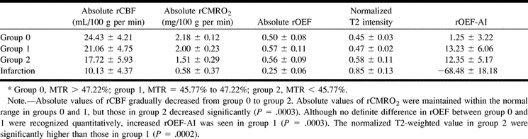
    Mean values of absolute regional cerebral blood flow (rCBF), absolute regional cerebral metabolic rate of oxygen (rCMRO2), absolute regional oxygen extraction fraction (rOEF), normalized T2-weighted images, and rOEF asymmetric index (AI) from the lesional side in 13 patients with unilateral severe stenosis of the internal carotid artery by group*

PreviousNext
Back to top

In this issue

American Journal of Neuroradiology
Vol. 22, Issue 5
1 May 2001
  • Table of Contents
  • Index by author
Advertisement
Print
Download PDF
Email Article

Thank you for your interest in spreading the word on American Journal of Neuroradiology.

NOTE: We only request your email address so that the person you are recommending the page to knows that you wanted them to see it, and that it is not junk mail. We do not capture any email address.

Enter multiple addresses on separate lines or separate them with commas.
Abnormal Magnetization Transfer Ratios in Normal-appearing White Matter on Conventional MR Images of Patients with Occlusive Cerebrovascular Disease
(Your Name) has sent you a message from American Journal of Neuroradiology
(Your Name) thought you would like to see the American Journal of Neuroradiology web site.
CAPTCHA
This question is for testing whether or not you are a human visitor and to prevent automated spam submissions.
Cite this article
Hirotsugu Kado, Hirohiko Kimura, Tatsuro Tsuchida, Yoshiharu Yonekura, Tomoo Tokime, Yasuhiko Tokuriki, Harumi Itoh
Abnormal Magnetization Transfer Ratios in Normal-appearing White Matter on Conventional MR Images of Patients with Occlusive Cerebrovascular Disease
American Journal of Neuroradiology May 2001, 22 (5) 922-927;

Citation Manager Formats

  • BibTeX
  • Bookends
  • EasyBib
  • EndNote (tagged)
  • EndNote 8 (xml)
  • Medlars
  • Mendeley
  • Papers
  • RefWorks Tagged
  • Ref Manager
  • RIS
  • Zotero
0 Responses
Respond to this article
Share
Bookmark this article
Abnormal Magnetization Transfer Ratios in Normal-appearing White Matter on Conventional MR Images of Patients with Occlusive Cerebrovascular Disease
Hirotsugu Kado, Hirohiko Kimura, Tatsuro Tsuchida, Yoshiharu Yonekura, Tomoo Tokime, Yasuhiko Tokuriki, Harumi Itoh
American Journal of Neuroradiology May 2001, 22 (5) 922-927;
del.icio.us logo Twitter logo Facebook logo Mendeley logo
  • Tweet Widget
  • Facebook Like
  • Google Plus One

Jump to section

  • Article
    • Abstract
    • Methods
    • Results
    • Discussion
    • Conclusion
    • Footnotes
    • References
  • Figures & Data
  • Info & Metrics
  • Responses
  • References
  • PDF

Related Articles

  • No related articles found.
  • PubMed
  • Google Scholar

Cited By...

  • Extent of Microstructural Tissue Damage Correlates with Hemodynamic Failure in High-Grade Carotid Occlusive Disease: An MRI Study Using Quantitative T2 and DSC Perfusion
  • Magnetization Transfer Imaging Shows Tissue Abnormalities in the Reversible Penumbra
  • Crossref
  • Google Scholar

This article has not yet been cited by articles in journals that are participating in Crossref Cited-by Linking.

More in this TOC Section

  • Progression of Microstructural Damage in Spinocerebellar Ataxia Type 2: A Longitudinal DTI Study
  • Usefulness of Quantitative Susceptibility Mapping for the Diagnosis of Parkinson Disease
  • Evaluating the Effects of White Matter Multiple Sclerosis Lesions on the Volume Estimation of 6 Brain Tissue Segmentation Methods
Show more Brain

Similar Articles

Advertisement

Indexed Content

  • Current Issue
  • Accepted Manuscripts
  • Article Preview
  • Past Issues
  • Editorials
  • Editors Choice
  • Fellow Journal Club
  • Letters to the Editor

Cases

  • Case Collection
  • Archive - Case of the Week
  • Archive - Case of the Month
  • Archive - Classic Case

Special Collections

  • Special Collections

Resources

  • News and Updates
  • Turn around Times
  • Submit a Manuscript
  • Author Policies
  • Manuscript Submission Guidelines
  • Evidence-Based Medicine Level Guide
  • Publishing Checklists
  • Graphical Abstract Preparation
  • Imaging Protocol Submission
  • Submit a Case
  • Become a Reviewer/Academy of Reviewers
  • Get Peer Review Credit from Publons

Multimedia

  • AJNR Podcast
  • AJNR SCANtastic
  • Video Articles

About Us

  • About AJNR
  • Editorial Board
  • Not an AJNR Subscriber? Join Now
  • Alerts
  • Feedback
  • Advertise with us
  • Librarian Resources
  • Permissions
  • Terms and Conditions

American Society of Neuroradiology

  • Not an ASNR Member? Join Now

© 2025 by the American Society of Neuroradiology All rights, including for text and data mining, AI training, and similar technologies, are reserved.
Print ISSN: 0195-6108 Online ISSN: 1936-959X

Powered by HighWire