
Article Figures & Data
Figures
- fig 1.
A 67-year-old patient with astrocytoma grade II. MR examination 36 months after stereotactic radiotherapy showed two contrast-enhancing lesions on T1-weighted spin-echo images.
A and C, A lesion with ring-shaped enhancement zone is seen in the left frontal lobe (A) and a lesion with homogeneous enhancement in seen in the left temporal lobe (C) (boxes indicate MRS voxels).
B and D, 1H MR spectra [double spin-echo sequence 1500/135/200 (TR/TE/excitations)] of both lesions show a peak attributed to free lipids, indicating necrosis. An intense tCho resonance is only seen in the lesion in the left temporal lobe. FDG-PET revealed high glucose uptake in this region, indicating tumor progression. Low tCho signal was found in the lesion in the left frontal lobe. Radionecrosis in this region was confirmed by biopsy.
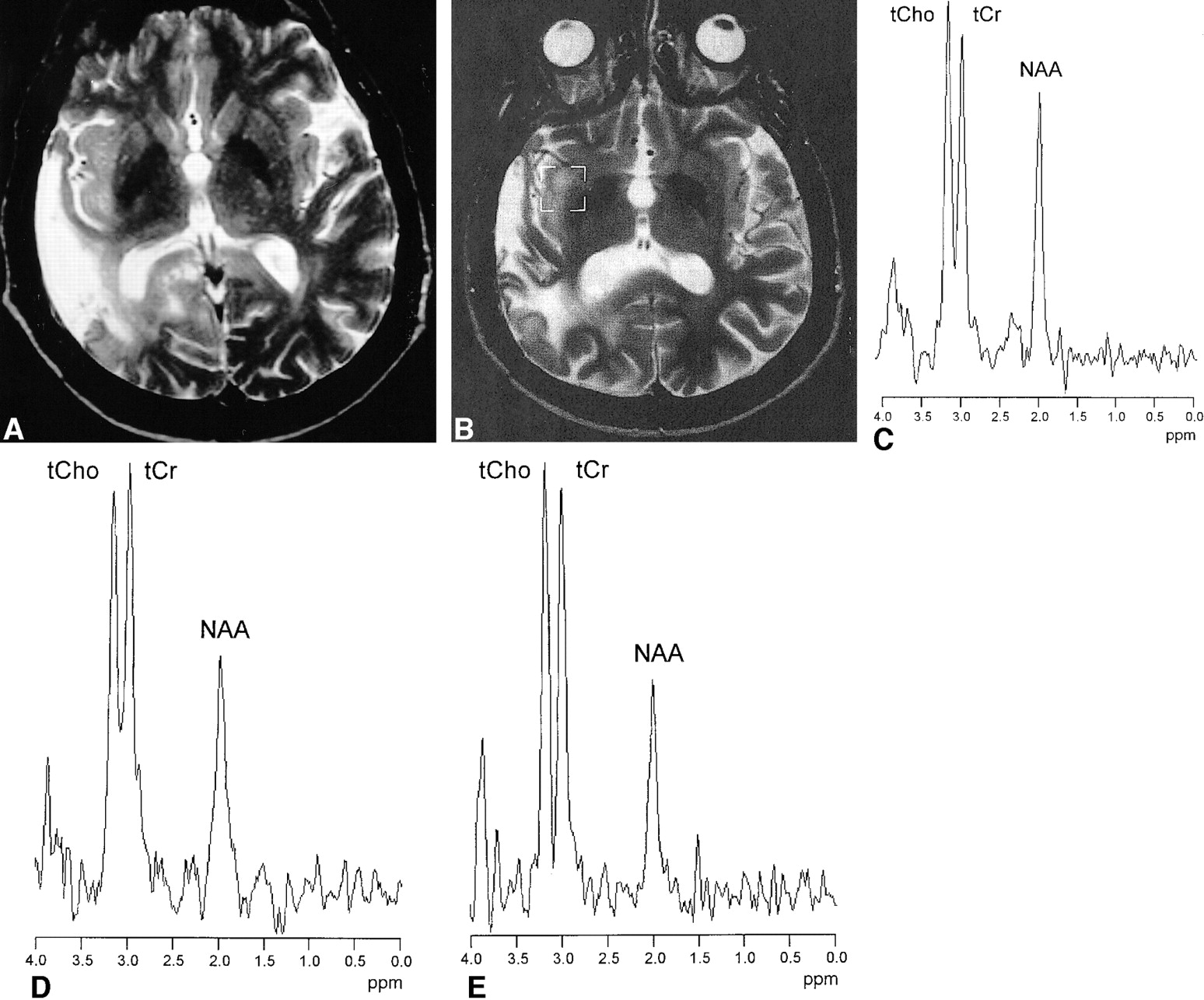
- fig 2.
A 36-year-old patient with astrocytoma II in the right temporo-parietal lobe, examined before (A) and 20 months after stereotactic radiotherapy (B). During follow-up, an irregular contrast-enhancing lesion appeared at the upper region of the irradiated volume (B) with complete involution 13 months later (D). 1H MR spectra (double spin-echo sequence 1500/135/200) (C) show initially elevated ItCho/ItCr and ItCho/INAA values and a strong lipid resonance. Thirteen months later, the spectrum indicates normal brain tissue (E)
- fig 3.
A 54-year-old patient with astrocytoma II of the right temporal lobe after surgery and stereotactic radiotherapy. T2-weighted turbo spin-echo images obtained before (A) and 9 months after radiotherapy (B) show a hyperintense lesion within the irradiated insula on the right hemisphere, a surgical tissue defect in the right temporal, and gliosis in the right occipital lobe (T1-weighted spin-echo images showed no uptake of contrast medium.) Corresponding 1H MR spectra (double spin-echo sequence 1500/135/200) acquired 9, 15, and 21 months after stereotactic radiotherapy (C–E) demonstrated similar signal intensities of tCho and tCr, with only minor changes during follow-up. MR imaging follow-up showed no further change of the lesion consistent with diagnosis of nonneoplastic lesion
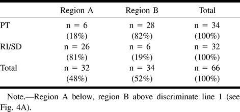
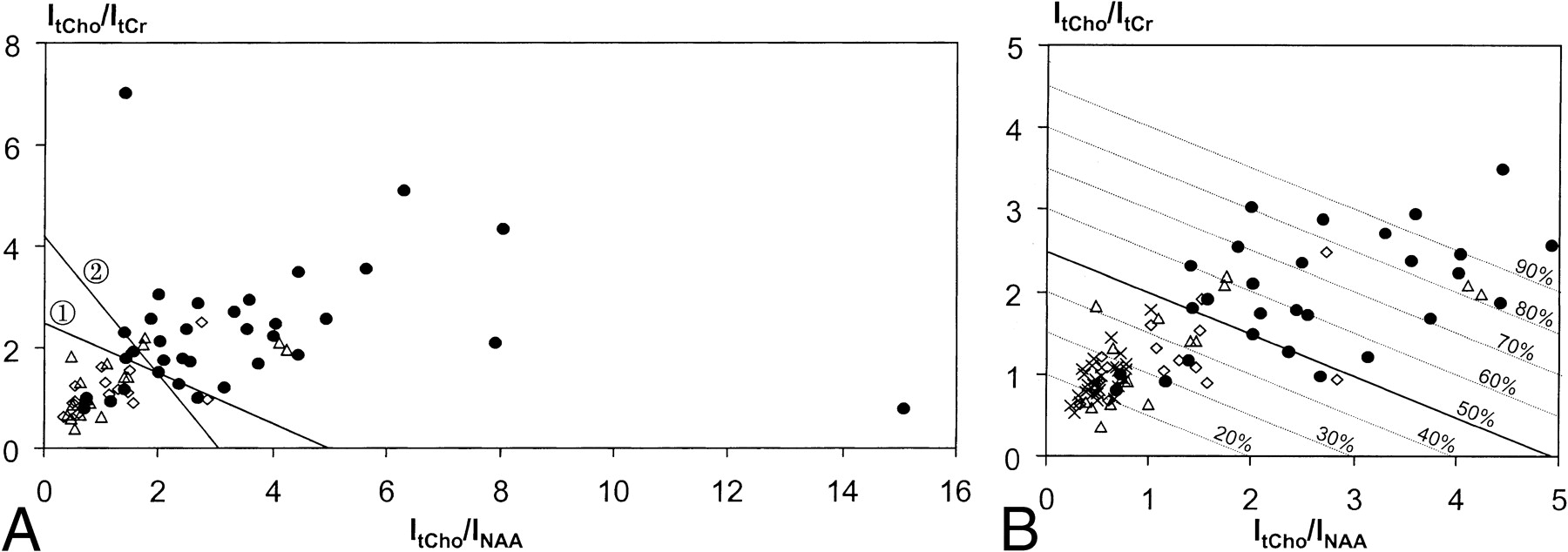
- fig 4.
a, Ratios of 1H MR signal intensities (double spin-echo sequence 1500/135/200–300) of total choline and total creatine (ItCho/ItCr) versus ratios of intensities of total choline and NAA (ItCho/INAA) observed in lesions classified as progressive tumor (●, n = 35), radiation injury (Δ, n = 17), and stable disease (⋄, n = 15). The straight lines: (ItCho/ItCr) = 2.5–0.5 × (ItCho/INAA) (line 1) and (ItCho/ItCr) = 4.2–1.35 × (ItCho/INAA) (line 2) obtained from linear discriminant analysis and 7-point neighbor method, respectively, differentiate neoplastic and nonneoplastic lesions.
b, Detail of (a) with ratios ≤ 5.0 together with ItCho/ItCr and ItCho/INAA values of normal brain (×, n = 33, voxel in the contralateral unaffected side). Parallel lines obtained from linear discriminant analysis are labeled with the probability of PT.


Tables
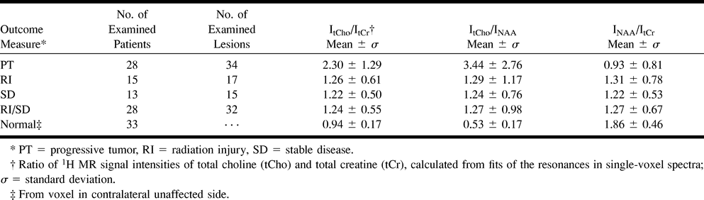
 TABLE 1:
Mean 1H MR signal intensity ratios from lesions and contralateral normal brain tissue
- TABLE 2:
P values from two-sample t test between 1H MR signal intensity ratios of different lesions and contralateral normal brain
- TABLE 3:
Retrospective classification of lesions using linear discriminant analysis

In this issue


Jump to section
Related Articles
Cited By...
- MR Spectroscopy in Radiation Injury
- Distinguishing Recurrent Intra-Axial Metastatic Tumor from Radiation Necrosis Following Gamma Knife Radiosurgery Using Dynamic Susceptibility-Weighted Contrast-Enhanced Perfusion MR Imaging
- MGMT Promoter Methylation Status Can Predict the Incidence and Outcome of Pseudoprogression After Concomitant Radiochemotherapy in Newly Diagnosed Glioblastoma Patients
- Diagnostic performance of spectroscopic and perfusion MRI for distinction of brain tumors
- Physiologic and Metabolic Magnetic Resonance Imaging in Gliomas
- PET and SPECT for Detection of Tumor Progression in Irradiated Low-Grade Astrocytoma: A Receiver-Operating-Characteristic Analysis