
Article Figures & Data
Figures
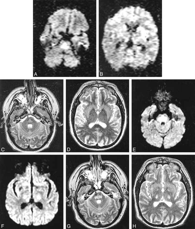
- fig 1.
Case 1. MR images obtained using a 1.5-T magnet. Images for study 1 (A–D) were taken in an oblique axial plane 6 days after development of tetraplegia. Images for study 2 (E–H) were taken in an axial plane 21 days after development of tetraplegia.
A, DW image shows increased signal intensity in pons due to restricted water diffusion.
B, DW image is normal in the internal capsules bilaterally.
C and D, In the pons, there is little hyperintensity on the T2-weighted images and low ADC values, with mean ADC in basis pontis being 0.39 ± 0.14 × 10⁻³ mm2/s (mean ± SD) compared with 0.90 ± 0.15 × 10⁻³ mm2/s in unaffected white matter.
E, DW image shows increased signal intensity still present in the pons.
F, The DW image shows increased signal intensity present in the internal capsules bilaterally.
G and H, The DW abnormality is accounted for by shine-through from the hyperintense T2-weighted images. Mean ADC in basis pontis (1.09 ± 0.10 × 10⁻³ mm2/s) is normal compared with that of unaffected white matter (1.05 ± 0.09 × 10⁻³ mm2/s).
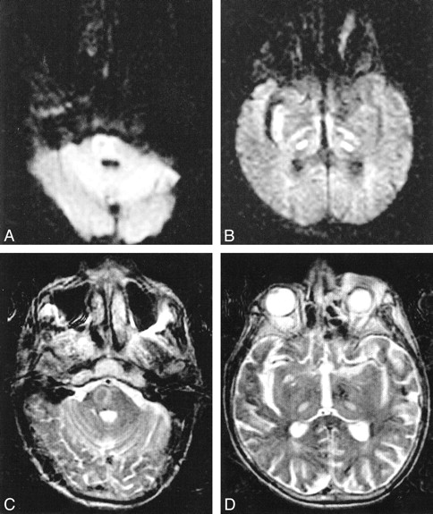
- fig 2.
Case 2. Images obtained in the axial plane 7 days after development of tetraplegia. DW (A and B) and T2-weighted (C and D) images show increased signal intensity within the pons and the thalami bilaterally, consistent with CPM with extrapontine involvement. ADC is decreased in affected pontine (0.62 ± 0.11 × 10⁻³ mm2/s) and thalamic (0.43 ± 0.09 × 10⁻³ mm2/s) regions compared with that of unaffected white matter (0.82 ± 0.12 × 10⁻³ mm2/s). The patient died before repeat images could be obtained


