Advertisement

Main menu

  • Home
  • Content
    • Current Issue
    • Accepted Manuscripts
    • Article Preview
    • Past Issue Archive
    • AJNR Case Collection
    • Case of the Week Archive
    • Classic Case Archive
    • Case of the Month Archive
  • Special Collections
    • Spinal CSF Leak Articles (Jan 2020-June 2024)
    • 2024 AJNR Journal Awards
    • Most Impactful AJNR Articles
  • Multimedia
    • AJNR Podcast
    • AJNR Scantastics
    • Video Articles
  • For Authors
    • Submit a Manuscript
    • Author Policies
    • Fast publishing of Accepted Manuscripts
    • Graphical Abstract Preparation
    • Manuscript Submission Guidelines
    • Imaging Protocol Submission
    • Submit a Case for the Case Collection
  • About Us
    • About AJNR
    • Editorial Board
  • More
    • Become a Reviewer/Academy of Reviewers
    • Subscribers
    • Permissions
    • Alerts
    • Feedback
    • Advertisers
    • ASNR Home
  • Other Publications
    • ajnr

User menu

  • Alerts
  • Log in

Search

  • Advanced search
American Journal of Neuroradiology

American Journal of Neuroradiology

ASHNR American Society of Functional Neuroradiology ASHNR American Society of Pediatric Neuroradiology ASSR
  • Alerts
  • Log in

Advanced Search

  • Home
  • Content
    • Current Issue
    • Accepted Manuscripts
    • Article Preview
    • Past Issue Archive
    • AJNR Case Collection
    • Case of the Week Archive
    • Classic Case Archive
    • Case of the Month Archive
  • Special Collections
    • Spinal CSF Leak Articles (Jan 2020-June 2024)
    • 2024 AJNR Journal Awards
    • Most Impactful AJNR Articles
  • Multimedia
    • AJNR Podcast
    • AJNR Scantastics
    • Video Articles
  • For Authors
    • Submit a Manuscript
    • Author Policies
    • Fast publishing of Accepted Manuscripts
    • Graphical Abstract Preparation
    • Manuscript Submission Guidelines
    • Imaging Protocol Submission
    • Submit a Case for the Case Collection
  • About Us
    • About AJNR
    • Editorial Board
  • More
    • Become a Reviewer/Academy of Reviewers
    • Subscribers
    • Permissions
    • Alerts
    • Feedback
    • Advertisers
    • ASNR Home
  • Follow AJNR on Twitter
  • Visit AJNR on Facebook
  • Follow AJNR on Instagram
  • Join AJNR on LinkedIn
  • RSS Feeds

Welcome to the new AJNR, Updated Hall of Fame, and more. Read the full announcements.


AJNR is seeking candidates for the position of Associate Section Editor, AJNR Case Collection. Read the full announcement.


Research ArticleBRAIN

Normalization of Cerebral Volumes by Use of Intracranial Volume: Implications for Longitudinal Quantitative MR Imaging

Jennifer L. Whitwell, William R. Crum, Hilary C. Watt and Nick C. Fox
American Journal of Neuroradiology September 2001, 22 (8) 1483-1489;
Jennifer L. Whitwell
  • Find this author on Google Scholar
  • Find this author on PubMed
  • Search for this author on this site
William R. Crum
  • Find this author on Google Scholar
  • Find this author on PubMed
  • Search for this author on this site
Hilary C. Watt
  • Find this author on Google Scholar
  • Find this author on PubMed
  • Search for this author on this site
Nick C. Fox
  • Find this author on Google Scholar
  • Find this author on PubMed
  • Search for this author on this site
  • Article
  • Figures & Data
  • Info & Metrics
  • Responses
  • References
  • PDF
Loading

Article Figures & Data

Figures

  • Tables
  • fig 1.
    • Download figure
    • Open in new tab
    • Download powerpoint
    fig 1.

    T1-weighted MR images with sagittal and axial views. The intensity windowing is as used for segmentation. The TIV is calculated by summation and linear interpolation of the segmented axial slices.

    A, The total intracranial area is shown on one axial section.

    B, The axial sections used to sample the total intracranial volume are marked on the sagittal view.

  • fig 2.
    • Download figure
    • Open in new tab
    • Download powerpoint
    fig 2.

    The relationships between TIV and age (A), brain volume and age (B), and normalized brain volume and age (C), in healthy controls

  • fig 3.
    • Download figure
    • Open in new tab
    • Download powerpoint
    fig 3.

    Brain volumes and normalized brain volumes in male (M) and female (F) controls with group averages marked, showing a reduction in sex-dependent differences after TIV normalization.fig 4. Comparison of TIV measurements from T1- and T2-weighted MR images in five controls and five AD patients.fig 5. Serial T1-weighted TIV measurements in five controls and five AD patients
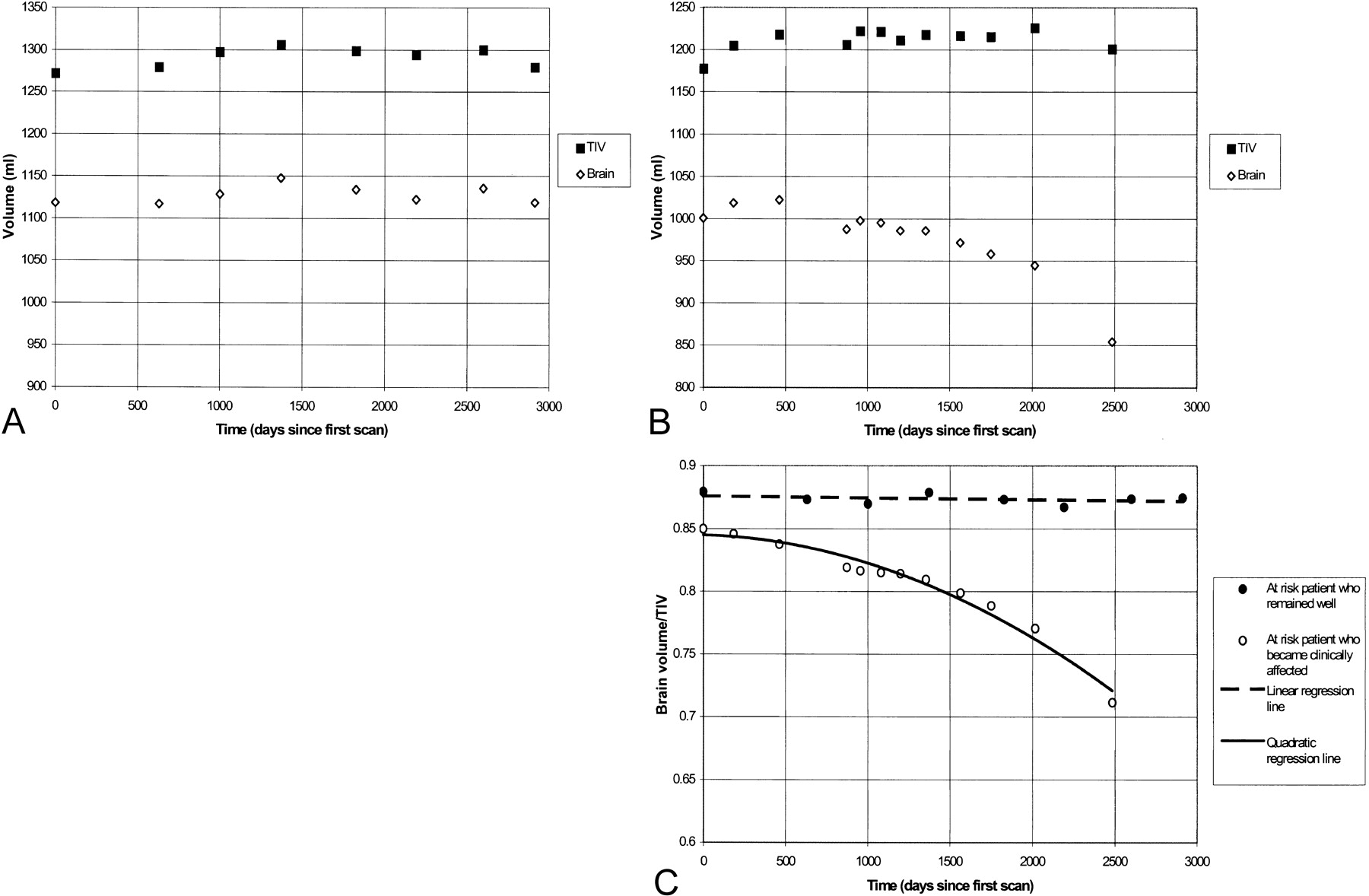
  • fig 6.
    • Download figure
    • Open in new tab
    • Download powerpoint
    fig 6.

    Serial TIV and brain volumes in an at-risk patient who remained well (A) and an at-risk patient who developed AD (B), compared with normalized brain volumes from serial images in both of the patients at risk (C)

Tables

  • Figures
  • Table1

    Reproducibilities and operator times

PreviousNext
Back to top

In this issue

American Journal of Neuroradiology: 22 (8)
American Journal of Neuroradiology
Vol. 22, Issue 8
1 Sep 2001
  • Table of Contents
  • Index by author
Advertisement
Print
Download PDF
Email Article

Thank you for your interest in spreading the word on American Journal of Neuroradiology.

NOTE: We only request your email address so that the person you are recommending the page to knows that you wanted them to see it, and that it is not junk mail. We do not capture any email address.

Enter multiple addresses on separate lines or separate them with commas.
Normalization of Cerebral Volumes by Use of Intracranial Volume: Implications for Longitudinal Quantitative MR Imaging
(Your Name) has sent you a message from American Journal of Neuroradiology
(Your Name) thought you would like to see the American Journal of Neuroradiology web site.
CAPTCHA
This question is for testing whether or not you are a human visitor and to prevent automated spam submissions.
Cite this article
Jennifer L. Whitwell, William R. Crum, Hilary C. Watt, Nick C. Fox
Normalization of Cerebral Volumes by Use of Intracranial Volume: Implications for Longitudinal Quantitative MR Imaging
American Journal of Neuroradiology Sep 2001, 22 (8) 1483-1489;

Citation Manager Formats

  • BibTeX
  • Bookends
  • EasyBib
  • EndNote (tagged)
  • EndNote 8 (xml)
  • Medlars
  • Mendeley
  • Papers
  • RefWorks Tagged
  • Ref Manager
  • RIS
  • Zotero
0 Responses
Respond to this article
Share
Bookmark this article
Normalization of Cerebral Volumes by Use of Intracranial Volume: Implications for Longitudinal Quantitative MR Imaging
Jennifer L. Whitwell, William R. Crum, Hilary C. Watt, Nick C. Fox
American Journal of Neuroradiology Sep 2001, 22 (8) 1483-1489;
del.icio.us logo Twitter logo Facebook logo Mendeley logo
  • Tweet Widget
  • Facebook Like
  • Google Plus One

Jump to section

  • Article
    • Abstract
    • Methods
    • Results
    • Discussion
    • Conclusion
    • Acknowledgments
    • Footnotes
    • References
  • Figures & Data
  • Info & Metrics
  • Responses
  • References
  • PDF

Related Articles

  • No related articles found.
  • PubMed
  • Google Scholar

Cited By...

  • Choroid Plexus Enlargement in Secondary Progressive MS: phenotype comparison
  • Lifetime brain atrophy estimated from a single MRI: measurement characteristics and genome-wide correlates
  • Anatomical Abnormalities Suggest a Compensatory Role of the Cerebellum in Early Parkinsons Disease
  • AutoMacq: an automatic pipeline to analyse macaque structural MRI data
  • Brain age gap in neuromyelitis optica spectrum disorders and multiple sclerosis
  • Comparison of Structural Changes in Nodding Syndrome and Other Epilepsies Associated With Onchocerca volvulus
  • A comparison of intracranial volume estimation methods and their cross-sectional and longitudinal associations with age
  • Hyper-adaptation in the Human Brain: Functional and structural changes in the foot section of the primary motor cortex in a top wheelchair racing Paralympian
  • Identifying neurocognitive outcomes and cerebral oxygenation in critically ill adults on acute kidney replacement therapy in the intensive care unit: the INCOGNITO-AKI study protocol
  • Network diffusion model predicts neurodegeneration in limb-onset amyotrophic lateral sclerosis
  • Association of Infarct Volume Before Hemicraniectomy and Outcome After Malignant Infarction
  • Functional plasticity coupled with structural predispositions in auditory cortex shape successful music category learning
  • Interoception Primes Emotional Processing: Multimodal Evidence from Neurodegeneration
  • Kynurenine pathway metabolites in cerebrospinal fluid and blood as potential biomarkers in Huntingtons disease
  • Neuroanatomical predictors of response to subcallosal cingulate deep brain stimulation for treatment-resistant depression
  • White Matter Connectivity Abnormalities in Prediabetes and Type 2 Diabetes: The Maastricht Study
  • Multimodal Hippocampal Subfield Grading For Alzheimers Disease Classification
  • Evaluation of mutant huntingtin and neurofilament proteins as potential markers in Huntingtons disease
  • Neurofilament light protein in blood predicts regional atrophy in Huntington disease
  • How Does the Accuracy of Intracranial Volume Measurements Affect Normalized Brain Volumes? Sample Size Estimates Based on 966 Subjects from the HUNT MRI Cohort
  • Effects of Gadolinium Contrast Agent Administration on Automatic Brain Tissue Classification of Patients with Multiple Sclerosis
  • Interhemispheric Functional Connectivity following Prenatal or Perinatal Brain Injury Predicts Receptive Language Outcome
  • Flavour identification in frontotemporal lobar degeneration
  • Quantitative Assessment of Brain Stem and Cerebellar Atrophy in Spinocerebellar Ataxia Types 3 and 6: Impact on Clinical Status
  • The progression of regional atrophy in premanifest and early Huntington's disease: a longitudinal voxel-based morphometry study
  • Onset and Progression of Pathologic Atrophy in Huntington Disease: A Longitudinal MR Imaging Study
  • Pitfalls in the Use of Voxel-Based Morphometry as a Biomarker: Examples from Huntington Disease
  • Magnetization Transfer Ratio in Alzheimer Disease: Comparison with Volumetric Measurements
  • Increased rate of whole-brain atrophy over 6 months in early Huntington disease.
  • Effects of A{beta} immunization (AN1792) on MRI measures of cerebral volume in Alzheimer disease
  • Parkinson disease, brain volumes, and subthalamic nucleus stimulation
  • Quantitative MRI measurement of superior cerebellar peduncle in progressive supranuclear palsy
  • Mapping the onset and progression of atrophy in familial frontotemporal lobar degeneration
  • Pontine atrophy precedes cerebellar degeneration in spinocerebellar ataxia 7: MRI-based volumetric analysis
  • Do cognitive patterns of brain magnetic activity correlate with hippocampal atrophy in Alzheimer's disease?
  • Crossref
  • Google Scholar

This article has not yet been cited by articles in journals that are participating in Crossref Cited-by Linking.

More in this TOC Section

  • Fast Contrast-Enhanced 4D MRA and 4D Flow MRI Using Constrained Reconstruction (HYPRFlow): Potential Applications for Brain Arteriovenous Malformations
  • Quiet PROPELLER MRI Techniques Match the Quality of Conventional PROPELLER Brain Imaging Techniques
  • Predictors of Reperfusion in Patients with Acute Ischemic Stroke
Show more Brain

Similar Articles

Advertisement

Indexed Content

  • Current Issue
  • Accepted Manuscripts
  • Article Preview
  • Past Issues
  • Editorials
  • Editors Choice
  • Fellow Journal Club
  • Letters to the Editor

Cases

  • Case Collection
  • Archive - Case of the Week
  • Archive - Case of the Month
  • Archive - Classic Case

Special Collections

  • Special Collections

Resources

  • News and Updates
  • Turn around Times
  • Submit a Manuscript
  • Author Policies
  • Manuscript Submission Guidelines
  • Evidence-Based Medicine Level Guide
  • Publishing Checklists
  • Graphical Abstract Preparation
  • Imaging Protocol Submission
  • Submit a Case
  • Become a Reviewer/Academy of Reviewers
  • Get Peer Review Credit from Publons

Multimedia

  • AJNR Podcast
  • AJNR SCANtastic
  • Video Articles

About Us

  • About AJNR
  • Editorial Board
  • Not an AJNR Subscriber? Join Now
  • Alerts
  • Feedback
  • Advertise with us
  • Librarian Resources
  • Permissions
  • Terms and Conditions

American Society of Neuroradiology

  • Not an ASNR Member? Join Now

© 2025 by the American Society of Neuroradiology All rights, including for text and data mining, AI training, and similar technologies, are reserved.
Print ISSN: 0195-6108 Online ISSN: 1936-959X

Powered by HighWire