
Article Figures & Data
Figures
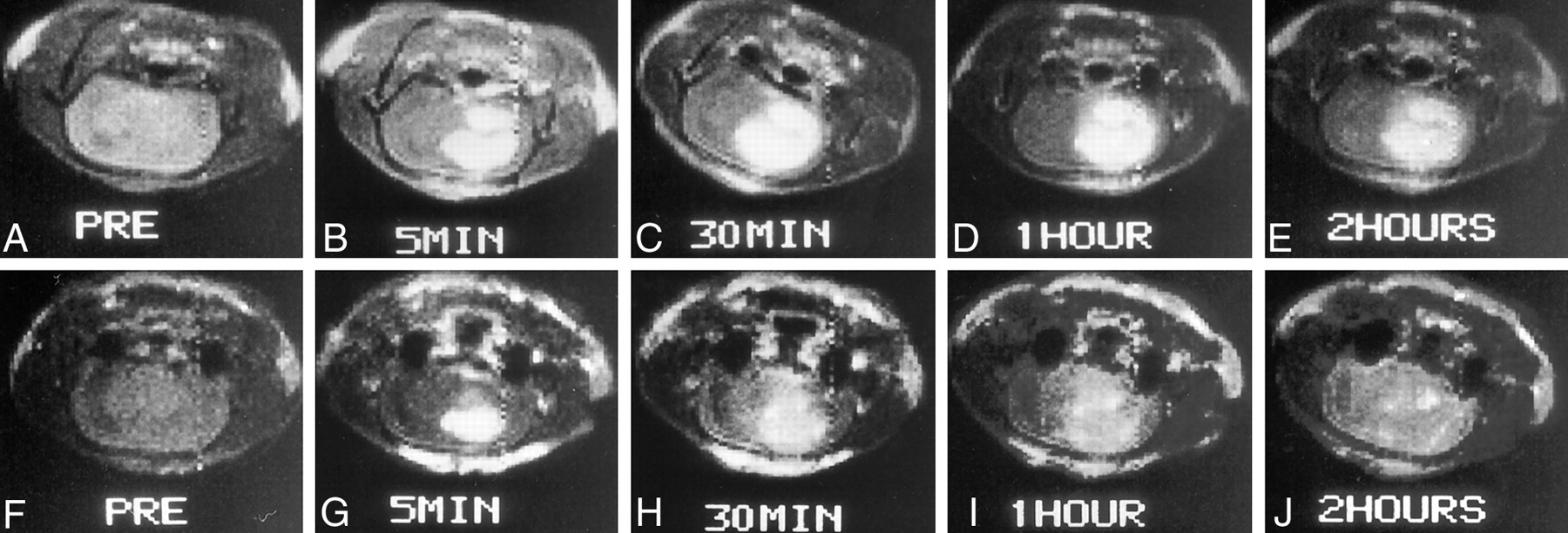
- Fig 1.
Serial T1-weighted spin-echo MR images (514/28) obtained before and at 5 minutes, 30 minutes, 1 hour, and 2 hours after contrast agent administration.
A–E, Gd-BOPTA was the contrast agent used.
F–J, Gd-DTPA was the contrast agent used.
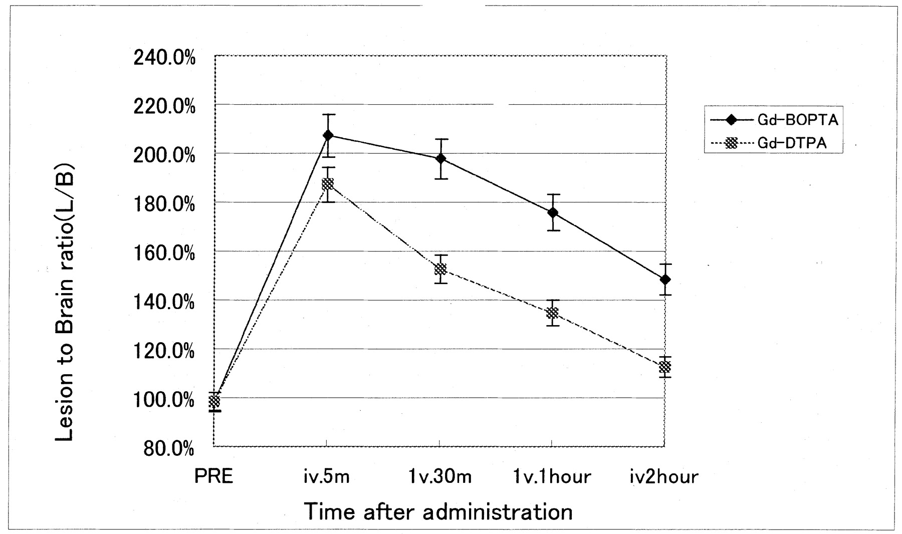
- Fig 2.
L/B time profiles after IV administration of 0.1 mmol/kg of Gd-BOPTA and Gd-DTPA during T1-weighted imaging (514/28) at 2.4 T. Data are mean ± SD of six animals.
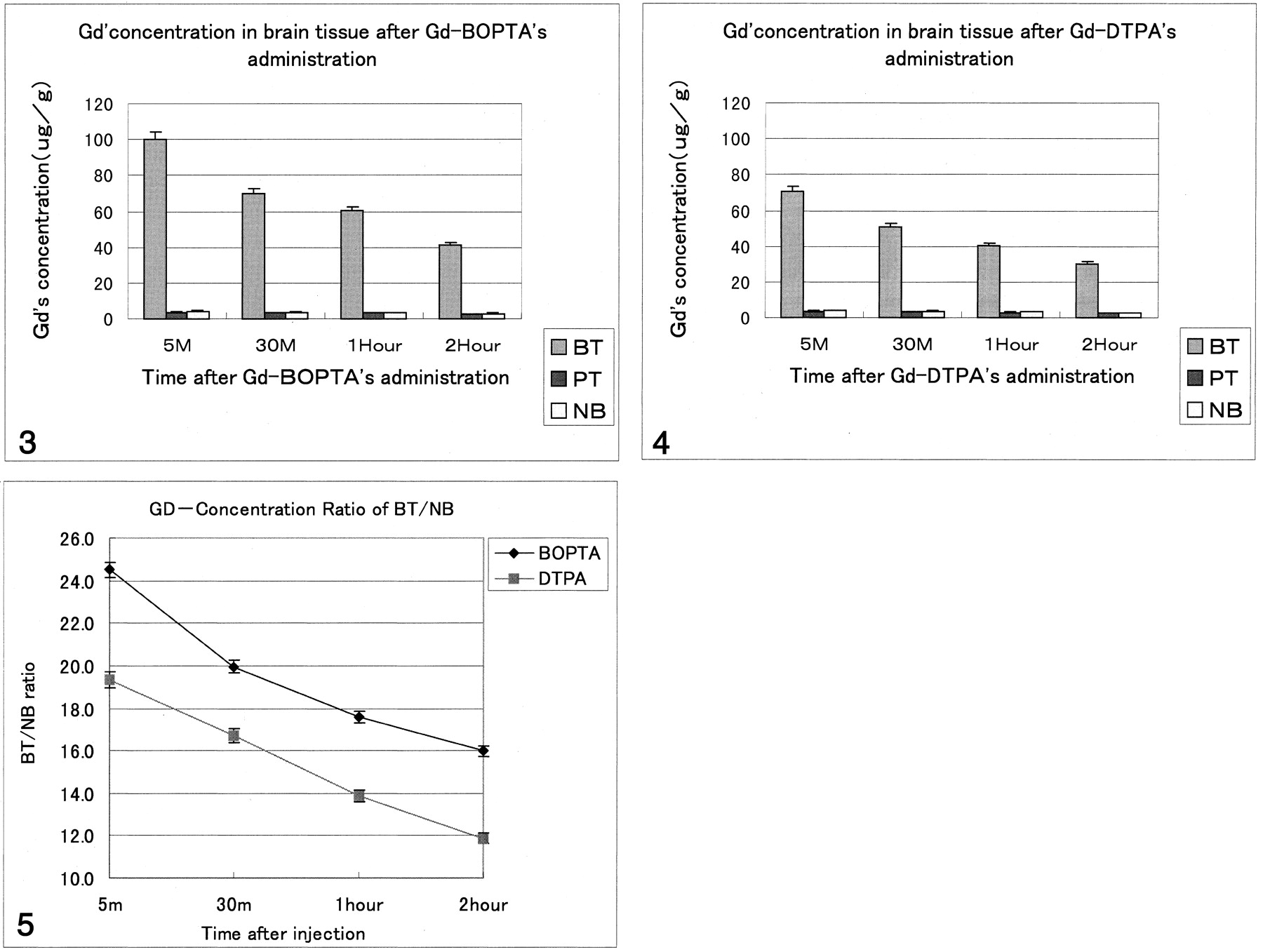
- Fig 3.
Graphic representation of the gadolinium concentration in brain tissue in rats that received Gd-BOPTA. (Gd indicates gadolinium; BT, brain tumor; PT, peritumoral tissue; NB, normal brain.)
- Fig 4.
Graphic representation of gadolinium concentration in brain tissue in rats that received Gd-DTPA.
- Fig 5.
Graphic comparison of BT/NB with Gd-BOPTA versus that of Gd-DTPA.


Tables
Gadolinium concentration (μg/g) in rat organ tissue after IV 0.1 mmol/mL/kg gadobenate dimeglumine and gadopentetate dimeglumine
Tissue Gd-BOPTA Gd-DTPA 5 Minutes 30 Minutes 1 Hour 2 Hours 5 Minutes 30 Minutes 1 Hour 2 Hours Brain tumor 100.33 ± 7.91 70.21 ± 1.22 60.18 ± 2.31 40.93 ± 1.83 70.39 ± 8.75 50.92 ± 6.37 40.31 ± 7.42 30.22 ± 4.91 Peritumoral tissue 3.63 ± 0.19 3.45 ± 0.29 3.40 ± 0.57 2.73 ± 0.31 3.51 ± 0.22 3.30 ± 0.46 2.97 ± 0.11 2.58 ± 0.27 Normal brain 4.21 ± 0.69 3.61 ± 0.77 3.51 ± 0.86 3.08 ± 0.82 3.82 ± 0.41 3.54 ± 0.52 3.10 ± 0.42 2.71 ± 0.62 Blood 270.13 ± 29.57 81.16 ± 12.31 40.49 ± 8.29 8.27 ± 0.76 229.61 ± 42.73 69.02 ± 10.32 34.42 ± 6.86 7.03 ± 1.87 Liver 570.12 ± 92.71 397.85 ± 49.29 149.71 ± 29.62 132.46 ± 44.29 480.46 ± 78.02 327.17 ± 62.34 123.37 ± 36.63 107.22 ± 46.29 Kidney 897.28 ± 62.01 730.65 ± 88.72 590.47 ± 70.91 420.16 ± 82.66 843.23 ± 97.34 793.17 ± 54.71 612.23 ± 82.67 452.17 ± 93.68 Spleen 88.38 ± 6.29 84.63 ± 9.29 60.71 ± 8.65 46.32 ± 6.23 86.58 ± 3.87 80.47 ± 6.28 59.23 ± 7.39 43.79 ± 5.99 Muscle 87.15 ± 11.87 57.68 ± 10.21 14.32 ± 2.76 5.37 ± 0.27 79.17 ± 2.38 48.79 ± 8.66 11.73 ± 2.83 4.86 ± 0.78
In this issue


Jump to section
Related Articles
Cited By...
- No citing articles found.