
Article Figures & Data
Figures
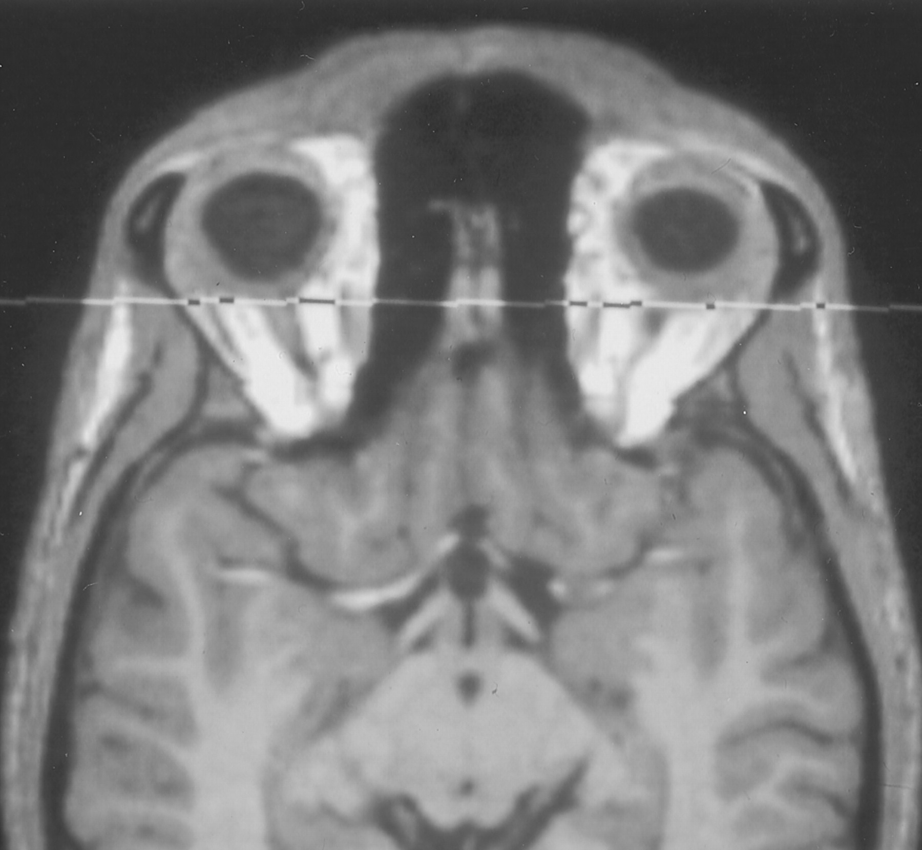
- Fig 1.
Transverse reformation of a 3D data set of an MP-RAGE image. The eyeballs and olfactory bulbs are visible. The coronal plane indicates position of the plane of the PPTE. Note that in normosmic subjects with normal olfactory bulb, this plane cuts through the olfactory bulb.
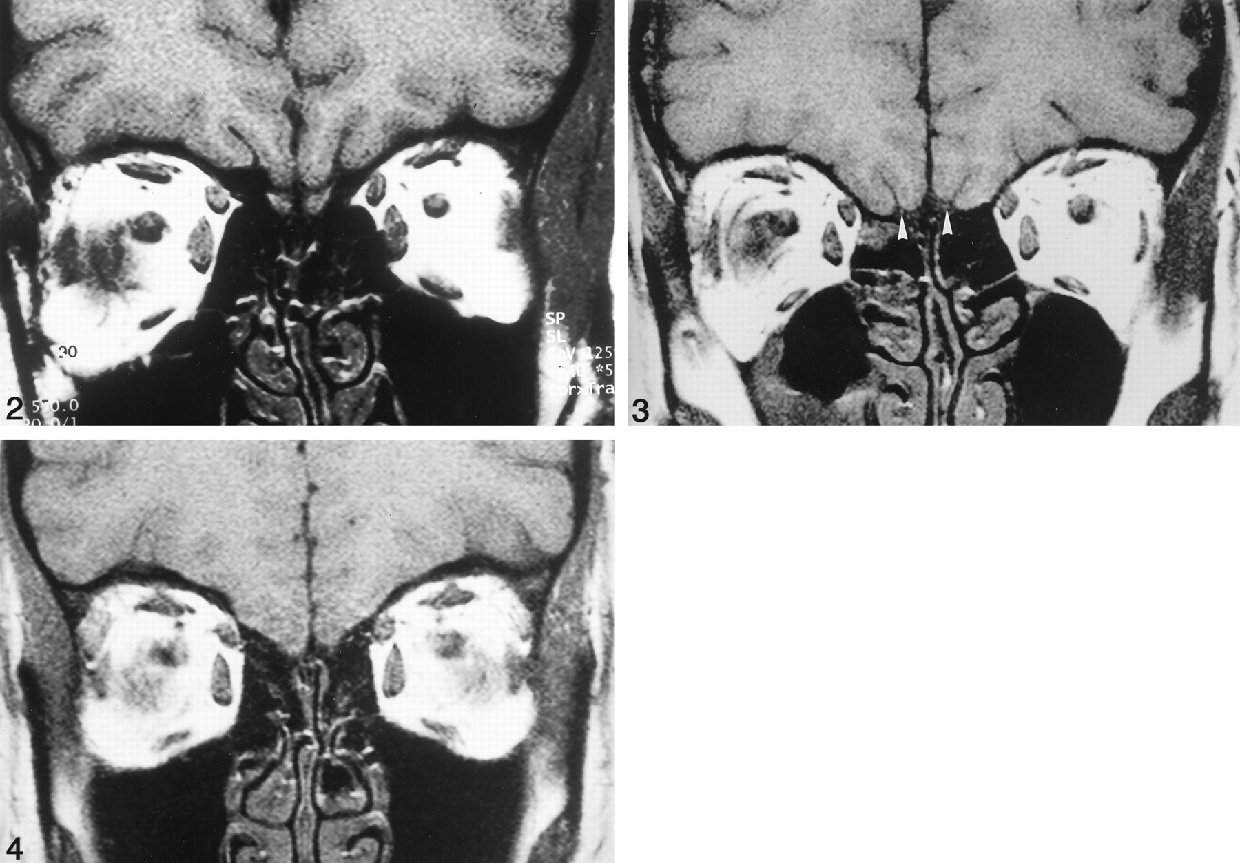
- Fig 2.
Coronal T1-weighted SE image in the PPTE in a healthy subject. Note visualization of the olfactory bulbs and normal development of the olfactory sulci.
- Fig 3.
Coronal T1-weighted SE image in the PPTE in a patient with bilateral aplasia of the olfactory bulb, visible olfactory tracts (arrowheads), and slightly flattened olfactory sulci. Note partial volume effect of the right eyeball.
- Fig 4.
Coronal T1-weighted SE image in the PPTE in a patient with absent olfactory tracts and sulci. Note partial volume effects of both eyeballs.
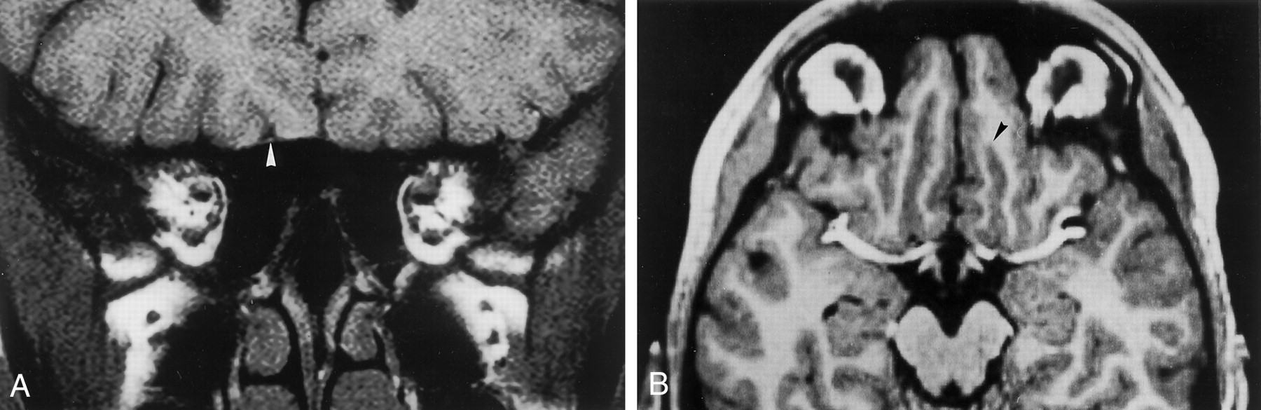
- Fig 5.
Patient with olfactory tract present only on the right.
A, Coronal T1-weighted SE dorsal image in the PPTE. Olfactory tract is visible on the right (arrowhead). There is accordingly different development of the olfactory sulcus, as shown in B.
B, Transverse reformation of a 3D data set of an MP-RAGE image in the same patient. Olfactory tract on the left is absent; olfactory tract on the right is visible. Accordingly the olfactory sulcus is shorter (arrowhead) on the left where the olfactory tract is not detectable. Note exact transverse reformation as identified by symmetrical display of periorbital fat and the middle cerebral arteries.
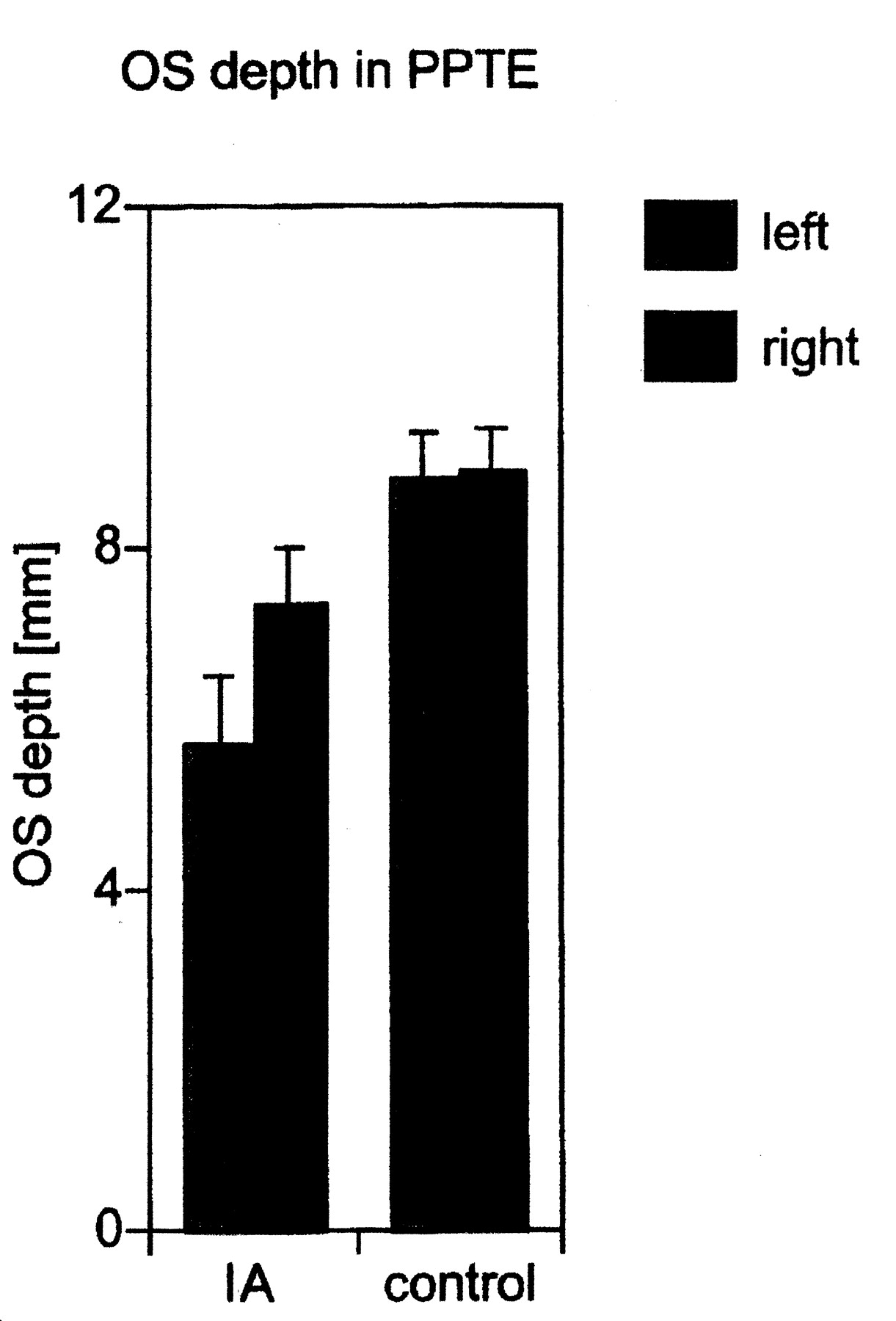
- Fig 6.
Olfactory sulcus (OS) depth in PPTE in patients with IA compared with that in healthy controls (means, SEM). Only olfactory sulcus depths greater than 0 were taken into consideration. The olfactory sulcus was significantly deeper on the right than on the left.
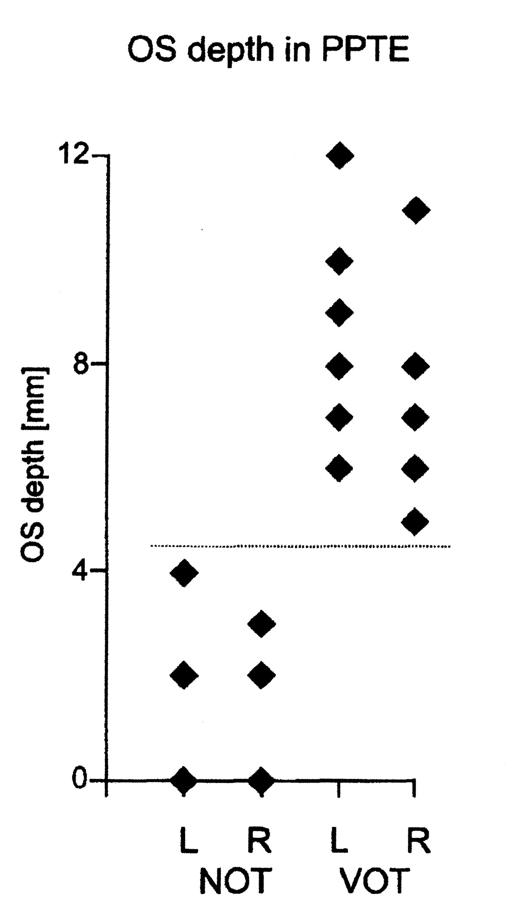
- Fig 7.
Olfactory sulcus (OS) depth in the PPTE in IA patients with visible olfactory tracts (VOT) and in those with nonvisible olfactory tracts (NOT). There is no overlap between the two groups in terms of olfactory sulcus depth.


Tables
- TABLE 1:
Descriptive statistics of olfactory sulcus measures, both as absolute measures and as measures in relation to the maximum inner coronal diameter of the anterior skull
Olfactory Sulcus Measure Controls (n = 8) IA Patients (n = 16) Left Right Left Right Absolute length (mm) 28.6 ± 5.0 28.9 ± 6.1 28.1 ± 6.9 30.6 ± 1.7 Relative length (%) 28.4 ± 5.0 28.9 ± 5.9 27.6 ± 6.7 29.9 ± 6.3 Maximum depth (mm) 9.9 ± 1.5 10.2 ± 1.1 9.1 ± 2.4 9.8 ± 2.1 Relative depth (%) 9.2 ± 1.9 10.1 ± 1.4 8.8 ± 2.4 9.6 ± 2.0 Depth in the PPTE (mm) 8.8 ± 1.5 8.9 ± 1.4 5.0 ± 3.3 6.4 ± 3.4 Relative depth in the PPTE (%) 9.3 ± 1.2 8.8 ± 1.4 4.9 ± 3.3 6.4 ± 3.4 Depth >0 in the PPTE (mm)* — — 5.7 ± 2.9 7.5 ± 2.4 Relative depth >0 in the PPTE (%)* — — 5.6 ± 2.8 7.3 ± 2.5 Note.—Data are the mean ±SD.
* Only the depth of the olfactory sulcus in the PPTE >0 was taken into consideration (in two IA patients, the olfactory sulcus in the PPTE was not visible).
- TABLE 2:
Depth of left and right olfactory sulci in control subjects and IA patients with a visible olfactory sulcus in the PPTE
Depth Control Subjects (n = 8) Patients with IA (n = 14) Deeper olfactory sulcus left 2 1 Deeper olfactory sulcus right 3 11 Equal depth on right and left 3 2 - TABLE 3:
Olfactory sulcus depth in the PPTE in patients with IA by presence of olfactory bulb and olfactory tract
Patient No. Sex Olfactory Bulb Olfactory Tract Right OS (mm) Left OS (mm) 1 M Aplastic Visible bilaterally 7 6 2 F Hypoplastic Visible bilaterally 12 11 3 F Aplastic Nonvisible bilaterally 0 0 4 F Hypoplastic Visible bilaterally 8 8 5 M Aplastic Visible bilaterally 9 8 6 F Hypoplastic Visible bilaterally 8 7 7 F Aplastic Nonvisible bilaterally 0 0 8 F Hypoplastic Visible bilaterally 7 8 9 F Aplastic Nonvisible bilaterally 4 2 10 F Hypoplastic right, aplastic left Visible right 9 3 Nonvisible left 11 F Aplastic Visible bilaterally 7 5 12 F Hypoplastic right, aplastic left Visible right 6 3 Nonvisible left 13 F Aplastic Visible bilaterally 9 8 14 F Hypoplastic right, aplastic left Visible right 7 2 Nonvisible left 15 F Hypoplastic Visible bilaterally 10 7 16 F Aplastic Nonvisible bilaterally 2 2
In this issue


Jump to section
Related Articles
Cited By...
- Acquired olfactory loss alters functional connectivity and morphology
- Bilateral transient olfactory bulb edema during COVID-19-related anosmia
- Structural changes in secondary, but not primary, sensory cortex in individuals with congenital olfactory sensory loss
- Brain Changes in Kallmann Syndrome
- The Depth of the Olfactory Sulcus Is an Indicator of Congenital Anosmia
- Association of Olfactory Bulb Volume and Olfactory Sulcus Depth with Olfactory Function in Patients with Parkinson Disease