
Article Figures & Data
Figures
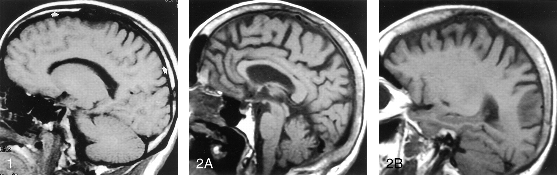
- Fig 1.
Off-midline non-contrast-enhanced sagittal T1-weighted MR image (500/11/1 [TR/TE/excitations]) in a 25-year-old healthy man shows the normal appearance of the diploic marrow in the calvarium. Usually by the time an individual is aged 21 years, conversion from hematopoietic marrow to fatty marrow is completed. The fat content is responsible for the high signal intensity of the diploic marrow on T1-weighted MR images (arrows). The area of hypointensity along the region of the frontal bone represents the neighboring suture.
- Fig 2.
Midline (A) and parasagittal (B) non-contrast-enhanced T1-weighted MR images (500/11/1) in a 73-year-old healthy woman show the normal high signal intensity of the diploic space due to fat, the major constituent of adult marrow. The mild heterogeneity and thickening of the diploic space is common in older patients. Also note the normal appearance of the clivus in A, which, in this case, is homogeneously hyperintense.
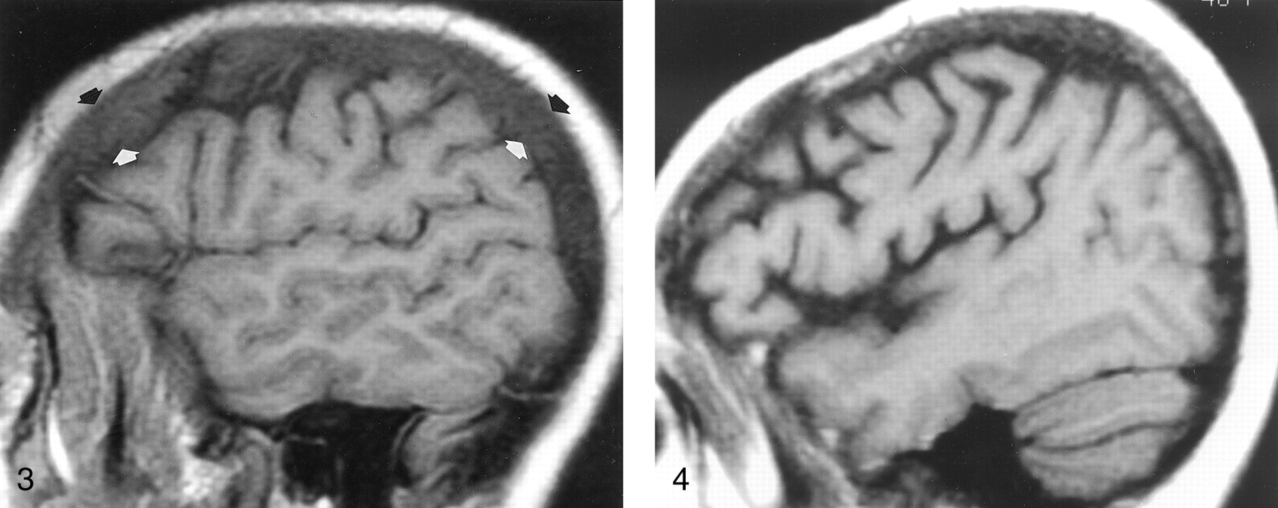
- Fig 3.
Parasagittal non-contrast-enhanced T1-weighted MR image (600/12/1) in a 35-year-old man with a history of thalassemia and a chronic hemoglobin level of 7.7 mg/dL. The marrow in the diploic space is diffusely abnormal (arrows), because the normal fat has been replaced by hematopoietic tissue, with resultant signal that is hypointense relative that of the adjacent WM and GM.
- Fig 4.
Off-midline non-contrast-enhanced sagittal T1-weighted MR image (600/12/1) in a 48-year-old woman with breast cancer who presented with headache and fatigue. Overall, heterogeneous marrow is hypointense relative to WM. A subsequent bone scan was diffusely abnormal. Pelvic marrow biopsy revealed metastatic adenocarcinoma. In this patient, alteration of the marrow signal intensity on T1-weighted images was the first indication of metastatic disease.
- Fig 5.
Nonenhanced sagittal T1-weighted MR image (500/11/1) in a 37-year-old woman with AIDS and chronic anemia (hemoglobin level, 8.2 mg/dL) shows diffuse abnormal signal intensity in the calvarial marrow, which is markedly hypointense relative to WM.
- Fig 6.
Sagittal T1-weighted MR image (600/12/1) in a 70-year-old woman with sarcoid proved at calvarial marrow biopsy shows diffusely abnormal calvarial marrow, which is hypointense relative to WM.
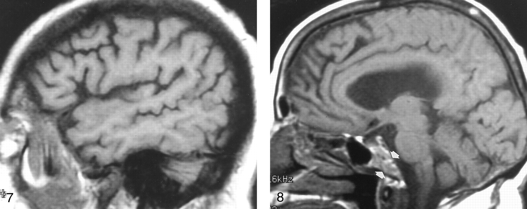
- Fig 7.
Off-midline sagittal T1-weighted MR image (600/12/1) in a 63-year-old man with newly diagnosed non-Hodgkin lymphoma shows diffusely abnormal diploic marrow. Lymphomatous cellular infiltration was found at bone marrow biopsy in the iliac crest.
- Fig 8.
Sagittal T1-weighted MR image (500/11/1) in a 74-year-old healthy woman shows heterogeneous clival (arrows) and diploic marrow patterns. While the marrow is heterogeneous or mottled in character, its overall signal is relatively isointense to that of WM.


Tables
Signal Characteristic Age (y) 21–30(n = 11) 31–40(n = 3) 41–50(n = 4) 51–60(n = 5) 61–70(n = 14) 71–78(n = 7) All*(n = 44) Isointense to fat 1 1 1 0 2 1 6 (14) Hypointense to fat and iso- or hyperintense to white matter 8 1 2 3 11 6 31 (70) Hypointense to white matter and iso- or hyperintense to gray matter 2 1 1 0 0 0 4 (9) Hypointense to gray matter 0 0 0 2 1 0 3 (7) * Data in parentheses are percentages.
Signal Characteristic Age (y) 21–30(n = 11) 31–40(n = 3) 41–50(n = 4) 51–60(n = 5) 61–70(n = 14) 71–78(n = 7) All*(n = 44) Isointense to fat 2 0 0 1 11 4 18 (41) Hypointense to fat and iso- or hyperintense to white matter 7 3 4 4 1 3 22 (50) Hypointense to white matter and iso- or hyperintense to gray matter 2 0 0 0 2 0 4 (9) * Data in parentheses are percentages.
- TABLE 3:
Calvarial diploic marrow signal intensity relative to that of fat, GM, and WM in patients with known systemic disorders
Finding Hypointense to Orbital Fat Hypointense to WM Hypointense to GM Subjective Sensitivity (%) 100 93 67 93 Specificity (%) 16 86 96 86 Accuracy (%) 37 88 88 88 - TABLE 4:
Clival marrow signal intensity relative to that of fat, GM, and WM in patients with known systemic disorders
Finding Hypointense to Orbital Fat Hypointense to WM Hypointense to GM Subjective Sensitivity (%) 100 60 7 67 Specificity (%) 43 89 100 91 Accuracy (%) 58 81 76 85


