Advertisement

Main menu

  • Home
  • Content
    • Current Issue
    • Accepted Manuscripts
    • Article Preview
    • Past Issue Archive
    • Video Articles
    • AJNR Case Collection
    • Case of the Week Archive
    • Case of the Month Archive
    • Classic Case Archive
  • Special Collections
    • AJNR Awards
    • Low-Field MRI
    • Alzheimer Disease
    • ASNR Foundation Special Collection
    • Photon-Counting CT
    • View All
  • Multimedia
    • AJNR Podcasts
    • AJNR SCANtastic
    • Trainee Corner
    • MRI Safety Corner
    • Imaging Protocols
  • For Authors
    • Submit a Manuscript
    • Submit a Video Article
    • Submit an eLetter to the Editor/Response
    • Manuscript Submission Guidelines
    • Statistical Tips
    • Fast Publishing of Accepted Manuscripts
    • Graphical Abstract Preparation
    • Imaging Protocol Submission
    • Author Policies
  • About Us
    • About AJNR
    • Editorial Board
    • Editorial Board Alumni
  • More
    • Become a Reviewer/Academy of Reviewers
    • Subscribers
    • Permissions
    • Alerts
    • Feedback
    • Advertisers
    • ASNR Home

User menu

  • Alerts
  • Log in

Search

  • Advanced search
American Journal of Neuroradiology

American Journal of Neuroradiology

ASHNR American Society of Functional Neuroradiology ASHNR American Society of Pediatric Neuroradiology ASSR
  • Alerts
  • Log in

Advanced Search

  • Home
  • Content
    • Current Issue
    • Accepted Manuscripts
    • Article Preview
    • Past Issue Archive
    • Video Articles
    • AJNR Case Collection
    • Case of the Week Archive
    • Case of the Month Archive
    • Classic Case Archive
  • Special Collections
    • AJNR Awards
    • Low-Field MRI
    • Alzheimer Disease
    • ASNR Foundation Special Collection
    • Photon-Counting CT
    • View All
  • Multimedia
    • AJNR Podcasts
    • AJNR SCANtastic
    • Trainee Corner
    • MRI Safety Corner
    • Imaging Protocols
  • For Authors
    • Submit a Manuscript
    • Submit a Video Article
    • Submit an eLetter to the Editor/Response
    • Manuscript Submission Guidelines
    • Statistical Tips
    • Fast Publishing of Accepted Manuscripts
    • Graphical Abstract Preparation
    • Imaging Protocol Submission
    • Author Policies
  • About Us
    • About AJNR
    • Editorial Board
    • Editorial Board Alumni
  • More
    • Become a Reviewer/Academy of Reviewers
    • Subscribers
    • Permissions
    • Alerts
    • Feedback
    • Advertisers
    • ASNR Home
  • Follow AJNR on Twitter
  • Visit AJNR on Facebook
  • Follow AJNR on Instagram
  • Join AJNR on LinkedIn
  • RSS Feeds

AJNR Awards, New Junior Editors, and more. Read the latest AJNR updates

Research ArticleBRAIN

Temporal Lobe Morphology in Normal Aging and Traumatic Brain Injury

Erin D. Bigler, Carol V. Andersob and Duane D. Blatter
American Journal of Neuroradiology February 2002, 23 (2) 255-266;
Erin D. Bigler
  • Find this author on Google Scholar
  • Find this author on PubMed
  • Search for this author on this site
Carol V. Andersob
  • Find this author on Google Scholar
  • Find this author on PubMed
  • Search for this author on this site
Duane D. Blatter
  • Find this author on Google Scholar
  • Find this author on PubMed
  • Search for this author on this site
  • Article
  • Figures & Data
  • Info & Metrics
  • Responses
  • References
  • PDF
Loading

Article Figures & Data

Figures

  • Tables
  • Fig 1.
    • Download figure
    • Open in new tab
    • Download powerpoint
    Fig 1.

    Images used in volume determination.

    A, Coronal T2-weighted MR image.

    B, Coronal intermediate-weighted MR image.

    C, Segmentation image from A and B.

    D, Feature space showing separation of CSF (blue), white matter (khaki), and gray matter (tan).

    E, Close-up segmented image of the right temporal lobe depicts the hippocampus and five temporal gyri.

    F, The red line indicates the length of WM from the base of the superior temporal sulcus to the base of rhinal sulcus and defines the temporal stem measurement. The linear distance (in centimeters) provided the basis for this measure, summed across all sections.

  • Fig 2.
    • Download figure
    • Open in new tab
    • Download powerpoint
    Fig 2.

    Bar graph shows hippocampal and temporal lobe gyral volumes, along with WM temporal stem measurements grouped by subjects’ ages (in years). The P values are based on analysis of variance comparisons across the decades in which significant changes may have occurred. Note the significant age effects on hippocampal volume and several gyral volumes, although considerable variability exists, as represented by the SD bars. All measures are in cubic centimeters3, with the exception of the temporal stem linear measure, which is in millimeters.

  • Fig 3.
    • Download figure
    • Open in new tab
    • Download powerpoint
    Fig 3.

    Bar graph shows sulcal CSF and temporal horn volumes grouped by subjects’ ages (in years). The P values indicate whether a significant change in volume by decade was present. The number on the bars are the SDs. Note the consistent and highly significant increases in CSF volumes (except for that of the left rhinal sulcus) with aging.
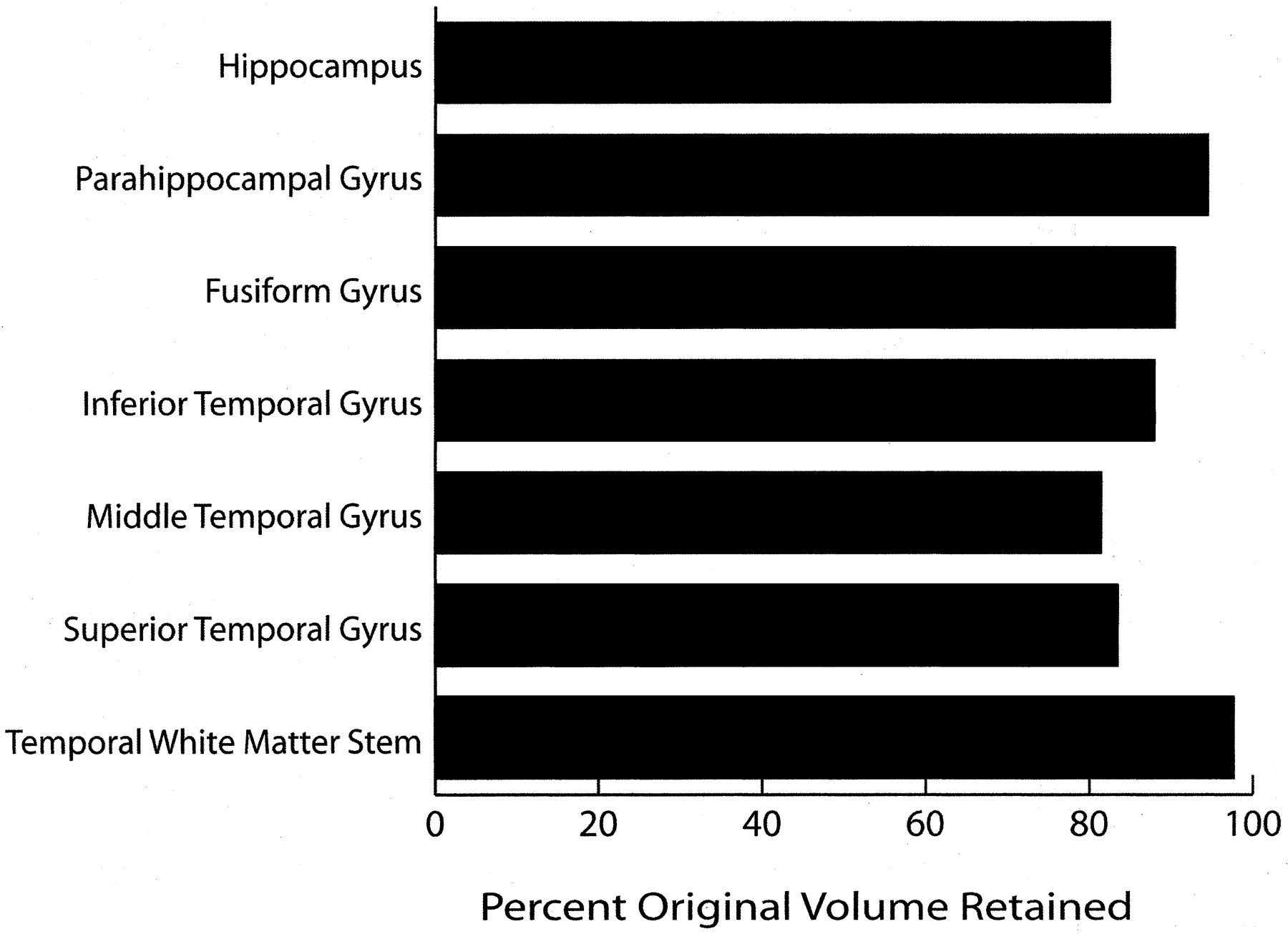
  • Fig 4.
    • Download figure
    • Open in new tab
    • Download powerpoint
    Fig 4.

    Bar graph shows the percentage of original volume retained in each temporal lobe structure, as determined by comparing the value in 16–25-year-old subjects with that in 56–72-year old subjects. Most structures, particularly the temporal WM stem, retain a large percentage of their original volume over time.
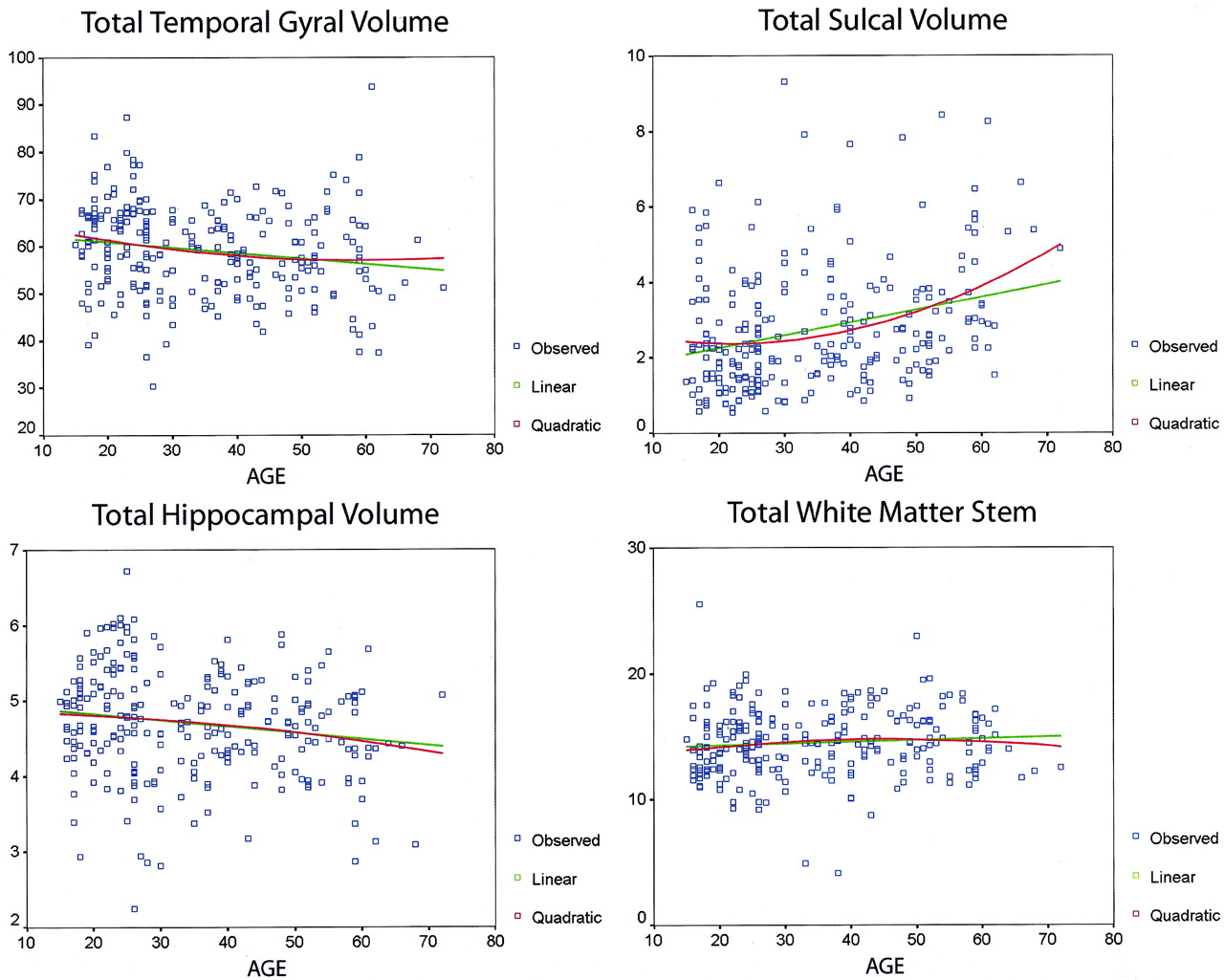
  • Fig 5.
    • Download figure
    • Open in new tab
    • Download powerpoint
    Fig 5.

    Scatterplots show total temporal gyral and sulcal volumes, hippocampal volumes, and temporal stem measurements, fitted with linear and quadratic functions. Note the greater variability in sulcal volume compared with the parenchymal measures. At statistical analysis, degrees of freedom for regression and residuals, respectively, were 2 and 251 for the linear function and 1 and 252 for the quadratic function. For each structure, values with the functions were as follows: total gyral volume, quadratic F = 4.35 and P ≤ .014, linear F = 7.97 and P ≤ .005; total sulcal volume, quadratic F = 14.09 and P ≤ .00001, linear F = 24.06 and P ≤ .00001; total hippocampal volume, quadratic F = 3.65 and P ≤ .03, linear F = 7.14 and P ≤ .008; and total WM, quadratic F = 1.2 and P ≤ .29, linear F = 1.41 and P ≤ .24. In each case, head size (total intracranial volume) and sex were used as covariates.
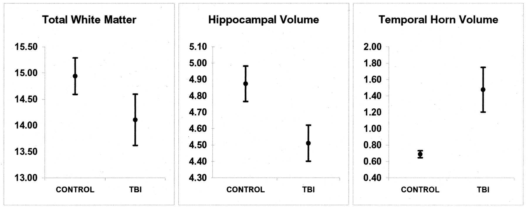
  • Fig 6.
    • Download figure
    • Open in new tab
    • Download powerpoint
    Fig 6.

    Bar graph shows the mean for volumes in the comparison of healthy control subjects and patients with TBI. In each case, TBI resulted in significant atrophy (P ≤ .01). The number on the bars are the SDs.

  • Fig 7.
    • Download figure
    • Open in new tab
    • Download powerpoint
    Fig 7.

    Graphs show the total WM measure in the temporal stem, hippocampal volume, and temporal horn volume. In each case, TBI resulted in significant atrophy (P ≤ .01). The bars indicate the SDs.

Tables

  • Figures
  • Pearson correlation values in patients with TBI

    StructureR HippocampusL HippocampusR Temporal HornLeft Temporal Horn
    R parahippocampal gyrus0.55*0.46*−0.04−0.07
    L parahippocampal gyrus0.55*0.60*0.07−0.08
    R fusiform gyrus0.55*0.45*0.04−0.08
    L fusiform gyrus0.39*0.39*0.01−0.13
    R inferior temporal gyrus0.54*0.56*−0.10−0.16
    L inferior temporal gyrus0.43*0.51*−0.17‡−0.25†
    R middle temporal gyrus0.47*0.41*−0.14−0.08
    L middle temporal gyrus0.55*0.56*−0.13−0.29*
    R superior temporal gyrus0.55*0.44*0.02−0.04
    L superior temporal gyrus0.54*0.57*−0.05−0.15
    R superior temporal lobe sulcus−0.13−0.110.41*0.31*
    L superior temporal lobe sulcus−0.14−0.160.160.31*
    R middle temporal lobe sulcus−0.18‡−0.160.45*0.39*
    L middle temporal lobe sulcus−0.26†−0.24†0.240.39*
    R inferior temporal lobe sulcus−0.11−0.150.51*0.48*
    L inferior temporal lobe sulcus−0.19‡−0.23†0.120.37*
    R rhinal sulcus−0.19‡−0.120.44*0.31*
    L rhinal sulcus−0.23†−0.28*0.27*0.35*
    R sylvian fissure−0.13−0.080.25†0.26†
    L sylvian fissure−0.26†−0.26†0.19‡0.27*
    • * P ≤ .001.

    • † P ≤ .01.

    • ‡ P ≤ .05.

PreviousNext
Back to top

In this issue

American Journal of Neuroradiology: 23 (2)
American Journal of Neuroradiology
Vol. 23, Issue 2
1 Feb 2002
  • Table of Contents
  • Index by author
Advertisement
Print
Download PDF
Email Article

Thank you for your interest in spreading the word on American Journal of Neuroradiology.

NOTE: We only request your email address so that the person you are recommending the page to knows that you wanted them to see it, and that it is not junk mail. We do not capture any email address.

Enter multiple addresses on separate lines or separate them with commas.
Temporal Lobe Morphology in Normal Aging and Traumatic Brain Injury
(Your Name) has sent you a message from American Journal of Neuroradiology
(Your Name) thought you would like to see the American Journal of Neuroradiology web site.
CAPTCHA
This question is for testing whether or not you are a human visitor and to prevent automated spam submissions.
Cite this article
Erin D. Bigler, Carol V. Andersob, Duane D. Blatter
Temporal Lobe Morphology in Normal Aging and Traumatic Brain Injury
American Journal of Neuroradiology Feb 2002, 23 (2) 255-266;

Citation Manager Formats

  • BibTeX
  • Bookends
  • EasyBib
  • EndNote (tagged)
  • EndNote 8 (xml)
  • Medlars
  • Mendeley
  • Papers
  • RefWorks Tagged
  • Ref Manager
  • RIS
  • Zotero
0 Responses
Respond to this article
Share
Bookmark this article
Temporal Lobe Morphology in Normal Aging and Traumatic Brain Injury
Erin D. Bigler, Carol V. Andersob, Duane D. Blatter
American Journal of Neuroradiology Feb 2002, 23 (2) 255-266;
del.icio.us logo Twitter logo Facebook logo Mendeley logo
  • Tweet Widget
  • Facebook Like
  • Google Plus One

Jump to section

  • Article
    • Abstract
    • Methods
    • Results
    • Discussion
    • Acknowledgments
    • Footnotes
    • References
  • Figures & Data
  • Info & Metrics
  • Responses
  • References
  • PDF

Related Articles

  • Erratum
  • PubMed
  • Google Scholar

Cited By...

  • Individualised quantitative susceptibility mapping reveals abnormal hippocampal iron markers in acute mild traumatic brain injury
  • Apolipoprotein E genotype modulates longitudinal atrophy at the temporal lobe after mild traumatic brain injury
  • Characterization and Preclinical Treatment of Rotational Force-Induced Brain Injury
  • The Presence of the Temporal Horn Exacerbates the Vulnerability of Hippocampus during Head Impacts
  • Age- and sex-related differences in baboon (Papio anubis) gray matter covariation
  • Chronic Cognitive Dysfunction after Traumatic Brain Injury Is Improved with a Phosphodiesterase 4B Inhibitor
  • Gross morphology and morphometric sequelae in the hippocampus, fornix, and corpus callosum of patients with severe non-missile traumatic brain injury without macroscopically detectable lesions: a T1 weighted MRI study
  • Differential aging of the medial temporal lobe: A study of a five-year change
  • Crossref
  • Google Scholar

This article has not yet been cited by articles in journals that are participating in Crossref Cited-by Linking.

More in this TOC Section

  • Fast Contrast-Enhanced 4D MRA and 4D Flow MRI Using Constrained Reconstruction (HYPRFlow): Potential Applications for Brain Arteriovenous Malformations
  • Multimodal CT Provides Improved Performance for Lacunar Infarct Detection
  • Optimal MRI Sequence for Identifying Occlusion Location in Acute Stroke: Which Value of Time-Resolved Contrast-Enhanced MRA?
Show more Brain

Similar Articles

Advertisement

Indexed Content

  • Current Issue
  • Accepted Manuscripts
  • Article Preview
  • Past Issues
  • Editorials
  • Editor's Choice
  • Fellows' Journal Club
  • Letters to the Editor
  • Video Articles

Cases

  • Case Collection
  • Archive - Case of the Week
  • Archive - Case of the Month
  • Archive - Classic Case

More from AJNR

  • Trainee Corner
  • Imaging Protocols
  • MRI Safety Corner
  • Book Reviews

Multimedia

  • AJNR Podcasts
  • AJNR Scantastics

Resources

  • Turnaround Time
  • Submit a Manuscript
  • Submit a Video Article
  • Submit an eLetter to the Editor/Response
  • Manuscript Submission Guidelines
  • Statistical Tips
  • Fast Publishing of Accepted Manuscripts
  • Graphical Abstract Preparation
  • Imaging Protocol Submission
  • Evidence-Based Medicine Level Guide
  • Publishing Checklists
  • Author Policies
  • Become a Reviewer/Academy of Reviewers
  • News and Updates

About Us

  • About AJNR
  • Editorial Board
  • Editorial Board Alumni
  • Alerts
  • Permissions
  • Not an AJNR Subscriber? Join Now
  • Advertise with Us
  • Librarian Resources
  • Feedback
  • Terms and Conditions
  • AJNR Editorial Board Alumni

American Society of Neuroradiology

  • Not an ASNR Member? Join Now

© 2025 by the American Society of Neuroradiology All rights, including for text and data mining, AI training, and similar technologies, are reserved.
Print ISSN: 0195-6108 Online ISSN: 1936-959X

Powered by HighWire