
Article Figures & Data
Figures
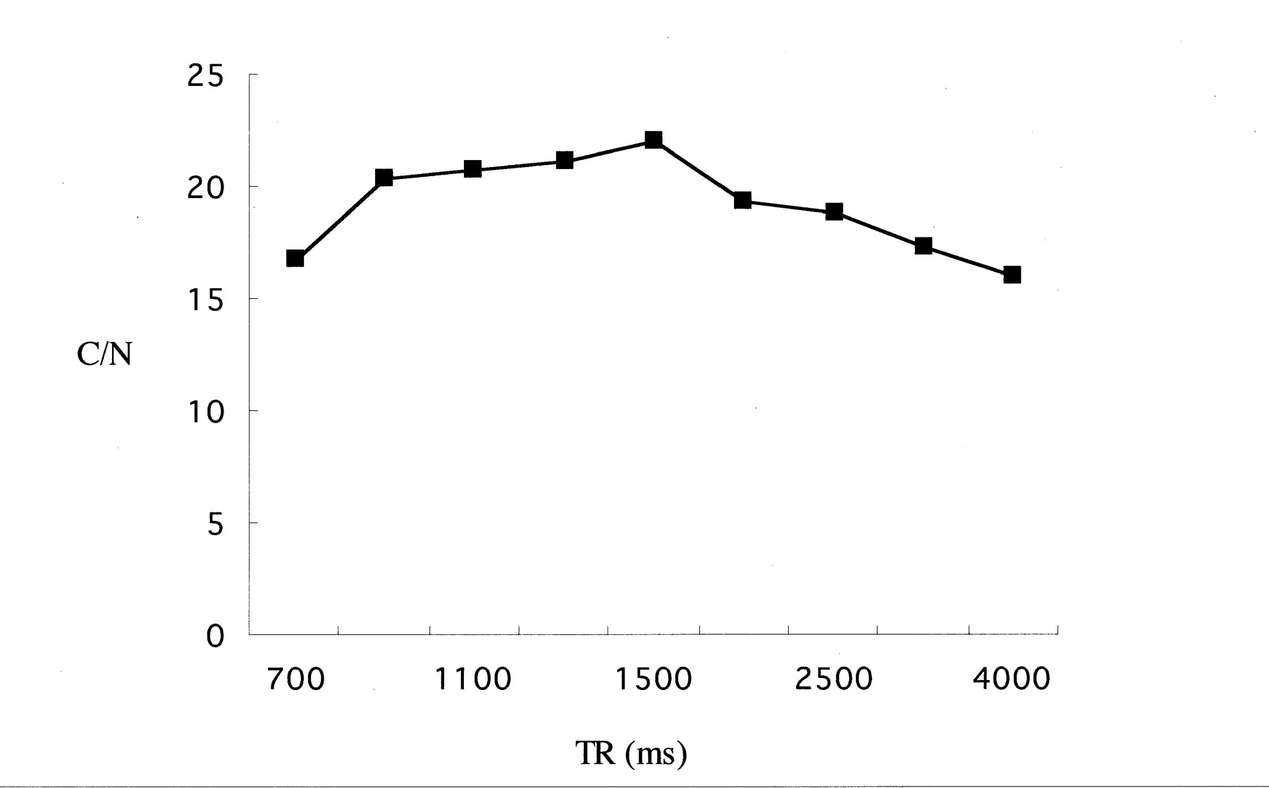
- Fig 1.
Contrast-to-noise (C/N) ratio between the polyvinyl alcohol phantom and water with various TR values. The highest contrast-to-noise ratio between polyvinyl alcohol and water was obtained at a TR of 1500 ms. A TE of 294 ms and an echo spacing of 18.1 ms were used in this experiment.
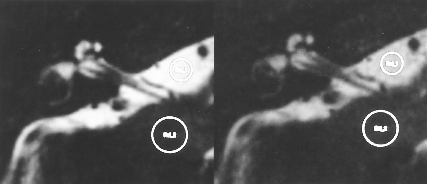
- Fig 2.
Comparison of images obtained with (left) and without (right) a fast recovery pulse in a volunteer. The contrast-to-noise ratio in the image with a fast recovery pulse is 41.1 and in the image without a fast recovery pulse is 24.3. This image was obtained with a TR of 1500 ms and a TE of 243.8 ms. The circles in the images indicate the regions of interest for CSF in the cistern and cerebellum.
- Fig 3.
Images with various TE values. Effective TE values were set at 72 (A), 144 (B), 217 (C), 294 (D), 325 (E), and 390 (F) ms. The TR was set at 1500 ms, and the echo spacing was set at 18.1 ms. The image with the effective TE of 294 ms, which was the center of the echo train length, provided the fewest ghost artifacts while maintaining a high contrast-to-noise ratio between neural tissue and CSF.
- Fig 4.
Contrast-to-noise (C/N) ratios at various bandwidths. The narrowest bandwidth compatible with the spatial resolution, an echo spacing of 18.1 ms, a TR of 1500 ms, and a TE of 294 ms, showed the highest contrast-to-noise ratios.
- Fig 5.
Contrast-to-noise (C/N) ratios at various echo spacing values. Wider echo spacing permits a narrower bandwidth, but the effective echo time is prolonged if the center of the echo train length is selected as the effective TE to prevent blurring and ghost artifacts. At a TR of 1500 ms, the highest contrast-to-noise ratio was obtained with an echo spacing of 18.1 ms and a bandwidth of 38 kHz.
- Fig 6.
Images obtained by using optimized parameters in a volunteer.
A, Magnified image of the labyrinth. Internal structures such as the osseous spiral lamina and modiolus are clearly visualized.
B, CSF in the prepontine cistern and cerebellopontine angle cistern shows high signal without significant signal loss.


