
Article Figures & Data
Figures
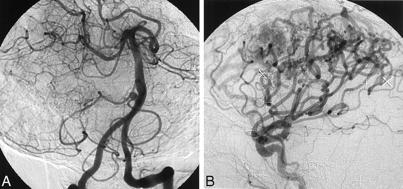
- Fig 1.
Arteriograms evaluated using a two-degree grading scale of good and poor.
A, Right anterior oblique arteriogram of the left vertebral artery was evaluated as good. A small aneurysm of the right vertebral-basilar junction can be seen. Note a retrograde flow to the opposite vertebral artery.
B, Lateral arteriogram of the common carotid artery in a patient with a high flow arteriovenous malformation was evaluated as poor. Peripheral branches of the cerebral arteries are not well visualized.

Tables
Characteristics Value Total number of participants 117 Age (y) 18–83 (56.9) Height (cm) 142–178 (158.5) Weight (kg) 37–100 (57.4) Body:weight ratio 1.68–4.05 (2.85) No. of patients receiving antiplatelet drugs 9 No. of patients receiving anticoagulant drugs 2 Prothrombin time (s) 9.5–12.7 (10.9) Partial thromboplastin time (s) 29.8–32.4 (30.8) Platelet count (1,000/mm3) 10.8–61.1 (26.8) No. of patients receiving IV administered heparin 15 Dose of heparin (U) 1000–2000 (1233) Blood pressure during procedure Systolic (mm Hg) 98–187 (137.2) Diastolic (mm Hg) 55–109 (81.1) Blood pressure after procedure Systolic (mm Hg) 94–174 (132.4) Diastolic (mm Hg) 42–103 (79.9) Note.—Data in parentheses are the mean.
Result Total No. No. of Successful Procedures (%) Procedures 117 115 (98.3) Intended arteries Right CCA 31 30 (96.8) Right ICA 59 59 (100) Right ECA 13 13 (100) Left CCA 30 29 (96.7) Left ICA 65 65 (100) Left ECA 9 9 (100) Right VA 17 16 (94.1) Left VA 60 59 (98.3) Total 284 280 (98.6) Note.—CCA indicates common carotid artery; ICA, internal carotid artery; ECA, external carotid artery; VA, vertebral artery.
Result Value Duration of procedure Totals (min) 6–62 (24.7) 1 or 2 vessels 6–32 (17.4) >3 vessels 15–62 (31.9) No. of catheters used 1–2 (1.05) Duration of groin compression (min) 3–7 (3.7) Time to ambulation after the procedure (hr) 2–3 (2.04) Complications No. of hematomas 0 of 115 No. of neurologic complications 0 of 115 No. of other complications 1 of 115* Note.—Data in parentheses are the means.
* This occurred 6 hours after the procedure.


