Advertisement

Main menu

  • Home
  • Content
    • Current Issue
    • Accepted Manuscripts
    • Article Preview
    • Past Issue Archive
    • AJNR Case Collection
    • Case of the Week Archive
    • Classic Case Archive
    • Case of the Month Archive
  • Special Collections
    • Spinal CSF Leak Articles (Jan 2020-June 2024)
    • 2024 AJNR Journal Awards
    • Most Impactful AJNR Articles
  • Multimedia
    • AJNR Podcast
    • AJNR Scantastics
    • Video Articles
  • For Authors
    • Submit a Manuscript
    • Author Policies
    • Fast publishing of Accepted Manuscripts
    • Graphical Abstract Preparation
    • Manuscript Submission Guidelines
    • Imaging Protocol Submission
    • Submit a Case for the Case Collection
  • About Us
    • About AJNR
    • Editorial Board
  • More
    • Become a Reviewer/Academy of Reviewers
    • Subscribers
    • Permissions
    • Alerts
    • Feedback
    • Advertisers
    • ASNR Home
  • Other Publications
    • ajnr

User menu

  • Alerts
  • Log in

Search

  • Advanced search
American Journal of Neuroradiology

American Journal of Neuroradiology

ASHNR American Society of Functional Neuroradiology ASHNR American Society of Pediatric Neuroradiology ASSR
  • Alerts
  • Log in

Advanced Search

  • Home
  • Content
    • Current Issue
    • Accepted Manuscripts
    • Article Preview
    • Past Issue Archive
    • AJNR Case Collection
    • Case of the Week Archive
    • Classic Case Archive
    • Case of the Month Archive
  • Special Collections
    • Spinal CSF Leak Articles (Jan 2020-June 2024)
    • 2024 AJNR Journal Awards
    • Most Impactful AJNR Articles
  • Multimedia
    • AJNR Podcast
    • AJNR Scantastics
    • Video Articles
  • For Authors
    • Submit a Manuscript
    • Author Policies
    • Fast publishing of Accepted Manuscripts
    • Graphical Abstract Preparation
    • Manuscript Submission Guidelines
    • Imaging Protocol Submission
    • Submit a Case for the Case Collection
  • About Us
    • About AJNR
    • Editorial Board
  • More
    • Become a Reviewer/Academy of Reviewers
    • Subscribers
    • Permissions
    • Alerts
    • Feedback
    • Advertisers
    • ASNR Home
  • Follow AJNR on Twitter
  • Visit AJNR on Facebook
  • Follow AJNR on Instagram
  • Join AJNR on LinkedIn
  • RSS Feeds

Welcome to the new AJNR, Updated Hall of Fame, and more. Read the full announcements.


AJNR is seeking candidates for the position of Associate Section Editor, AJNR Case Collection. Read the full announcement.


OtherPEDIATRICS

Metachromatic Leukodystrophy: Diffusion MR Imaging Findings

R. Nuri Sener
American Journal of Neuroradiology September 2002, 23 (8) 1424-1426;
R. Nuri Sener
  • Find this author on Google Scholar
  • Find this author on PubMed
  • Search for this author on this site
  • Article
  • Figures & Data
  • Info & Metrics
  • Responses
  • References
  • PDF
Loading

Abstract

Summary: Herein the case of a 10-month-old boy with metachromatic leukodystrophy is reported. Diffusion MR imaging performed with an echo-planar trace sequence revealed a cytotoxic edema-like pattern (high signal intensity on b = 1000 s/mm2 images and low apparent diffusion coefficient values) in the affected white matter in the absence of an ischemic condition. This finding was unchanged at a 6-month follow-up, as revealed by diffusion MR imaging. A gradient-echo diffusion sequence, reverse fast imaging in steady-state precession, revealed hyperintense changes at the corresponding regions. It is likely that the cytotoxic edematous pattern (restricted diffusion pattern) reflected restriction of mobility of the water molecules within abnormal portions of the myelin sheath, because impaired myelin breakdown and reutilization are known features of metachromatic leukodystrophy.

Metachromatic leukodystrophy is caused by a deficiency of arylsulfatase-A resulting in impaired myelination (1–8). Diffusion MR imaging features of the condition have not been reported previously. We report diffusion MR imaging of metachromatic leukodystrophy performed by using the echo-planar and gradient-echo sequences.

Case Report

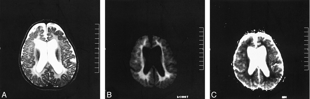
The patient was a 10-month-old boy presenting with progressive spasticity. An MR imaging examination was performed on a 1.5-T MR unit (Magnetom Vision; Siemens, Erlangen, Germany) with a gradient strength of 30mT/m. T2-weighted images revealed hyperintense changes, mainly involving the deep white matter, and atrophy (Fig 1A). Diffusion MR imaging was performed by using the echo-planar trace sequence (5700/139 [TR/TE]; acquisition time, 22 s) with averaging of the three gradients. On b = 1000 mm2/s images, which were heavily diffusion-weighted images, hyperintense changes were evident in the deep white matter and had a signal intensity pattern similar to that of cytotoxic edema (Fig 1B). On automatically generated apparent diffusion coefficient (ADC) maps, the ADC values obtained by region of interest evaluations were low at the corresponding regions (eg, 0.55 × 10⁻³ mm2/s), compared with the peripheral regions of the parenchyma (eg, 0.95 × 10⁻³ mm2/s) (Fig 1C).

Fig 1.
  • Download figure
  • Open in new tab
  • Download powerpoint
Fig 1.

Images obtained at initial examination, performed when the patient was 10 months old.

A, T2-weighted image reveals hyperintense changes in the deep white matter and atrophy.

B, A b = 1000 mm2/s (heavily diffusion-weighted) image from an echo-planar trace sequence reveals hyperintense changes similar to those of cytotoxic edema.

C, Corresponding ADC map reveals a low signal intensity and low ADC value (0.55 × 10⁻³ mm2/s) in the deep white matter compared with the peripheral regions of the parenchyma, including peripheral white matter and the cortex (0.95 × 10⁻³ mm2/s).

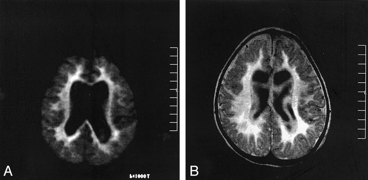
A diagnosis of metachromatic leukodystrophy was confirmed by revealing decreased activity of arylsulfatase-A in leukocytes and increased urinary sulfatides. After 6 months, the patient was reexamined by diffusion MR imaging. The same echo-planar trace sequence was used, and hyperintense changes were again observed on b = 1000 mm2/s images (Fig 2A), as were low ADC values in the deep white matter. A gradient-echo diffusion sequence, the reverse fast imaging in steady-state precession sequence, was obtained at the same session. For the reverse fast imaging in steady-state precession sequence (21.6/5 [TR/TE]; acquisition time, 2 min 48 s), high pixel values were evident in the deep white matter, compared with the peripheral regions of the parenchyma (Fig 2B). (ADC value calculation is not possible with reverse fast imaging in steady-state precession.)

Fig 2.
  • Download figure
  • Open in new tab
  • Download powerpoint
Fig 2.

Images obtained during follow-up, when the patient was 16 months old.

A, A b = 1000 mm2/s (heavily diffusion-weighted) image from echo-planar trace sequence reveals same hyperintense lesions.

B, Gradient-echo diffusion sequence (reverse fast imaging in steady-state precession) reveals high signal intensity and high pixel values (165) in the deep white matter, compared with the peripheral regions of the parenchyma (99).

Discussion

It has been reported that in cases of metachromatic leukodystrophy, the decreased activity of arylsulfatase-A accounts for failure of myelin breakdown and reutilization: hence, dysmyelination (1–8). On the diffusion MR images in the current case of metachromatic leukodystrophy, a cytotoxic, edematous pattern (restricted diffusion pattern) was evident in the deep white matter in the absence of an ischemic condition. The distribution of the lesions (Fig 1B) and their unchanging pattern on 6-month follow-up diffusion MR images (Fig 2A) strongly suggested that the lesions were directly related to the disease process. Restricted diffusion was manifested by hyperintense changes on heavily diffusion-weighted (b = 1000 s/mm2) images (Fig 1B) and low ADC values (eg, 0.55 ×10⁻³ mm2/s) on ADC maps (Fig 1C) of the echo-planar trace sequence. In addition, on the gradient-echo diffusion-weighted images, obtained using reverse fast imaging in steady-state precession, high signal intensity and high pixel values were evident in the deep white matter (Fig 2B) and were similar to those seen on b = 1000 s/mm2 images, which again indicated definite abnormality in these regions. However, absolute ADC value calculation is currently not possible with the reverse fast imaging in steady-state precession sequence.

A similar diffusion MR imaging pattern on echo-planar diffusion images of patients with phenylketonuria was recently reported by Phillips et al (9). In that study, patients with phenylketonuria had highly restricted diffusion of water molecules with high signal intensity of the white matter on b = 1000 s/mm2 images in association with low ADC values on ADC maps (between 0.56 and 0.63 × 10⁻³ mm2/s (9). The authors concluded that this restricted diffusion pattern in cases of phenylketonuria reflected impaired myelination, and protons within partially that destroyed portions of the myelin sheath may not be as mobile as those in normal parenchyma (9). On the other hand, in a recent experiment, Branco (10) showed that transition of water from the solid to the gel state contributes to the signal intensity on diffusion MR images, and in the gel state, high signal intensity is observed on b = 1000 s/mm2 images (this should result in low ADC values). Considering the observations by Phillips et al (9) and the experiment by Branco (10), in the current case of metachromatic leukodystrophy, the restricted diffusion pattern probably was consistent with some disintegration of the white matter secondary to impaired myelination, and restriction of mobility of the water molecules within abnormal portions of myelin sheath resulted in a diffusion MR imaging pattern similar to that of cytotoxic edema.

Conclusion

In a case of metachromatic leukodystrophy, an echo-planar trace sequence revealed a cytotoxic edematous pattern (restricted diffusion pattern) in the deep white matter in the absence of an ischemic condition. This pattern, manifested by high signal intensity on b = 1000 s/mm2 images and low ADC values, probably reflected restriction of mobility of the water molecules within abnormal portions of the myelin sheath, considering that impaired myelin breakdown and reutilization are known features of metachromatic leukodystrophy. Hence, dysmyelination is a feature of metachromatic leukodystrophy. A gradient-echo diffusion sequence, reverse fast imaging in steady-state precession, revealed hyperintense changes at the corresponding regions similar to those seen on b = 1000 s/mm2 images. Further diffusion MR imaging studies of dysmyelinating disorders could contribute to our understanding of the imaging properties of these diseases.

References

  1. ↵
    Barkovich AJ. Concepts of myelin and myelination in neuroradiology. AJNR Am J Neuroradiol 2000;21:1099–1109
    FREE Full Text
  2. Faerber EN, Melvin J, Smergel EM. MRI appearances of metachromatic leukodystrophy. Pediatr Radiol 1999;29:669–672
    CrossRefPubMed
  3. Kim TS, Kim IO, Kim WS, et al. MR of childhood metachromatic leukodystrophy. AJNR Am J Neuroradiol 1997;18:733–738
    Abstract/FREE Full Text
  4. Kaye EM. Update on genetic disorders affecting white matter. Pediatr Neurol 2001;24:11–24
    CrossRefPubMed
  5. Johannsen P, Ehlers L, Hansen HJ. Dementia with impaired temporal glucose metabolism in late-onset metachromatic leukodystrophy. Dement Geriatr Cogn Disord 2001;12:85–88
    CrossRefPubMed
  6. Felice KJ, Gome Zlira M, Natowicz M, et al. Adult-onset MLD: a gene mutation with isolated polyneuropathy. Neurology 2000;55:1036–1039
    Abstract/FREE Full Text
  7. Bostantjopoulou S, Katsarou Z, Michelakaki H, Kazis A. Seizures as a presenting feature of late onset metachromatic leukodystrophy. Acta Neurol Scand 2000;102:192–195
    CrossRefPubMed
  8. ↵
    Mancini GM, van Diggelen OP, Huijmans JG, Stroink H, de Coo RF. Pitfalls in the diagnosis of multiple sulfatase deficiency. Neuropediatrics 2001;32:38–40
    CrossRefPubMed
  9. ↵
    Phillips MD, McGraw P, Lowe MJ, Mathews VP, Hainline BE. Diffusion-weighted imaging of white matter abnormalities in patients with phenylketonuria. AJNR Am J Neuroradiol 2001;22:1583–1586
    Abstract/FREE Full Text
  10. ↵
    Branco G. An alternative explanation of the origin of the signal in diffusion-weighted MRI. Neuroradiology 2000;42:96–98
    CrossRefPubMed
  • Received December 5, 2001.
  • Accepted after revision May 1, 2002.
  • Copyright © American Society of Neuroradiology
View Abstract
PreviousNext
Back to top

In this issue

American Journal of Neuroradiology: 23 (8)
American Journal of Neuroradiology
Vol. 23, Issue 8
1 Sep 2002
  • Table of Contents
  • Index by author
Advertisement
Print
Download PDF
Email Article

Thank you for your interest in spreading the word on American Journal of Neuroradiology.

NOTE: We only request your email address so that the person you are recommending the page to knows that you wanted them to see it, and that it is not junk mail. We do not capture any email address.

Enter multiple addresses on separate lines or separate them with commas.
Metachromatic Leukodystrophy: Diffusion MR Imaging Findings
(Your Name) has sent you a message from American Journal of Neuroradiology
(Your Name) thought you would like to see the American Journal of Neuroradiology web site.
CAPTCHA
This question is for testing whether or not you are a human visitor and to prevent automated spam submissions.
Cite this article
R. Nuri Sener
Metachromatic Leukodystrophy: Diffusion MR Imaging Findings
American Journal of Neuroradiology Sep 2002, 23 (8) 1424-1426;

Citation Manager Formats

  • BibTeX
  • Bookends
  • EasyBib
  • EndNote (tagged)
  • EndNote 8 (xml)
  • Medlars
  • Mendeley
  • Papers
  • RefWorks Tagged
  • Ref Manager
  • RIS
  • Zotero
0 Responses
Respond to this article
Share
Bookmark this article
Metachromatic Leukodystrophy: Diffusion MR Imaging Findings
R. Nuri Sener
American Journal of Neuroradiology Sep 2002, 23 (8) 1424-1426;
del.icio.us logo Twitter logo Facebook logo Mendeley logo
  • Tweet Widget
  • Facebook Like
  • Google Plus One

Jump to section

  • Article
    • Abstract
    • Case Report
    • Discussion
    • Conclusion
    • References
  • Figures & Data
  • Info & Metrics
  • Responses
  • References
  • PDF

Related Articles

  • No related articles found.
  • PubMed
  • Google Scholar

Cited By...

  • Toward a Better Understanding of Brain Lesions during Metachromatic Leukodystrophy Evolution
  • Sporadic leucodystrophy with neuroaxonal spheroids: persistence of DWI changes and neurocognitive profiles: a case study
  • Crossref
  • Google Scholar

This article has not yet been cited by articles in journals that are participating in Crossref Cited-by Linking.

More in this TOC Section

  • Comparison of Image Quality and Radiation Dose in Pediatric Temporal Bone CT Using Photon-Counting Detector CT and Energy-Integrating Detector CT
  • SyMRI & MR Fingerprinting in Brainstem Myelination
  • Gad Contrast in PEDS Seizure Evaluation
Show more Pediatrics

Similar Articles

Advertisement

Indexed Content

  • Current Issue
  • Accepted Manuscripts
  • Article Preview
  • Past Issues
  • Editorials
  • Editors Choice
  • Fellow Journal Club
  • Letters to the Editor

Cases

  • Case Collection
  • Archive - Case of the Week
  • Archive - Case of the Month
  • Archive - Classic Case

Special Collections

  • Special Collections

Resources

  • News and Updates
  • Turn around Times
  • Submit a Manuscript
  • Author Policies
  • Manuscript Submission Guidelines
  • Evidence-Based Medicine Level Guide
  • Publishing Checklists
  • Graphical Abstract Preparation
  • Imaging Protocol Submission
  • Submit a Case
  • Become a Reviewer/Academy of Reviewers
  • Get Peer Review Credit from Publons

Multimedia

  • AJNR Podcast
  • AJNR SCANtastic
  • Video Articles

About Us

  • About AJNR
  • Editorial Board
  • Not an AJNR Subscriber? Join Now
  • Alerts
  • Feedback
  • Advertise with us
  • Librarian Resources
  • Permissions
  • Terms and Conditions

American Society of Neuroradiology

  • Not an ASNR Member? Join Now

© 2025 by the American Society of Neuroradiology All rights, including for text and data mining, AI training, and similar technologies, are reserved.
Print ISSN: 0195-6108 Online ISSN: 1936-959X

Powered by HighWire