
Article Figures & Data
Figures
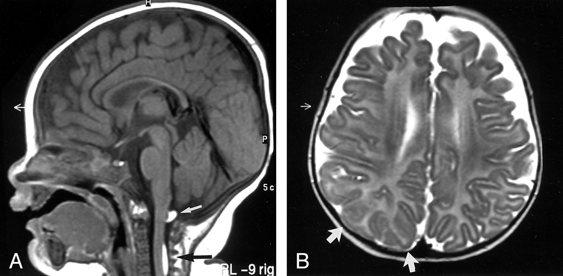
- Fig 1.
Case 1. A 5-month-old female infant with a right-sided epidermal nevus (nevus unius lateralis) involving her right torso, arm, neck, and face.
A, Sagittal T1-weighted (425/15/2 [TR/TE/NEX]) MR image shows two subpial lesions that are dorsal to the cervical cord (arrows). Both lesions are homogeneously hyperintense. The signal intensity was suppressed with chemical fat-saturation techniques (not shown).
B, Axial T2-weighted (4500/150/3) MR image shows subtle thickening and poor definition of the right parietal cortex (arrows), which likely represent a focal cortical dysplasia.
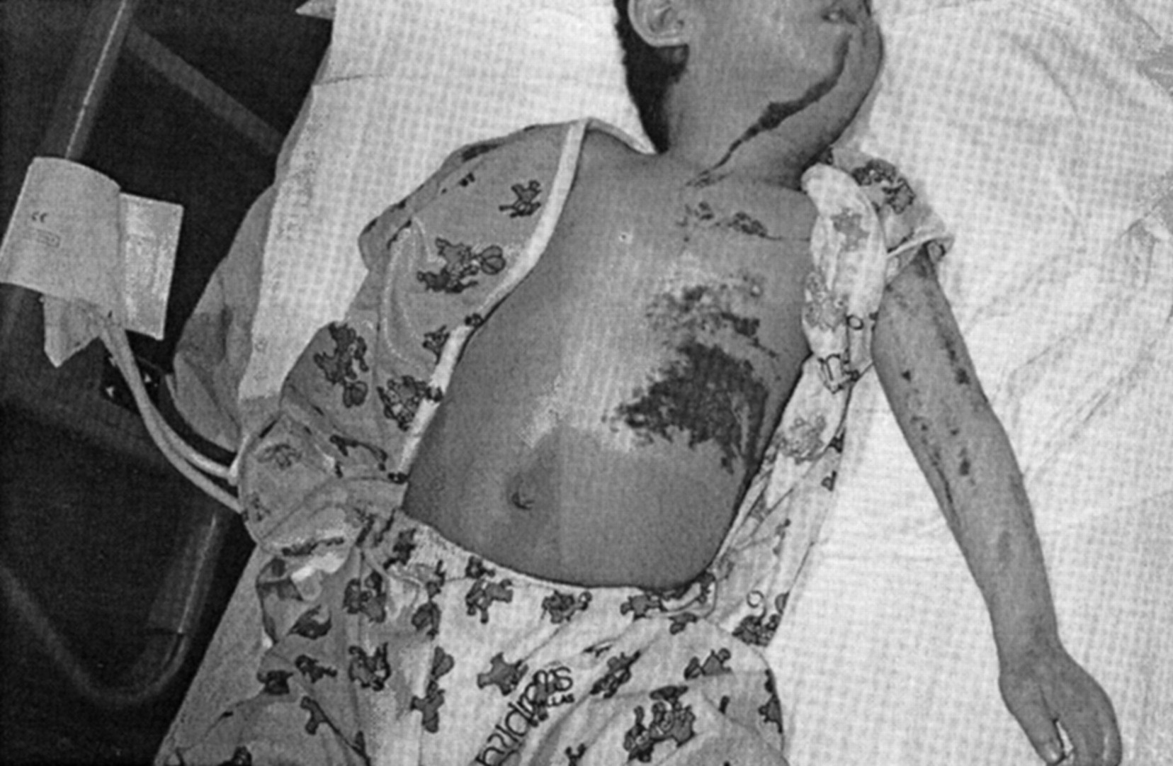
- Fig 2.
Case 2. A 3-year-old boy presented with an epidermal nevus involving the left side of his torso, extremities, neck, face, and head. Linear, brown, verrucous, plaquelike epidermal nevus involves the left side of the neck, face, and body.
- Fig 3.
Case 2.
A, Axial contrast-enhanced CT scan obtained at the level of the distal femur shows marked enlargement of the leg with a lipomatous and soft-tissue component (arrow).
B, Delayed axial CT scan obtained at the midpelvis shows that the nerve roots in the spinal canal and sacral foramina are enlarged (arrows).
C, Nonenhanced midline sagittal T1-weighted (450/20/2) MR image shows two subpial lipomas dorsal to the cord (arrows).
D, Nonenhanced off-midline T1-weighted (450/20/2) MR image shows that the cervical nerve roots are enlarged at the level of the neuroforamina (arrowheads).
E, Axial fat-suppressed contrast-enhanced T1-weighted (450/20/3) MR image at the C2-C3 level shows that the enlarged nerves are abnormally enhancing. Mass effect on the adjacent thecal sac is present.
F, Coronal T1-weighted (450/20/2) MR image of the lumbar spine shows thickened lumbar nerve roots. No lipomatous component is present.
G and H, Sagittal contrast-enhanced fat-suppressed T1-weighted (450/20/3) MR images show thickening and abnormal enhancement of the lumbar and sacral nerves within both the thecal sac (arrows in H) and the foramina (arrowheads in I).
I, Coronal T2-weighted (3000/120/3) MR image obtained through the temporal lobes shows slight thickening of the left temporal cortex with poor arborization of the inferior white matter tracts (arrow).


