
Article Figures & Data
Figures
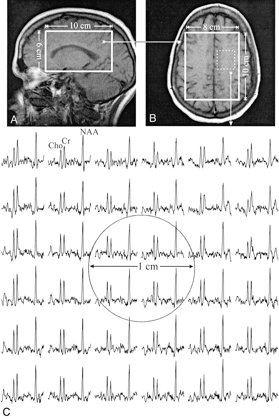
- Fig 1.
T1-weighted MR images of a 43-year-old female patient with multiple sclerosis, superimposed with the size and location of the 8LR ×10AP cm2 MR spectroscopy volumes of interest. The volumes of interest contained an approximately 1-cm diameter T1-hypointense lesion (arrow); the metabolite concentration ratios were NAA = 0.18, Cr = 0.21, and Cho = 0.25. The image contrast ratio was 0.76.
A, Sagittal view.
B, Axial view.
C, Real part (1.7–3.7 ppm range) of a 6 × 6 proton spectra matrix (0.2 cm3 voxels) from the 3 × 3 cm2 dashed box shown in B, containing the lesion (circle).
- Fig 2.
Corresponding T1-weighted MR images of a matched control participant, superimposed with the outline of the 8LR ×10AP cm2 proton MR spectroscopy volumes of interest.
A, Sagittal view.
B, Axial view.
C, Real part of a 6 × 6 proton spectra matrix (0.2 cm3 voxels) from the 3 × 3 cm2 dashed box shown in B. Circle indicates the region corresponding to the lesion shown in Figure 1C. The spectra span the same chemical shift (1.7–3.7 ppm) and intensity range as those in Figure 1C.
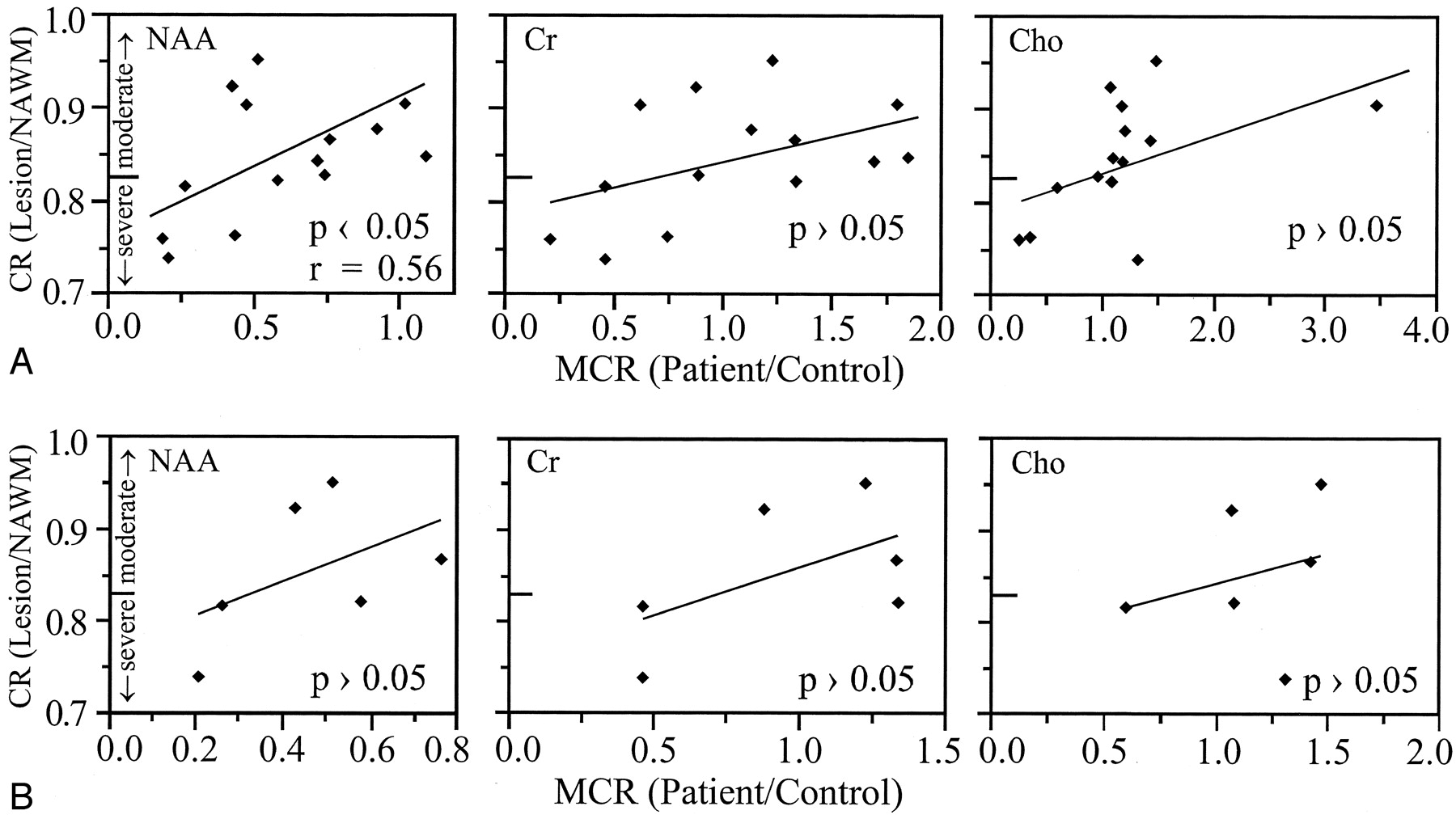
- Fig 3.
Contrast ratio (CR) of the lesions’ signal intensity versus metabolite concentration ratios (MCR) of NAA, Cr, and Cho for A, the 14 persistent T1-hypointense lesions in all patients, and B, six persistent T1-hypointense in the same 41-year-old male patient in whom images and spectra were obtained during the MR imaging–MR spectroscopy session. In both figures, hypointensity was characterized as severe if it was darker than the contrast ratio of normal-appearing gray matter (< 0.83), in accordance with van Walderveen et al (15).
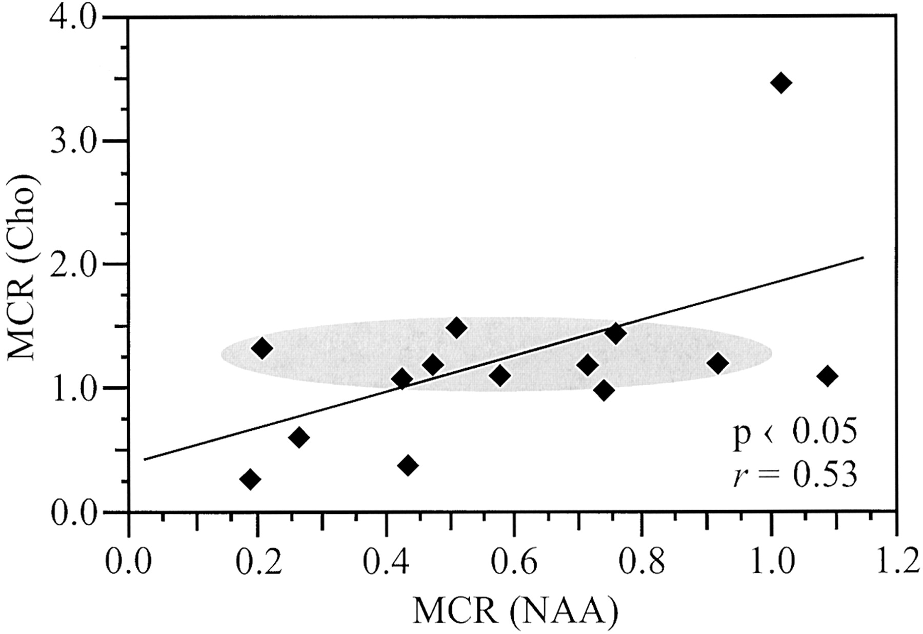
- Fig 4.
Cho versus NAA metabolite concentration ratios (MCR) for the 14 T1-hypointense lesions from all patients. Shaded oval highlights a region with a narrow 50% range (1.0–1.5) of elevated Cho levels, corresponding to a broad ±450% range (0.2–0.9) of low NAA levels.


In this issue


Jump to section
Related Articles
Cited By...
- No citing articles found.