
Article Figures & Data
Figures
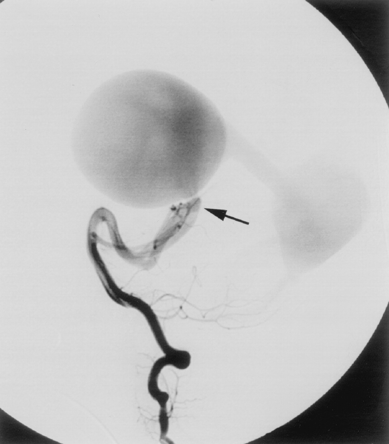
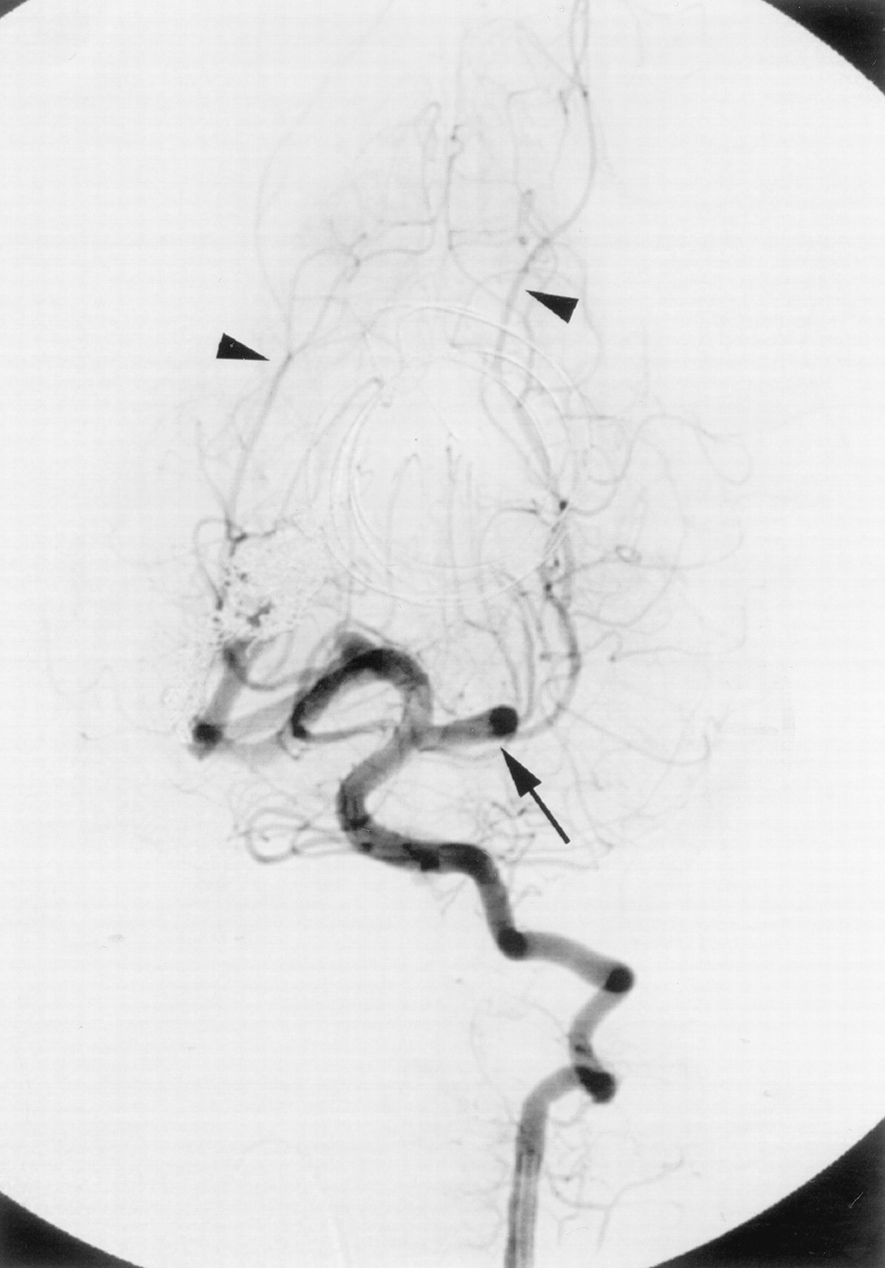
- Fig 1.
Anteroposterior projection left vertebral catheter injection digital subtraction angiogram shows hypertrophy of several posterior choroidal branches (arrow) arising from the right posterior cerebral artery and supplying a mural-type vein of Galen malformation. The malformation is also fed in a retrograde fashion from the left posterior communicating artery. Note wash-in of unopacified blood (arrowhead) retrograde from the P1 segment of the left posterior cerebral artery.
- Fig 2.
As in Figure 1, except lateral projection.
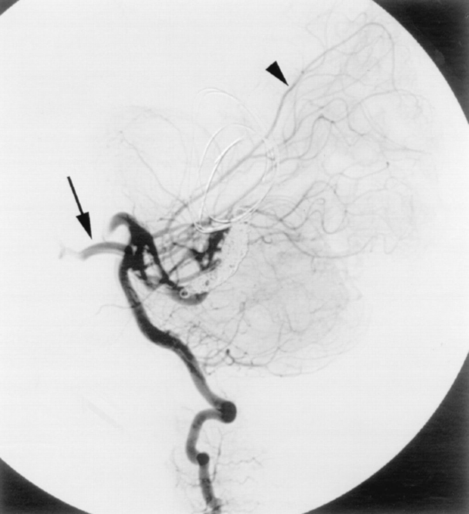
- Fig 3.
Lateral projection of the fluoroscopy fade image generated from the digital subtraction angiogram shown in Figure 2. The microcatheter (arrow on proximal marker) has passed through the fistulous communication into the venous pouch. Four vein of Galen coils have been deployed within the venous pouch but have migrated to cover the venous outflow (arrowhead).
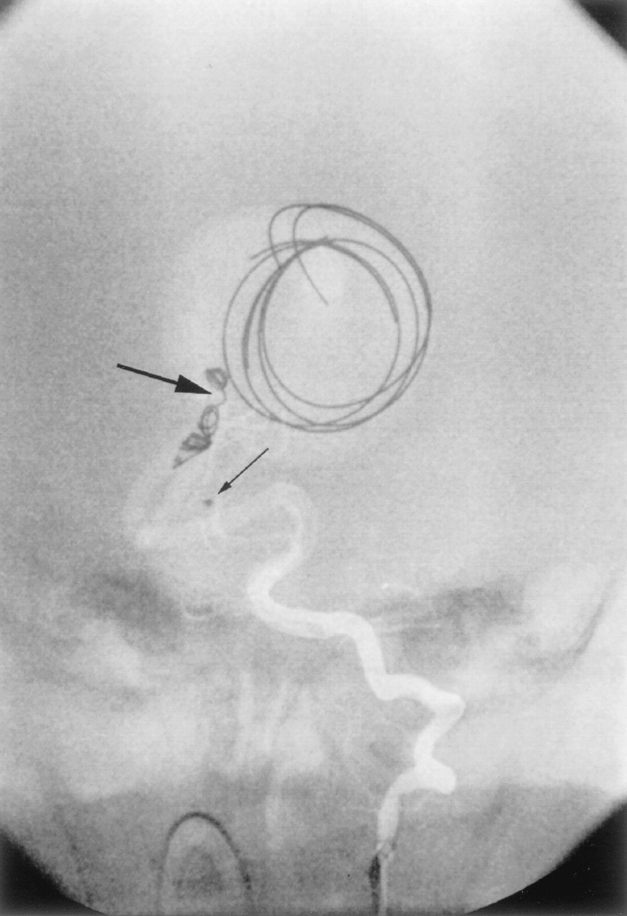
- Fig 4.
Anteroposterior projection of the fluoroscopy fade image generated from the digital subtraction angiogram shown in Figure 1. The microcatheter has been retracted (small arrow on proximal marker), with deployment of a coil partially within the venous pouch and partially within the feeding artery “bridging” the fistulous communication (large arrow).
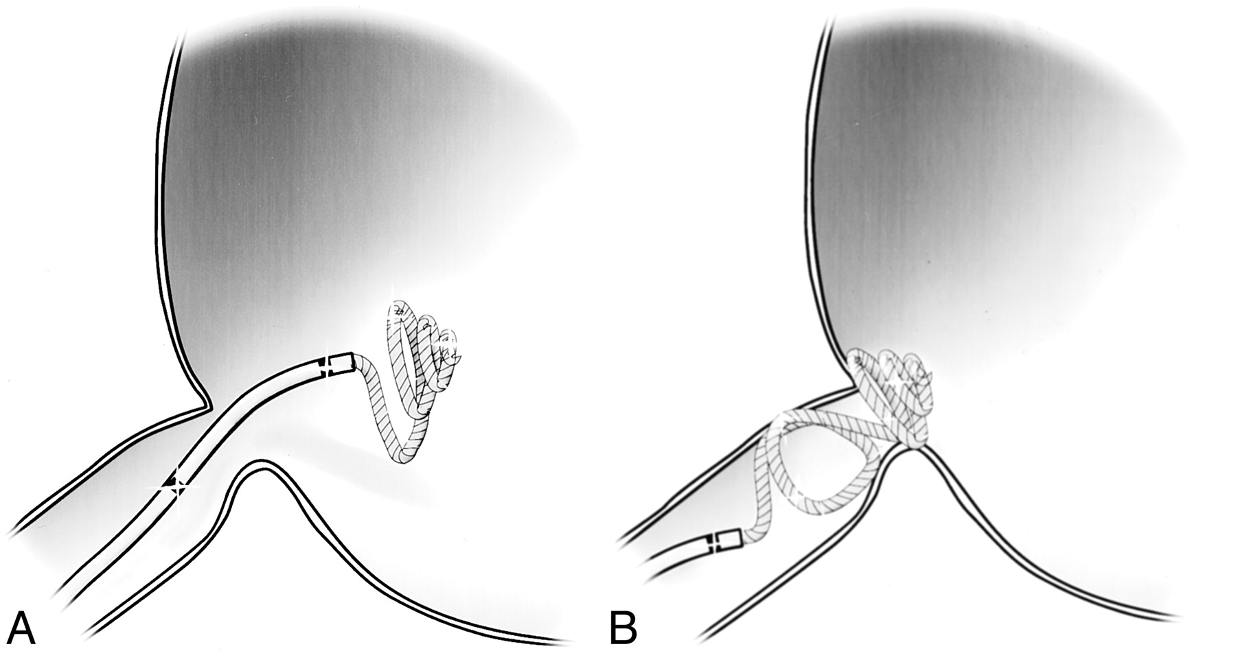
- Fig 5.
Illustrations of bridging technique used for coil embolization of the fistulous communication.
A, Several loops of coil were initially introduced into the venous pouch.
B, Microcatheter was then withdrawn into the arterial side of the fistula for deployment of the remaining coil.
- Fig 6.
Anteroposterior projection left vertebral catheter injection digital subtraction angiogram shows obliteration of the vein of Galen malformation. Restoration of normal antegrade flow from the basilar artery to the distal posterior cerebral arterial territories has been achieved (arrowheads). Note the hypertrophied left P1 segment of the posterior cerebral artery and left posterior communicating artery (arrow), resulting from the previous hemodynamic alteration and vascular steal induced by the malformation (see Fig 1).
- Fig 7.
As in Figure 6, except lateral projection.


In this issue


Jump to section
Related Articles
Cited By...
- No citing articles found.