
Article Figures & Data
Figures
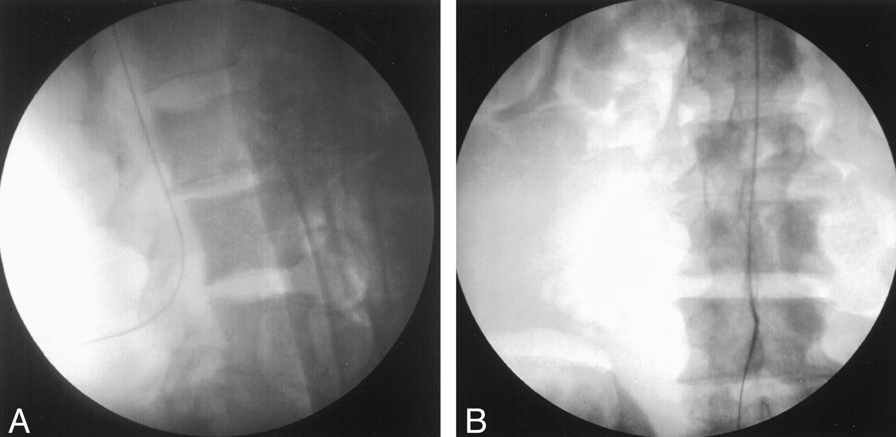
- Fig 1.
Radiographic images in cadaver 1 obtained with fluoroscopic guidance.
A, Lateral view of the lumbar spine shows the guidewire entering the spinal canal and ascending it.
B, Anteroposterior view shows the guidewire ascending in the canal from the lumbar entry point.
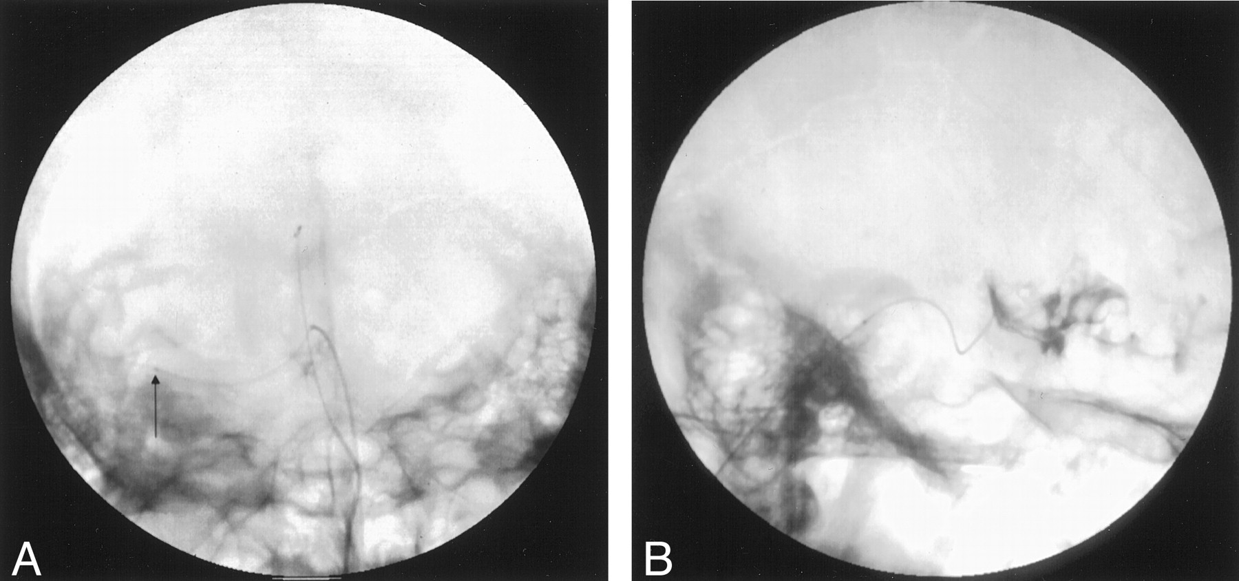
- Fig 2.
Placement of the guidewire under fluoroscopic guidance.
A, Lateral view in cadaver 1 shows the dorsal catheter (small arrow) posterior to the spinal cord and the ventral catheter (large arrow) anterior to the spinal cord. Diamond shows the tip of the ventral catheter in the sylvian fissure.
B, In cadaver 2, contrast material fills the third ventricle and spills through the foramina of Munro (arrows) into the lateral ventricles.
- Fig 3.
Images in cadaver 1.
A, Anteroposterior view shows the tip of the microcatheter in the sylvian fissure (arrow). The other (dorsal) catheter was traversing the cerebellum at the time this image is obtained.
B, Lateral digital subtraction angiogram obtained during the injection of contrast material through the catheter in the sylvian fissure. The subject is prone. Note the flow of contrast material over the gyri and sulci in the sylvian fissure as it falls away from the catheter tip.
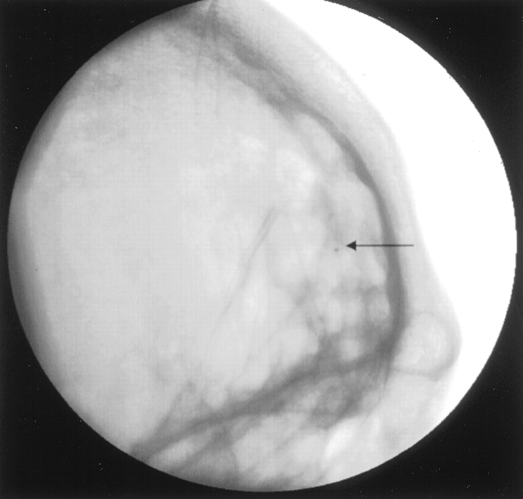
- Fig 4.
Lateral view in cadaver 2 shows the tip of the microcatheter anterior to the frontal lobe (arrow) along the inner table of the skull. The catheter passed along the orbital roof and turned superiorly, following the contour of the calvaria.
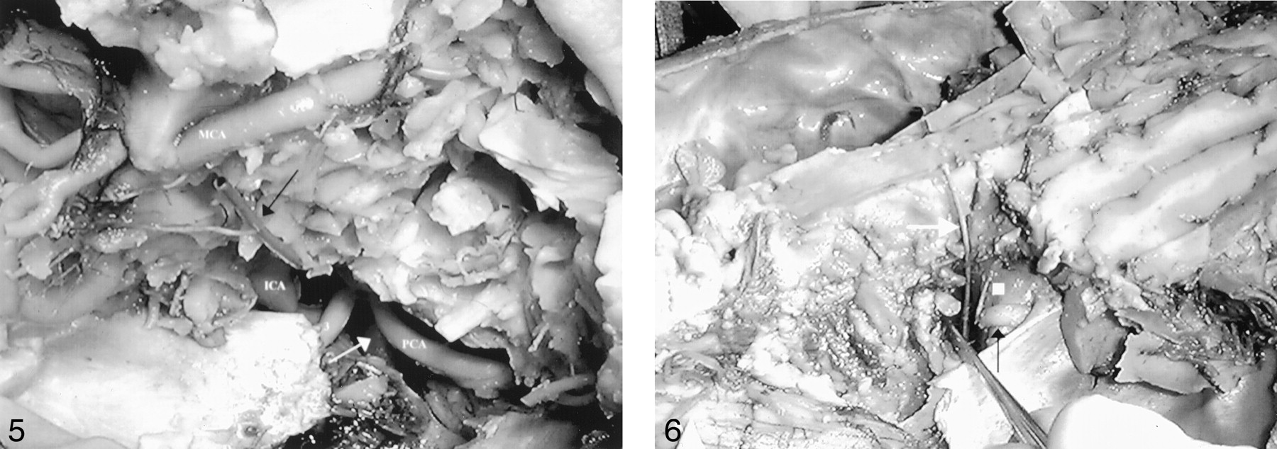
- Fig 5.
Image from the dissection in cadaver 1 shows the view into the sylvian fissure from an extensive frontotemporal craniotomy. The temporal lobe was removed. The catheter (black arrow) is seen in the sylvian fissure. Also seen are the middle cerebral artery (MCA), posterior cerebral artery (PCA), superior cerebellar artery (white arrow), internal carotid artery (ICA), and third cranial nerve (3).
- Fig 6.
View in cadaver 2 was obtained after the sagittal removal of the right hemisphere in the plane of the third ventricle. Image shows the course of the microcatheter (white arrow) that penetrates the floor of the ventricle. The dorsum sella (white square) and third cranial nerve (black arrow) are also shown. The brain stem and basilar artery are reflected posteriorly, and the course of the catheter in the subarachnoid space is shown.


