
Article Figures & Data
Figures
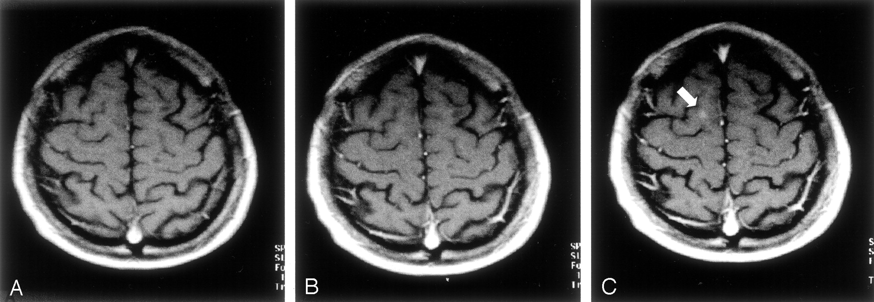
- Fig 1.
Para-axial T1-weighted spin-echo MR images.
A, Five minutes after the first intravenous administration of an SD (0.1 mmol/kg) of the paramagnetic contrast agent, no contrast enhancement attributable to a white matter lesions is visible.
B, Five minutes after the administration of a second SD of the paramagnetic contrast agent (for a total of 0.2 mmol/kg), no contrast enhancement attributable to a white matter lesion is detectable.
C, Five minutes after the administration of a third SD of the paramagnetic contrast agent (for a total of 0.3 mmol/kg), a small area of contrast enhancement is clearly visible in the right hemisphere (arrow).
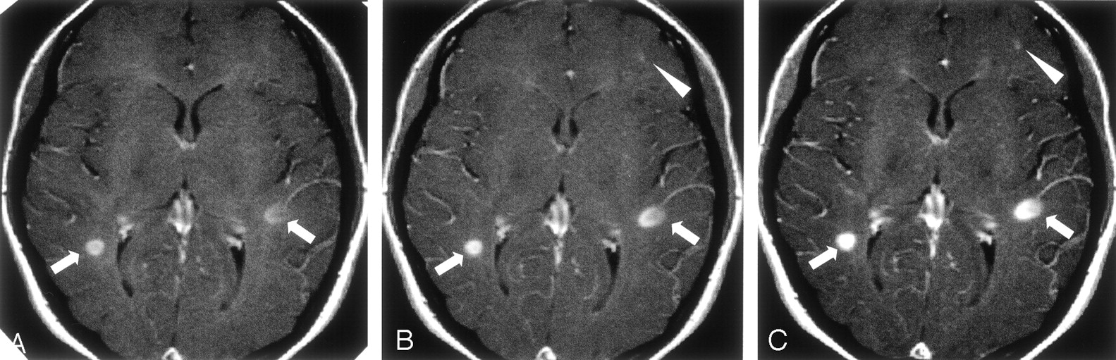
- Fig 2.
Para-axial T1-weighted spin-echo MR images.
A, Five minutes after the first intravenous administration of an SD (0.1 mmol/kg) of the paramagnetic contrast agent, two enhancing lesions are visible: one in the right and the other in the left hemisphere (arrows).
B, Five minutes after the administration of a second SD of the paramagnetic contrast agent (for a total of 0.2 mmol/kg), the enhancement in these two lesions is more conspicuous (arrows). A third area of focal contrast enhancement is barely detectable in the frontal left lobe (arrowhead).
C, Five minutes after the administration of a third SD of the paramagnetic contrast agent (for a total of 0.3 mmol/kg), the enhancement in all the three lesions becomes more conspicuous so that even the small enhancement in the frontal left lobe can be detected with higher confidence.
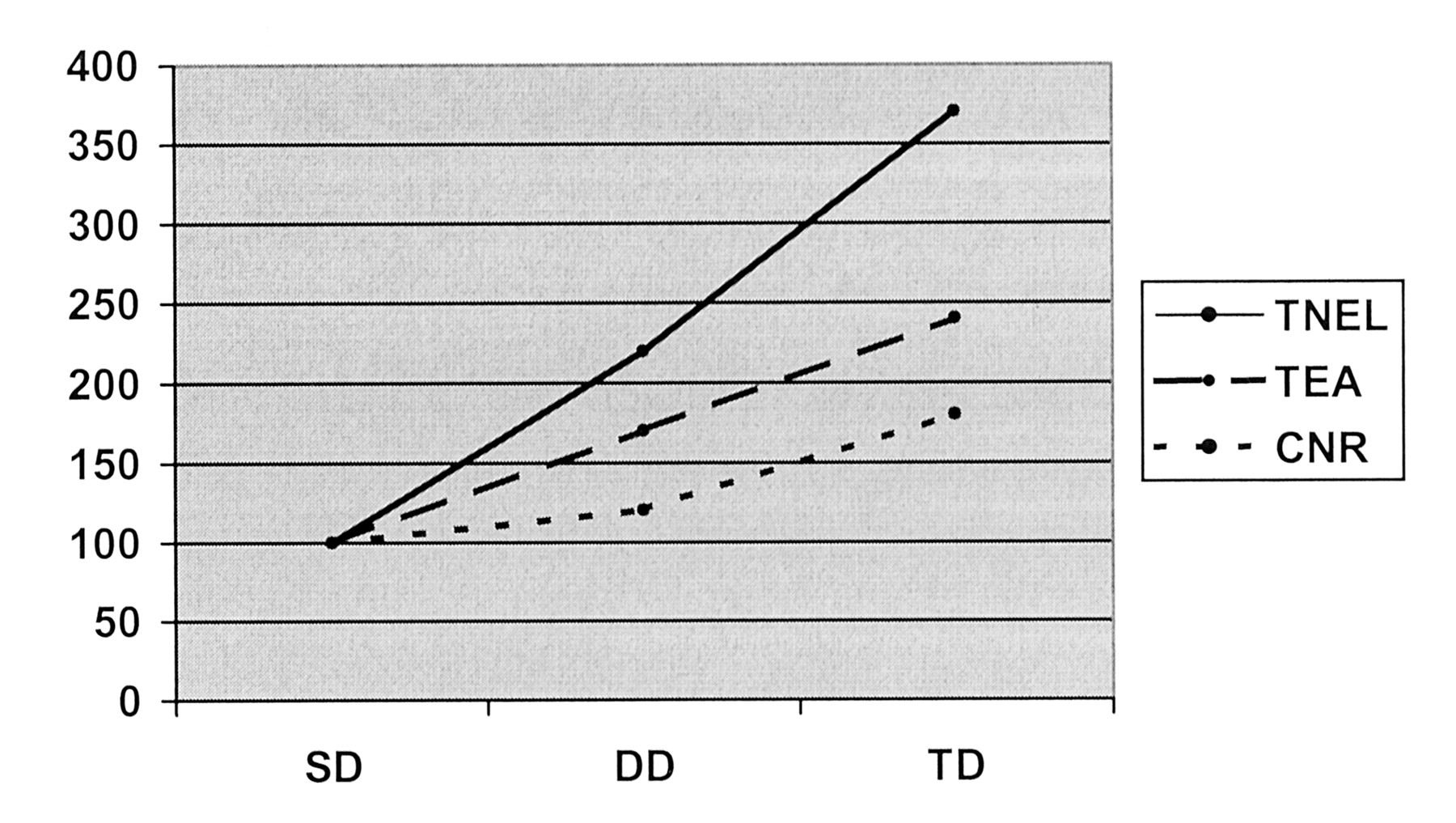
- Fig 3.
Plot shows the progressive percentage increase in the total number of enhancing lesions (TNEL), the total enhancing area (TEA), and the mean CNR (CNR) with an SD (SD), fractionated DD (DD), and fractionated TD (TD) of the paramagnetic contrast agent in 10 patients with MS.


Tables
Patient/Age, y/Sex No. of Enhancing Lesions Total Enhancing Area, cm2 Mean Lesion CNR SD DD TD SD DD TD SD DD TD 1/35/M 0 1 3 0 0.3 0.7 0 0.5 4.9 2/42/F 0 0 1 0 0 0.2 0 0 16.0 3/38/F 3 6 7 2.0 2.6 3.0 17.8 24.7 48.4 4/52/F 1 1 3 0.5 0.5 0.7 4.1 7.6 15.0 5/46/M 1 1 2 0.3 0.3 0.6 10.1 19.1 13.7 6/34/F 0 1 2 0 0.7 0.9 0 3.1 5.0 7/42/M 0 0 1 0 0 0.5 0 0 15.2 8/34/F 0 2 2 0 0.4 0.5 0 10.8 26.9 9/41/M 1 1 1 0.4 0.5 0.5 13.4 10.2 12.9 10/26/F 0 0 0 0 0 0 0 0 0 Total or mean 6 13 22 3.2 5.3 7.6 13.5 16.2 24.3 Mean per patient 0.6 1.3 2.2 0.3 0.5 0.8 NA NA NA Note—CNR indicates the contrast-to-noise ratio; NA, not applicable; SD, single dose (0.1 mmol/kg) of gadodiamide; DD, double dose (0.2 mmol/kg) of gadodiamide; and TD, triple dose (0.3 mmol/kg) of gadodiamide.
Comparison P Value No. of enhancing lesions Between SD and DD .068* Between SD and TD .012 Between DD and TD .018 Total enhancing area Between SD and DD .043 Between SD and TD .008 Between DD and TD .012 Mean CNR Between SD and DD .063* Between SD and TD .011 Between DD and TD .028 Note—SD indicates a single dose (0.1 mmol/kg) of gadodiamide; DD, double dose (0.2 mmol/kg) of gadodiamide; and TD, triple dose (0.3 mmol/kg) of gadodiamide.
* Not significant.


