
Article Figures & Data
Figures
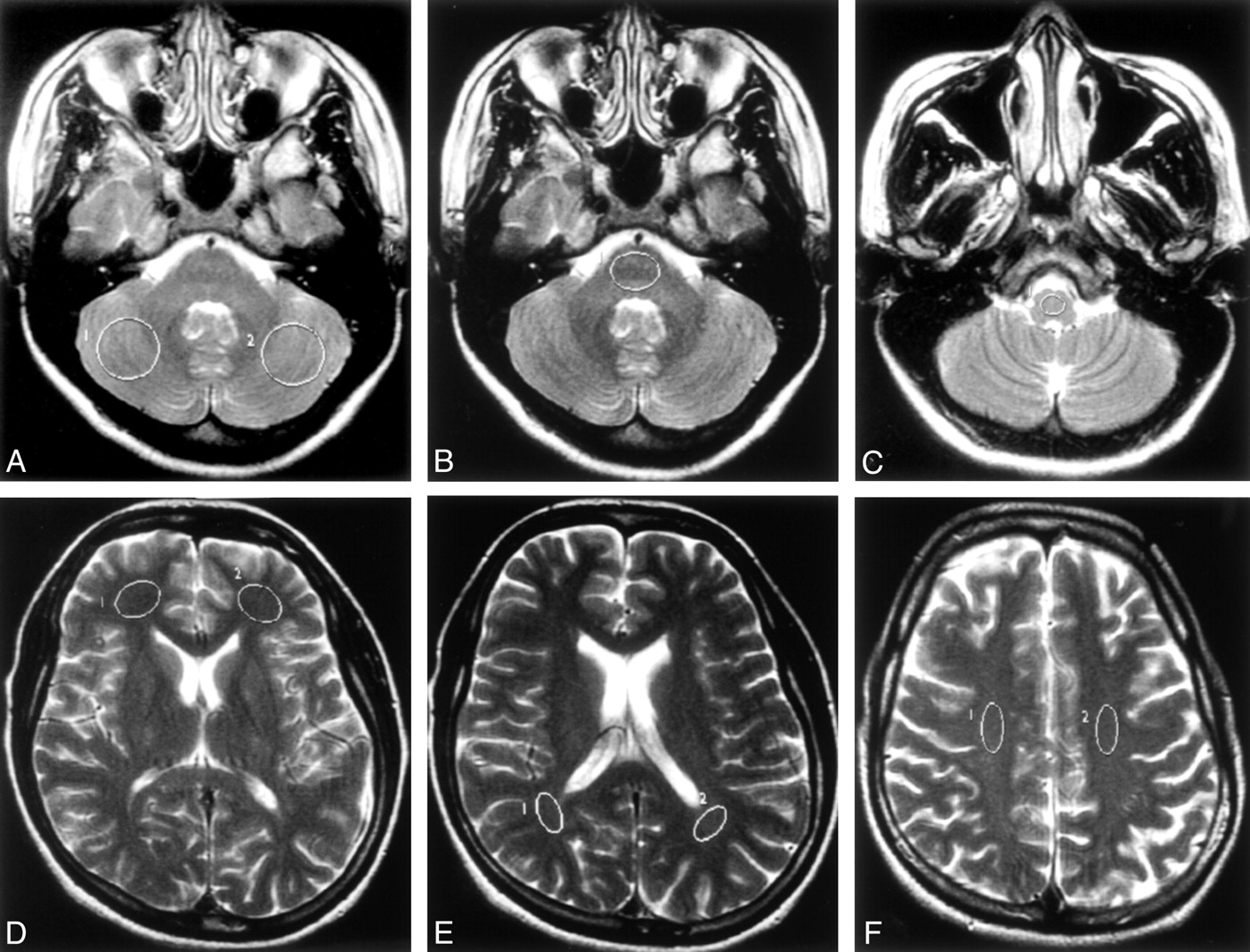
- Fig 1.
Axial T2-weighted images (100/4000/2 [TE/TR/NEX]) showing the locations of typical ROIs used in the study.
A, Bilateral cerebellar hemispheres.
B, Pons.
C, Medulla oblongata.
D, Bilateral frontal periventricular WM.
E, Bilateral parietal periventricular WM.
F, Bilateral corona radiata.
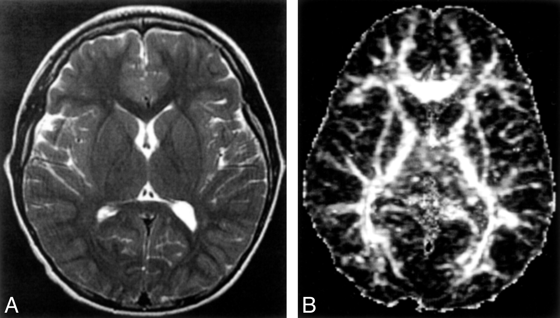
- Fig 2.
Healthy 10-year-old control showing MR images at the level of the basal ganglia
A, Axial T2-weighted images (100/4000/2 [TE/TR/NEX]).
B, Axial echo-planar spin-echo DT imaging-derived FA maps (minimum/10000/1200/1 [TE/TR/b factor/NEX]; b = 1200 s/mm2 × 25 directions and b = 0).
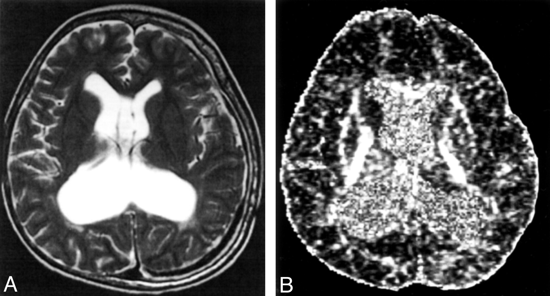
- Fig 3.
A 10-year-old medulloblastoma survivor with treatment-induced WM injury and postsurgical complications of hydrocephalus and shunt infection.
A, Axial T2-weighted images (100/4000/2 [TE/TR/NEX]).
B, Axial echo-planar spin-echo DT imaging-derived FA maps (minimum/10000/1200/1 [TE/TR/b factor/NEX]; b = 1200 s/mm2 × 25 directions and b = 0) showing (a) grade 3 white matter changes and (b) reduced signal intensity in the WM compared with the healthy age-matched control, in keeping with reduced FA.
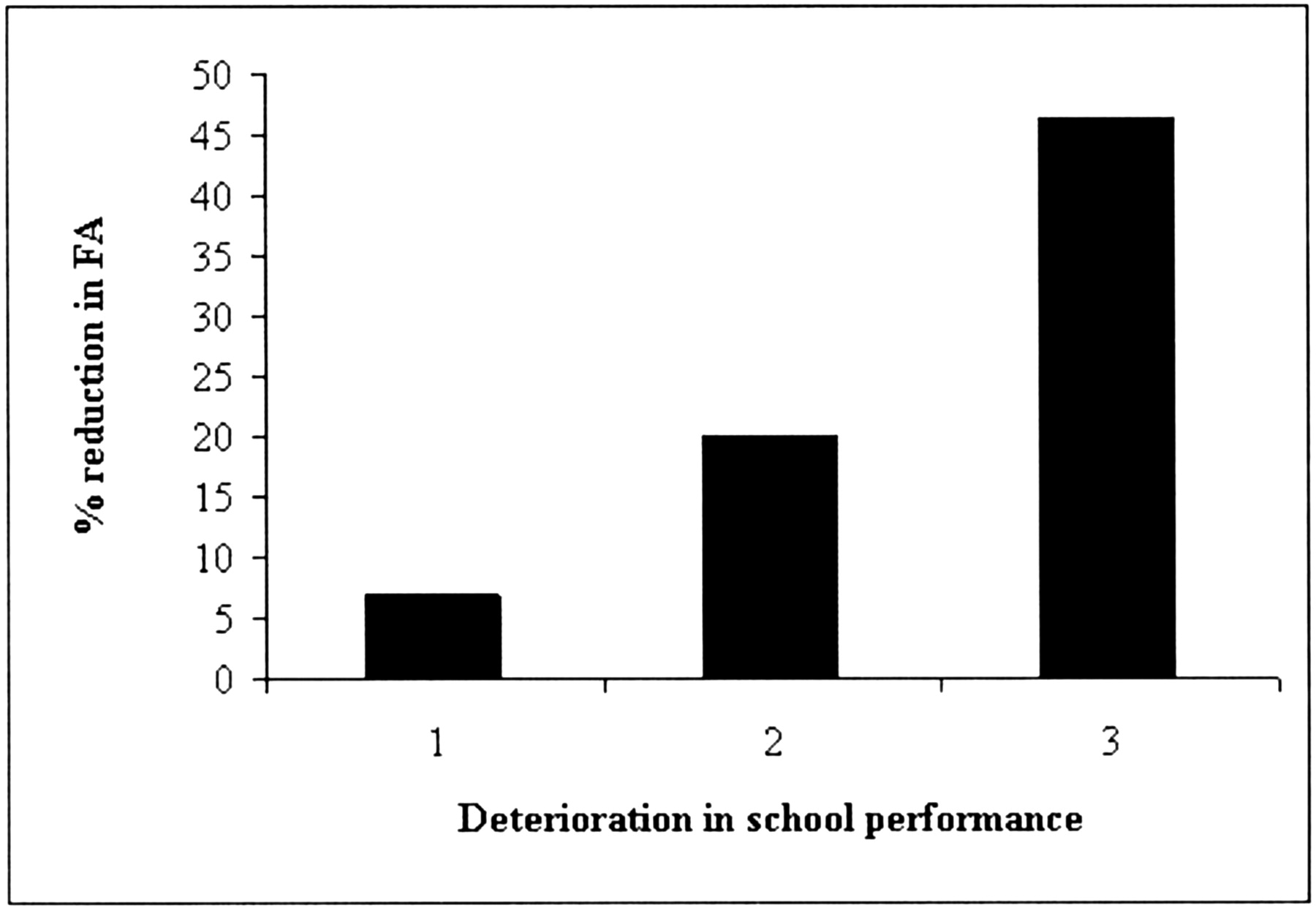
- Fig 4.
Graph showing relationship between severity of deterioration of school performance of the nine medulloblastoma survivors, and percentage reduction in supratentorial FA of the medulloblastoma survivors compared with healthy age-matched controls. 1, mild deterioration; 2, moderate deterioration; 3, severe deterioration.


Tables
Patient (No.) Age (y) Age (y) at Treatment Interval (y) Total CRT Dose (Gy) Chemo WM VI FA ▵ (%) Postoperative Complications Deterioration in School Performance 1 19 13 6 54 babyPOG 0 0.31 19.2 None Moderate 2 16 14 2 54.6 CCV 0 0.22 21.9 None Moderate 3 13 12 1 50.4 CCV 0 0.27 9.9 None Mild 4 13 7 6 54 babyPOG 0 0.24 11.6 Cerebellar mutism Moderate 5 10 4 6 54 babyPOG 3 0.33 46.2 Hydrocephalus, shunt infection Severe 6 9 3 6 54 babyPOG 1 0.31 15.8 None Moderate 7 7 5 2 50.4 babyPOG 0 0.25 21.1 None Moderate 8 7 5 2 54 CCV 0 0.24 4 Bleeding during surgery Mild 9 3 3 1 50.4 babyPOG 2 0.30 18.2 Cerebellar mutism Moderate* Note.—CRT signifies cranial radiation therapy; Chemo, chemotherapy regimen; WM, white matter grade determined by MR imaging (4-point scale [see Methods]); VI, ventricular index (maximum width of the frontal horns divided by the maximum internal skull diameter); FA ▵ (%), percentage of reduction in the supratentorial white matter fractional anisotropy of patients versus controls.
* Delayed developmental milestones.
- TABLE 2:
Fractional anisotropy and mean diffusivity at different anatomic sites in nine medulloblastoma survivors (patients) and age-matched controls
Fractional anisotropy (FA) Mean Diffusivity (MD) Patient (SD) Control (SD) Δ (%) P Value Patient (SD) Control (SD) Δ (%) P Value Pons 0.30 (0.04) 0.35 (0.02) −12.4 0.004* 1.11 (0.14) 1.14 (0.05) −2.8 0.422 Cereb 0.15 (0.03) 0.17 (0.04) −15.7 0.042† 1.37 (0.25) 1.09 (0.04) 25.9 0.010† Medulla 0.31 (0.06) 0.36 (0.06) −15.7 0.030† 1.28 (0.21) 1.17 (0.13) 9.3 0.270 FWM 0.24 (0.08) 0.30 (0.06) −18.4 0.079 1.30 (0.15) 1.22 (0.06) 6.7 0.066 PWM 0.33 (0.07) 0.41 (0.05) −19 0.028† 1.24 (0.10) 1.17 (0.08) 5.7 0.090 CR 0.38 (0.10) 0.47 (0.10) −17.9 0.008* 0.95 (0.12) 0.92 (0.12) 7.1 0.300 Note.—Cereb indicates cerebellar hemisphere; FWM, frontal white matter; PWM, parietal white matter; and CR, corona radiata.
Δ (%) = Percentage of reduction in FA of patients compared with controls.
* P < .01
† P < .05.
In this issue


Jump to section
Related Articles
Cited By...
- Processing Speed, Attention, and Working Memory After Treatment for Medulloblastoma: An International, Prospective, and Longitudinal Study
- Accelerated Aging, Decreased White Matter Integrity, and Associated Neuropsychological Dysfunction 25 Years After Pediatric Lymphoid Malignancies
- Longitudinal Diffusion Tensor Magnetic Resonance Imaging Study of Radiation-Induced White Matter Damage in a Rat Model
- White Matter Anisotropy in Post-Treatment Childhood Cancer Survivors: Preliminary Evidence of Association With Neurocognitive Function