
Article Figures & Data
Figures
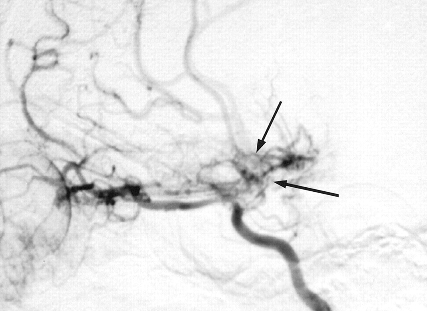
- Fig 1.
Selective angiogram of the right carotid artery shows occlusion of the terminal branches of the carotid artery (case 1, internal carotid artery grade IV). Collateral vessels are seen around the base of the brain (arrows), and the right ophthalmic artery is dilated and also supplies collaterals to the cerebral circulation.
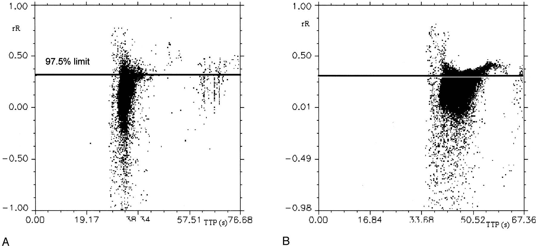
- Fig 2.
Scattergrams of rR versus TTP contrast medium concentration. Horizontal reference lines on both graphs indicate the 97.5th percentile of rR values in the normal participant.
A, Normal participant.
B, Patient with moyamoya disease (case 1). Note the increased values of TTP in the patient and the increased numbers of voxels above the reference line.
- Fig 3.
Parametric images of a normal participant. Color charts explain color coding for each image type.
A, Distribution of rCBV and TTP. Colors attempt to identify the distribution of large blood vessels (red indicates rCBV >50% and TTP <10 s), areas of increased vascular attenuation and delayed flow (new vessel formation) (white indicates rCBV >50% and TTP >10 s), areas of normal capillary flow (orchid indicates rCBV <50% and TTP <6 s), and areas of delayed flow in capillary beds (blue indicates rCBV <50% and TTP from 6 to 10 s, green indicates rCBV <50% and TTP from 10 to 14 s, and yellow indicates rCBV <50% and TTP >14 s).
3B and C, Distribution of abnormal TTP. Colors identify the distribution as follows: red, areas of normal TTP and elevated rR (TTP <6 s and rR >0.35); blue, areas of elevated TTP but normal rR (TTP >6 s and rR <0.35); yellow, areas of delayed flow and elevated rR (TTP >6 s and rR >0.35)
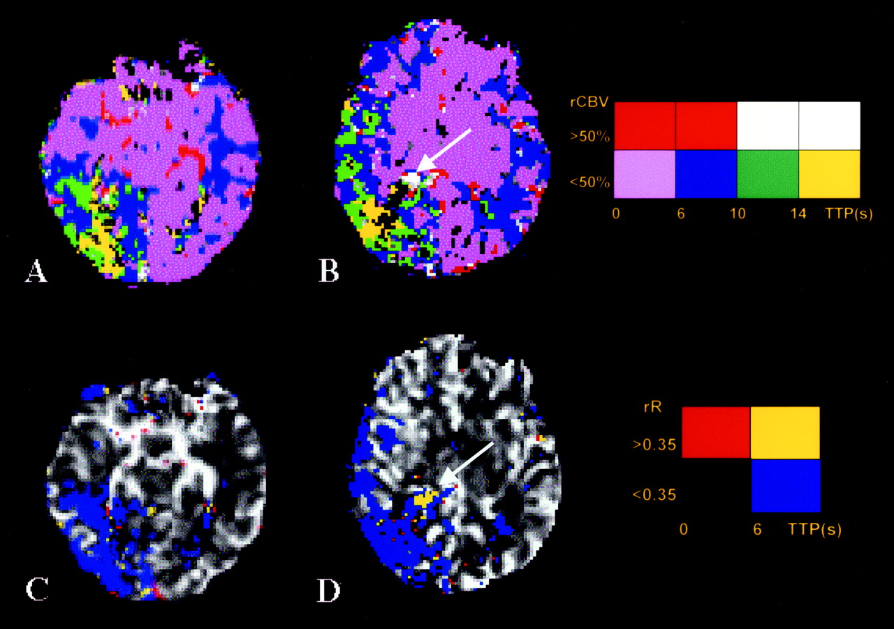
- Fig 4.
Parametric images of distribution of rCBV and TTP (A and B) and rR and TTP (C and D) in a patient (case 4) with minimal transient symptomatology. Angiogram shows only mild stenosis of the left internal carotid artery and that the right internal carotid artery was normal (internal carotid artery grade 1), but severe stenosis of the right posterior cerebral artery is shown with well-developed posterior cerebral artery moyamoya (posterior cerebral artery grade 3). Color coding is as described in the legend to Figure 3; Color charts explain color coding for each image type. Note the presence of abnormal areas of neovasculature (arrows) indicated by prolonged TTP, high rCBV, and abnormal prolongation of rR. The presence of moyamoya vessels was confirmed on digital subtraction angiograms. Distal to the abnormal vessels, a wedge-shaped area of prolonged TTP is seen; values of rR throughout this area are normal.
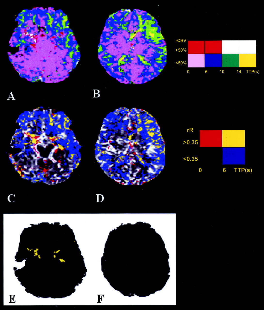
- Fig 5.
Parametric images of distribution of rCBV and TTP (A and B) and rR and TTP (C and D) in a patient presenting with expressive dysphasia and an established left-sided cortical laminar stroke (case 1). Angiogram shows advanced steno-occlusive changes in the left internal carotid artery (see Fig 1, internal carotid artery grade IV) with associated moyamoya changes. Color coding is as described in the legend to Figure 3; Color charts explain color coding for each image type. Areas of increased TTP and rR (E and F) where rCBV is >50%. Note the presence of abnormal areas of neovasculature around the middle cerebral arteries, indicated by prolonged TTP, high rCBV, and abnormal prolongation of rR. The presence of neovascularization was confirmed on digital subtraction angiograms (Fig 1). Extensive areas of prolonged TTP can be seen throughout both hemispheres. Abnormal values of rR are seen in the central portions of these areas (yellow on E and F). The rCBV in these areas is low in distinction to the areas of prolonged rR seen in areas of neovascularization.


Tables
- TABLE 1:
Classification of the severity of steno-occlusive change in the internal carotid artery circulation
ICA Stage Angiographic Description I Narrowing of carotid bifurcation II Dilation of ACA and MCA with appearance of ICA moyamoya III Partial disappearance of ACA and MCA with intensification of moyamoya IV Advanced stenoocclusive changes in ICA (ACA and MCA are traced very dimly or in a completely different shape) with small amount of ICA moyamoya V Absence of the ACA and MCA with further reduction of moyamoya VI Blood supply only from the ECA with almost complete disappearance of ICA moyamoya Note.—Classification is according to Mugikura et al (12). ACA indicates anterior cerebral artery; MCA, middle cerebral artery; ICA, internal carotid artery; ECA, external carotid artery.
- TABLE 2:
Classification of the severity of steno-occlusive change in the posterior cerebral artery
PCA Stage Angiographic Findings 1 No occlusive changes in the PCA 2 Stenosis in the PCA with or without slightly developed PCA moyamoya 3 Severe stenosis or virtually complete occlusion of the PCA with well-developed PCA moyamoya 4 Occlusion of the PCA with decreased PCA moyamoya Note.—Classification is according to Mugikura et al (12). PCA indicates posterior cerebral artery.
Patient Age (yrs) Sex ICA Stage PCA Stage Stroke 1 17 M IV 1 L MCA 2 16 F II 1 3 12 M III 2 R Post MCA 4 8 M I 3 5 5 M II 1 6 3 F III 1 Note.—Data are based on the scoring system presented by Mugikura et al (12) for all patients.
In this issue


Jump to section
Related Articles
Cited By...
- Quantitative Assessment of Neovascularization after Indirect Bypass Surgery: Color-Coded Digital Subtraction Angiography in Pediatric Moyamoya Disease
- Deregulation of Retinaldehyde Dehydrogenase 2 Leads to Defective Angiogenic Function of Endothelial Colony-Forming Cells in Pediatric Moyamoya Disease
- Normal-Appearing White Matter Permeability Distinguishes Poor Cognitive Performance in Processing Speed and Working Memory
- Characterization of Cortical Microvascularization in Adult Moyamoya Disease