
Article Figures & Data
Figures
- Fig 1.
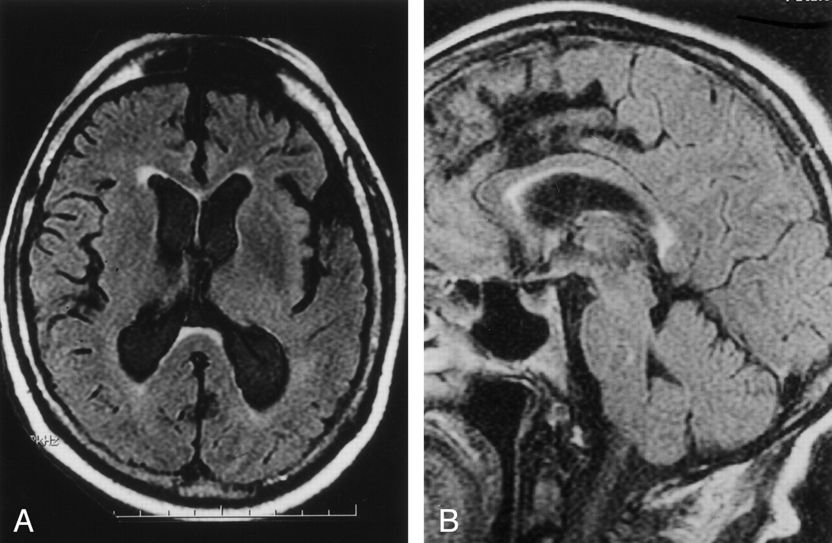
Case of a 67-year-old patient imaged to rule out aneurysm.
A, Axial and B, sagittal FLAIR MR images (10,000/156/1) show high signal intensity in the anterior subependymal region of the splenium of the corpus callosum, involving more than half the thickness (grade 2).
- Fig 2.
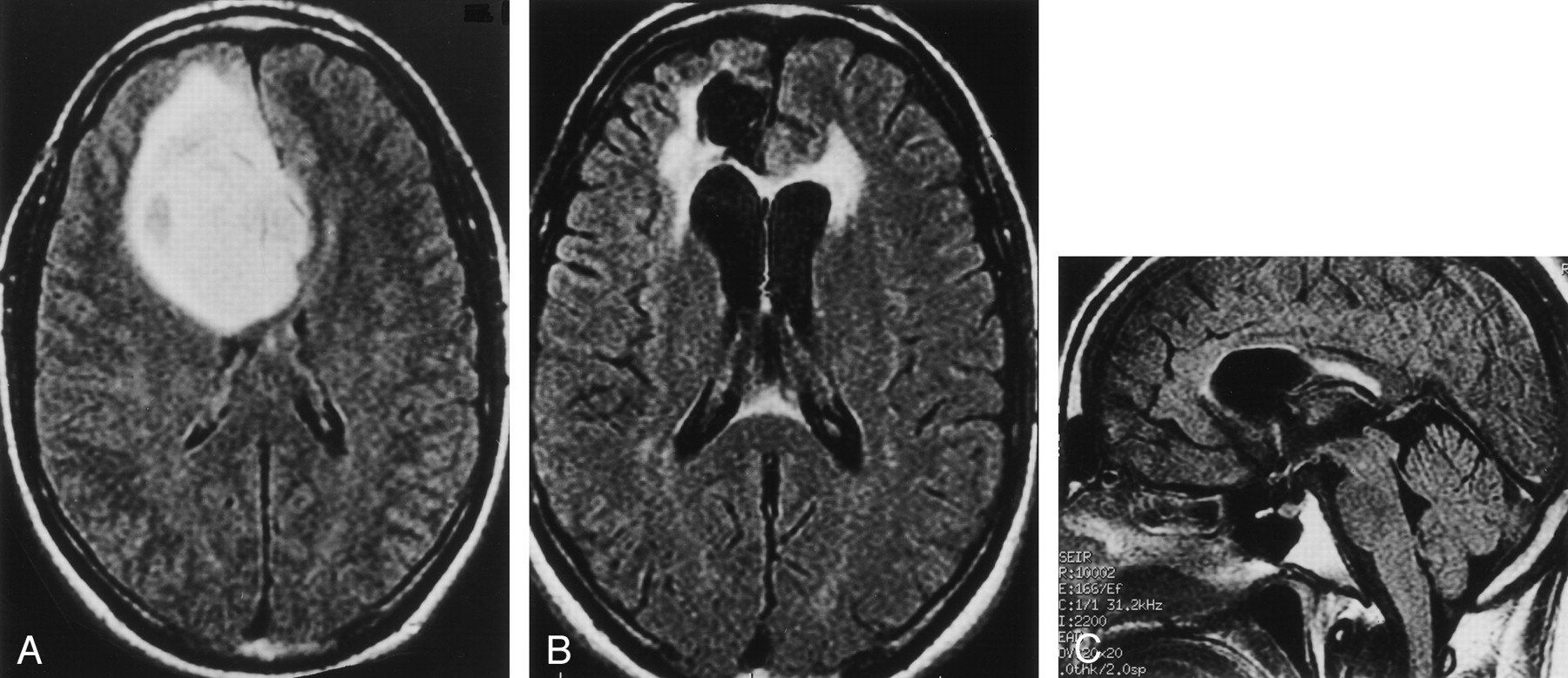
Case of a 32-year-old man (patient 12 in Table) with geminocystic astrocytoma.
A, Axial FLAIR MR image (10,002/166/1) obtained 0.8 months before radiation therapy shows normal signal intensity in the splenium.
B, Axial and C, sagittal FLAIR images (10,002/166/1) obtained 5.8 months after radiation therapy demonstrate a bright focus of abnormal signal intensity in the anterior subependymal region of the splenium of the corpus callosum (grade 2).
- Fig 3.
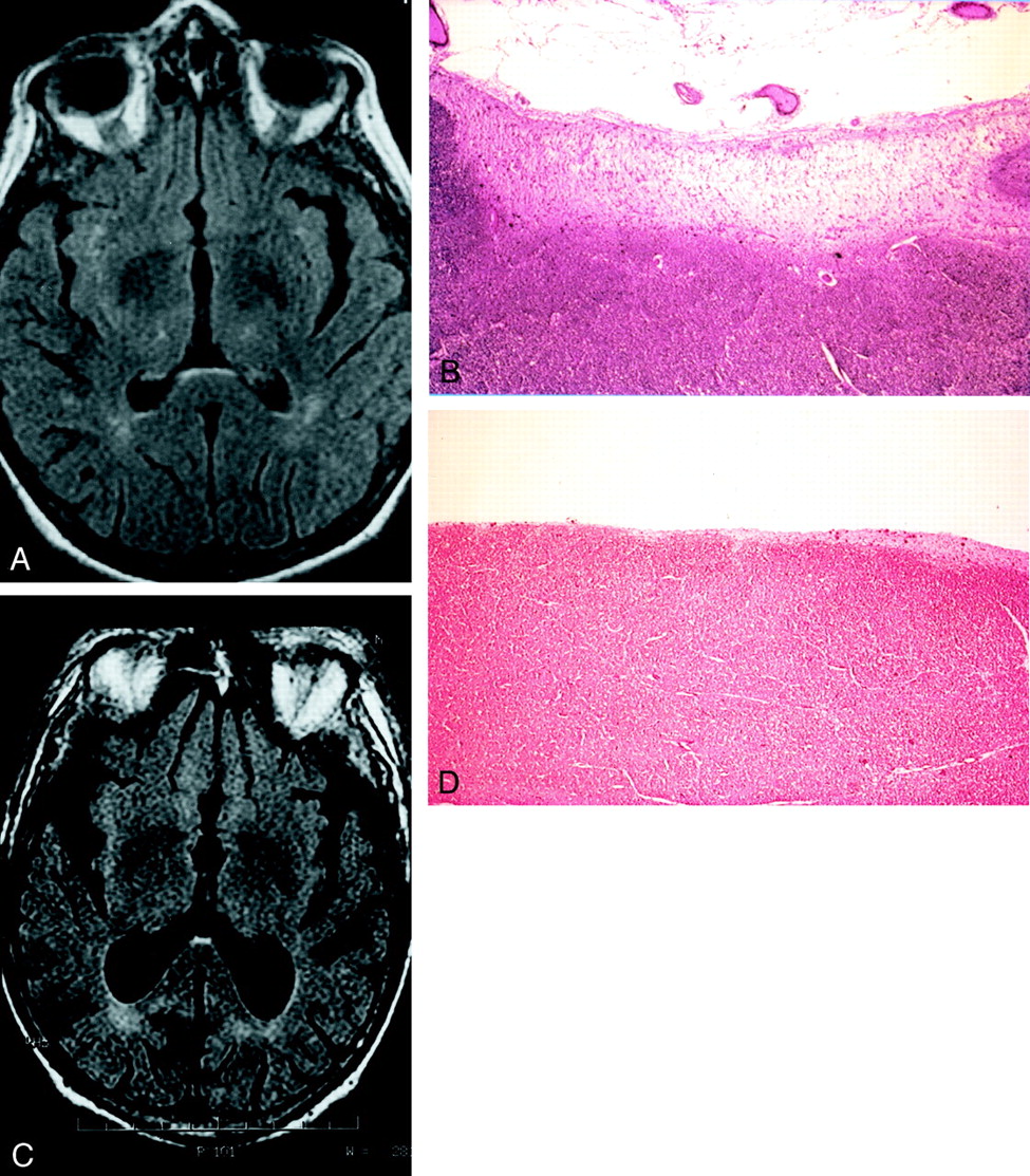
Two autopsy cases.
A, Axial FLAIR (10,000/156/1) image obtained in a 63-year-old man shows grade 1 signal intensity abnormality in the anterior subependymal region of the splenium.
B, Photomicrograph of histologic specimen of the splenium (same patient as in A) demonstrates a band of isomorphic gliosis (hematoxylin and eosin stain; original magnification, x60).
C, Axial FLAIR (10,000/156/1) image obtained in an 80-year-old man shows grade 1 signal intensity abnormality in the anterior subependymal region of the splenium.
D, Photomicrograph of histologic specimen of the splenium (same patient as in C) shows no gliosis (hematoxylin and eosin stain; original magnification, x60).


Tables
Patient No. Age (y)/Sex Pretherapy FLAIR Images Posttherapy FLAIR Images Date (mo)* Splenium Grade WMD Grade Date (mo)* Splenium Grade WMD Grade 1 53/F NA — — 4.8 2 1 2 5/F NA — — 5.0 2 0 3 35/F 1.3 0 0 8.2 2 0 4 34/F 1.0 0 0 6.2 0 0 5 64/M 1.1 2 1 6.2 2 1 6 46/M NA — — 5.4 0 1 7 55/M 7 0 1 8.2 2 1 8 52/M 1.1 0 0 11.0 2 1 9 54/M NA — — 11.3 2 1 10 43/M 1.2 0 0 9.4 2 1 11 50/M 1.2 0 0 8.5 2 0 12 32/M 0.8 0 0 5.8 2 1 13 50/M NA — — 10.0 2 1 14 62/M 0.9 1 0 11.8 2 1 15 53/M NA — — 3.2 2 1 16 57/F NA — — 4.0 2 1 17 53/M 2.0 0 0 5.2 2 1 18 57/F NA — — 5.9 2 1 Note.—WMD indicates white matter disease; NA, images not available.
* Indicates the number of months before or after radiation therapy that the FLAIR images were obtained.
In this issue


Jump to section
Related Articles
Cited By...
- No citing articles found.