
Article Figures & Data
Figures
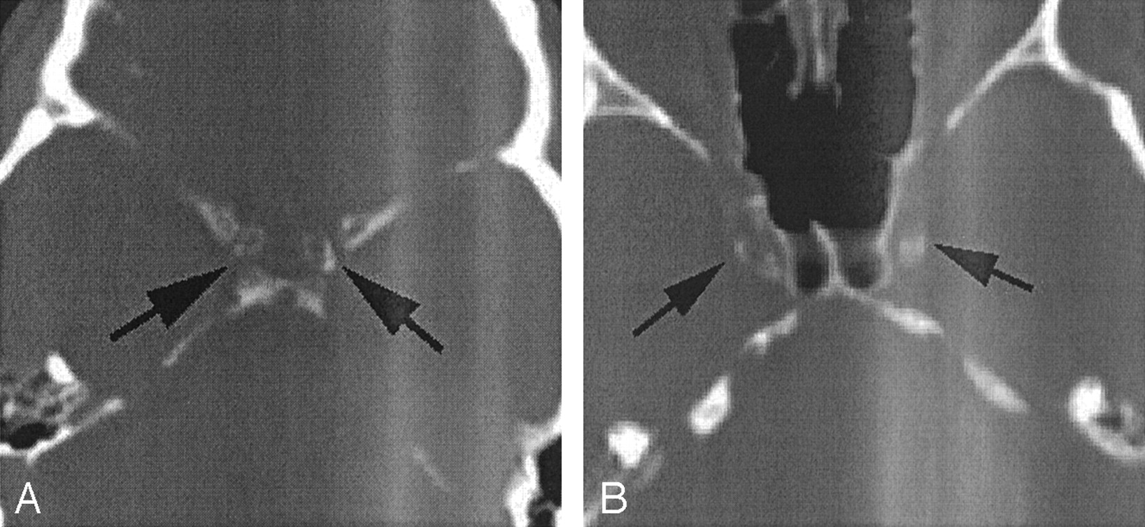
- Fig 1.
CT calcification scores.
A, Circumferential nature of cavernous carotid artery calcification on this CT scan, filmed in a bone window, warrants a grade of 4 (360 degrees of involvement).
B, If the calcification is between 90 and 270 degrees of involvement, it is graded as a 3. The thickness of the calcification, judged by the centimeter scale, was 2 mm in this case.
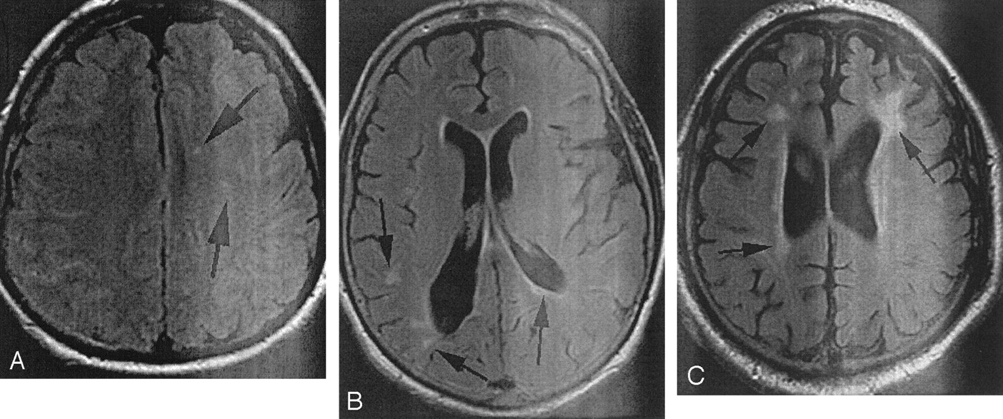
- Fig 2.
MR imaging white matter disease (arrows) scores. All images obtained through white matter were reviewed by using fluid-attenuated inversion recovery imaging before grades were assigned.
A, This patient was judged to have minimal disease (CHS grade 1).
B, This patients was assigned a CHS score of 4.
C, This patient was assigned a CHS score of 7.


Tables
Extent of calcification of cavernous carotid artery Grade 0 No calcification 1 Dot of calcification 2 Crescentic area of calcification <90 degrees of carotid wall circumference 3 Calcification from 90–270 degrees circumference 4 Calcification 270–360 degrees around carotid circumference Thickness of calcification of cavernous carotid artery Grade 0 No calcification 1 Calcification 1 mm thick 2 Calcification 2 mm thick 3 Calcification 3 mm thick 4 Calcification >3 mm thick White matter disease on fluid-attenuated inversion recovery MR images Grade 0 No white matter signal abnormalities 1 Discontinuous PV rim or minimal “dot” of SC disease 2 Thin continuous PV rim or few patches of SC disease 3 Thicker continuous PV rim with scattered patches of SC disease 4 Thicker shaggier periventricular rim and mild SC disease 5 Mild PV confluence surrounding the frontal and occipital horns 6 Moderate PV confluence surrounding the frontal and occipital horns 7 PV confluence with moderate involvement of the centrum semiovale 8 PV confluence involving most of the centrum semiovale 9 All supratentorial white matter involved (excluding the corpus callosum) Note.—PV indicates periventricular; SC, subcortical.
CT Score CT Thickness Number of Participants Median Age (yr) of Participants 25th Percentile of Age, 75th Percentile of Age 0 N/A 68 51.5 45.0, 60.0 1 1 38 62.0 52.0, 73.0 1 2 5 73.0 52.0, 75.0 1 3 2 70.5 67.0, 74.0 1 All 45 64.0 52.0, 73.0 2 1 14 67.0 56.0, 74.0 2 2 10 62.5 53.0, 72.0 2 3 3 65.5 65.0, 66.0 2 All 27 66.0 53.0, 72.0 3 1 16 68.0 54.5, 75.0 3 2 7 69.0 65.0, 75.0 3 3 6 74.5 70.0, 79.0 3 All 29 70.0 64.0, 75.0 4 1 8 70.5 65.5, 76.0 4 2 7 79.0 72.0, 88.0 4 3 1 83.0 N/A 4 4 2 85.0 79.0, 91.0 4 All 18 77.5 69.0, 82.0 All N/A 187 62.0 49.0, 73.0 MR Imaging Score Number of Participants Median Age (yr) of Participants 25th Percentile of Age, 75th Percentile of Age 0 40 46.5 43.0, 55.0 1 26 59.5 53.0, 67.5 2 32 59.0 50.0, 75.0 3 22 63.0 49.0, 72.0 4 10 64.5 62.0, 73.0 5 19 65.0 59.0, 74.0 6 20 73.0 66.5, 79.0 7 17 73.0 67.0, 79.0 8 1 76.0 N/A All 187 62.0 49.0, 73.0 Test Spearman Correlation, No Age Adjustment Spearman Correlation, with Age Adjustment CT grade and MR imaging score 0.33 (P < .001) 0.016 (P = .83) CT thickness and MR imaging score 0.27 (P < .001) 0.045 (0 = .53) Composite grade and thickness and MR imaging score 0.31 (P < .0001) 0.013 (P = .86)


