
Article Figures & Data
Figures
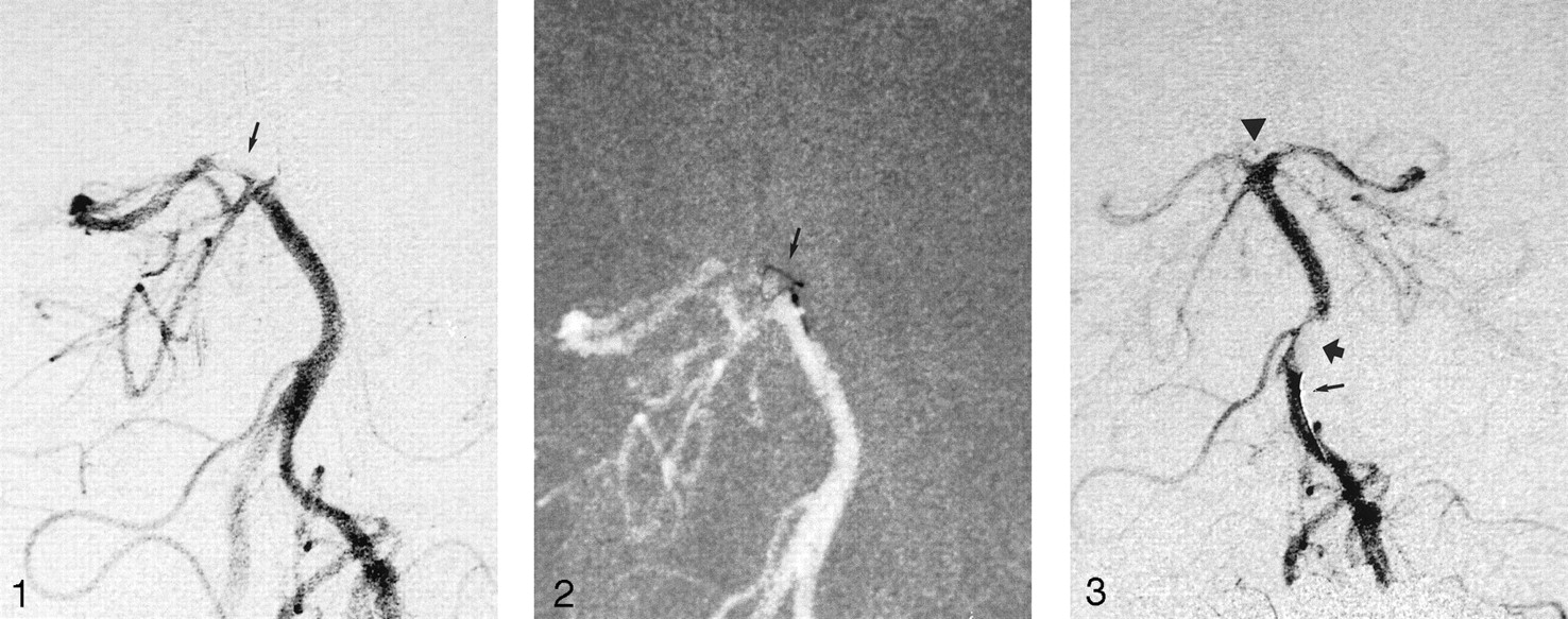
- Fig 1.
Diagnostic angiogram (left vertebral artery injection, anteroposterior projection of the basilar artery) shows the basilar tip embolus (arrow) occluding the left posterior cerebral artery, the left superior cerebellar artery, and the basilar tip perforators going to the thalamus.
- Fig 2.
The snare (arrow) has been pushed out of the microcatheter just enough to open fully, and together with the microcatheter, it has been further pushed into the embolus. A minor buckling of the loop is seen.
- Fig 3.
The snare has been partially withdrawn into the microcatheter, leaving a small eye outside the tip (bottom arrow) of the microcatheter. After this, the microcatheter with the snare is pulled back a few centimeters, and contrast material is injected. The angiogram shows the clot (top arrow) as a lucency in the contrast material hanging from the tip of the microcatheter. The now-open basilar tip is clearly seen with all of the branches filling (arrowhead).


Tables
Patient No. Age, y/Sex Affected Vessel Symptoms Time from Ictus, hours:minutes Angiographic Time, minutes† Symptoms after First Admission‡ CT and/or MR Imaging Findings 1/37/M Basilar artery Unconscious, extending legs 3:00 20 Fully alert with only minor eye muscle palsy Small ischemic areas in thalamus and brainstem 2/70/M Basilar artery Unconscious, variable for the first 4 hours 10:00 15 Alert and orientated, dysarthria and severe eye muscle palsy Ischemia of thalamus and left cerebral peduncle 3/53/F Middle cerebral artery Left-sided paralysis, somnolence 2:20 20 Arm palsy and slight facial palsy Infarction (early signs of ischemia seen before treatment) 4/69/F Middle cerebral artery Right-sided paralysis, dens aphasia 4:40 35 Walks with walking frame, slight remaining aphasia Not available 5/50/F Posterior cerebral artery None, patient under general anesthesia 1:00 20 Upper relative field cut No ischemia seen on day 1 * Time from ictus indicates the time from the appearance of the first symptoms to when the embolus was extracted.
† Angiographic time indicates the time for obtaining an angiographic diagnosis and extracting the embolus.
‡ At 1–2 weeks.
In this issue


Jump to section
Related Articles
Cited By...
- Mechanical Embolectomy for Acute Ischemic Stroke in the Anterior Cerebral Circulation: The Gothenburg Experience during 2000-2011
- Mechanical thrombectomy as the primary treatment for acute basilar artery occlusion: experience from 5 years of practice
- Mechanical Thromboembolectomy for Acute Ischemic Stroke: Comparison of the Catch Thromboectomy Device and the Merci Retriever In Vivo
- Debunking 7 Myths That Hamper the Realization of Randomized Controlled Trials on Intra-Arterial Thrombolysis for Acute Ischemic Stroke
- Mechanical Thrombectomy for Acute Ischemic Stroke: Thrombus-Device Interaction, Efficiency, and Complications In Vivo
- Analysis of Thrombi Retrieved From Cerebral Arteries of Patients With Acute Ischemic Stroke
- Mechanical Thrombolysis in Ischemic Stroke Attributable to Basilar Artery Occlusion as First-Line Treatment
- Reasons for exclusion from thrombolytic therapy following acute ischemic stroke
- Use of mechanical extraction devices in basilar artery occlusion
- Extending Reperfusion Therapy for Acute Ischemic Stroke: Emerging Pharmacological, Mechanical, and Imaging Strategies
- MERCI 1: A Phase 1 Study of Mechanical Embolus Removal in Cerebral Ischemia
- Endovascular embolectomy of acute basilar artery occlusion