
Article Figures & Data
Figures
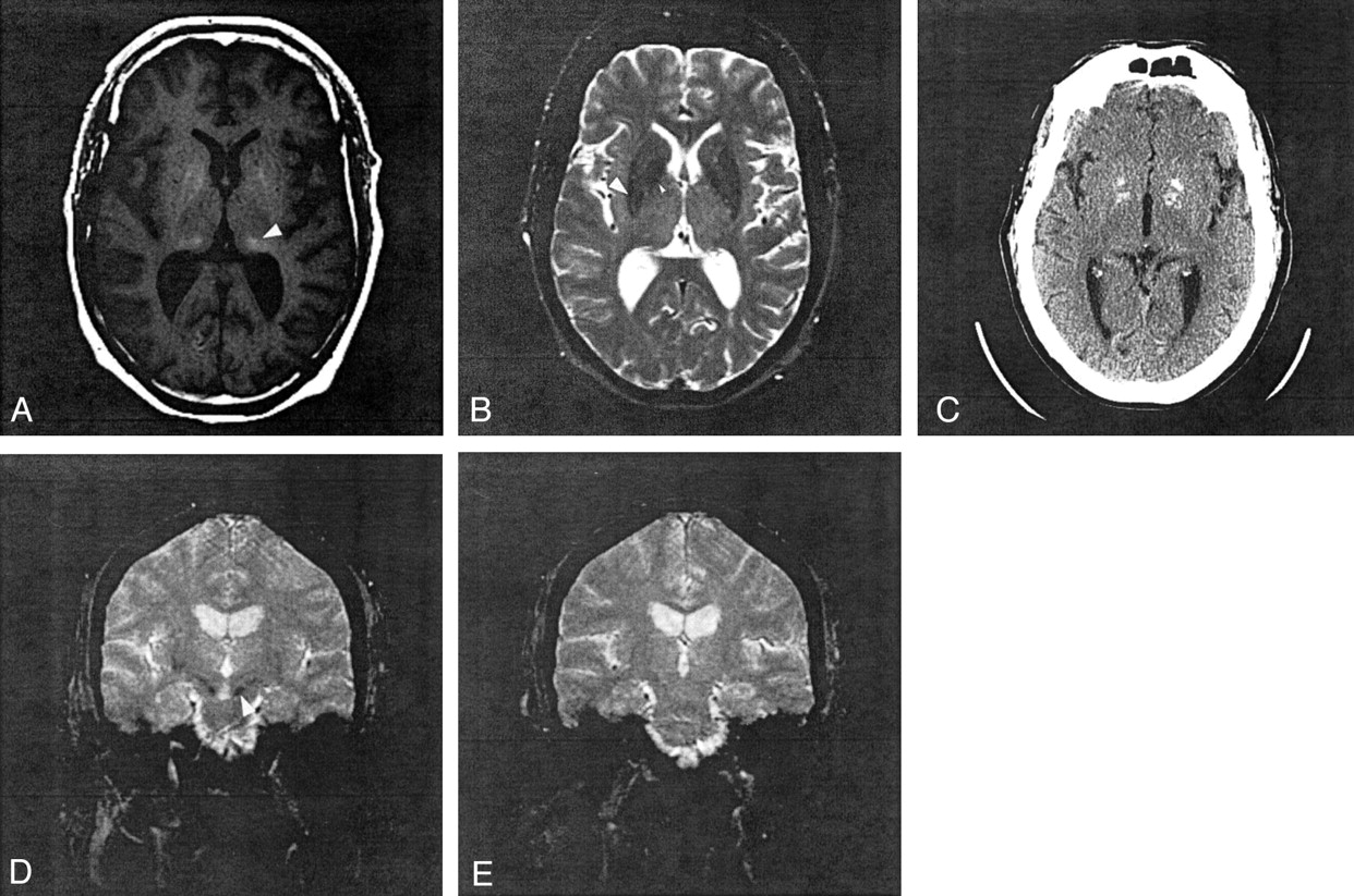
- Fig 1.
Patient 10.
A, Axial T1-weighted spin-echo image at the level of basal ganglia shows hyperintense lesions in the bilateral peripheral globus pallidus, lateral pulvinar (arrowhead), and medial occipital cortex.
B, Axial T2-weighted image spin-echo showing hypointensity in the bilateral globus pallidus (small arrowhead) and putamen (arrowhead).
C, Noncontrast axial CT scan reveals calcification in the bilateral globus pallidus and medial occipital cortex; however, no abnormal attenuation exists in the pulvinar.
D and E, Coronal gradient echo images show hypointensity in the putamen and substantia nigra (D, arrowhead) but normal SI in pulvinar (E).
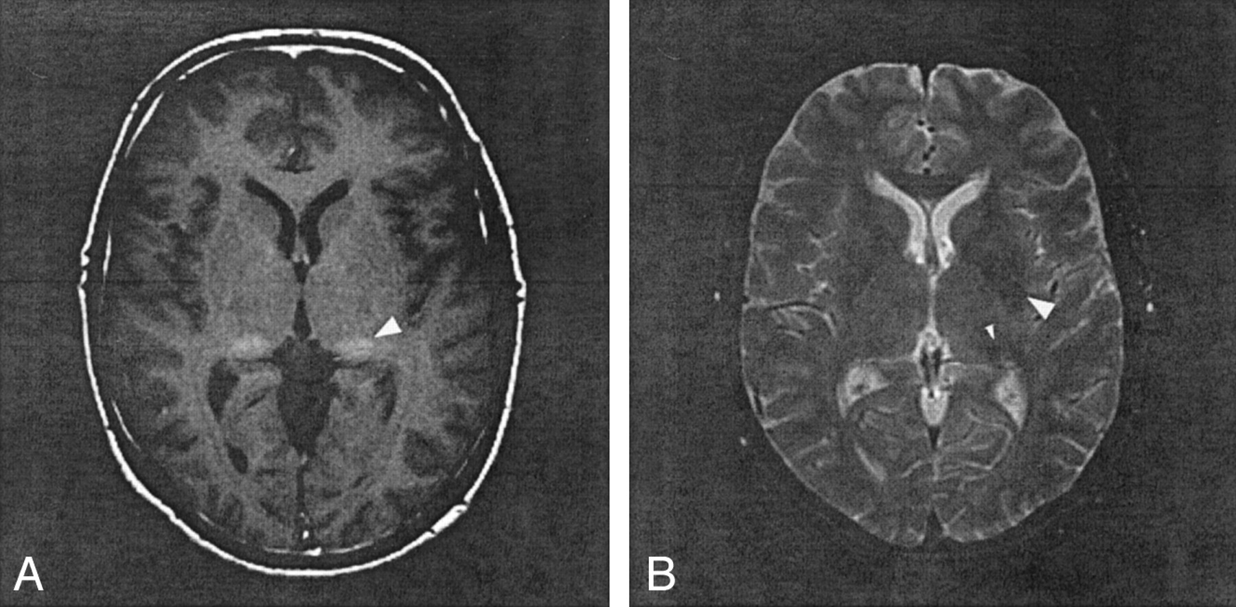
- Fig 2.
Patient 8.
A, T1-weighted spin-echo image at the level of basal ganglia shows hyperintensity in right globus pallidus and bilateral lateral pulvinar (arrowhead).
B, T2-weighted spin-echo image shows a small hyperintense foci in right frontal white matter and hypointensity in the bilateral lateral putamen (arrowhead) and pulvinar (small arrowhead).


Tables
Patient (No.) Age (y)/Sex CNS Symptoms Ischemic Lesion MR Imaging Findings Small Foci of WM DGM Lacunes 1 19/M Dizziness, syncope None None None 2 28/M Vertigo, vision loss None None None 3 30/F None None None None 4 31/M Dizziness, decreased vision None D, P 5 34/M Vertigo, double vision None S, D, P Left medial Th 6 36/M Visual loss, hemiplegia, seizure None D, P Bilateral BG 7 42/M None None S, D None 8 46/M None None S, D, P None 9 50/M Visual disturbance None D None 10 59/M Aphasia Bilateral F, O, left P S, D, P None Note.—Pt signifies patient; WM, white matter; DGM, deep gray matter; M, male; F, female; TIA, transient ischemic attack; Th, thalamus; BG, basal ganglia; S, subcortical; D, deep; P, periventricular; F, frontal; O, occipital; P, parietal.
Patient (No.) GP Put Th SN RN DN Q SI/CSF SI/CC Q SI/CSF SI/CC Q SI/CSF SI/CC Q SI/CSF SI/CC Q /CSF /CC Q /CSF /CC A. On T1WI 1 N N N N N N N N N N N N N N N N N N 2 H N N N N N H N N N N N N N N H N N 3 N N N N N N N N N N N N N N N N N N 4 N N N N N N H H H N N N N N N N N N 5 H H N N N H N H H H H H N N H N H H 6 N N N N N H H H N N N N H H N N N N 7 H H H N H N H H H N H N N H N N H N 8 H N N N N N H H H N N N H N N N N N 9 N N N N N N H H H H N N N N N H N N 10 H H N N N N H H H H N N H H H H N N B. On T2WI 1 2 L L L N N N N L L L L N N L L L L N 3 N L N N N N N N N N L N N L N L L N 4 N L N N L N N N N L L N N L N L L N 5 N L N N L N N L N N N N N N N L L N 6 N L L L L N N L N L L N N L L L N N 7 L L L N L N L L L L L L N N N L L L 8 N L L L L N L L N L L N N N N L N N 9 N L L N L N N L N N N N N N N L N N 10 L L L L L L L N N L L L N N N N N N Note.—Pt signifies patient; T1WI, T1-weighted image; T2WI, T2-weighted image; GP, globus pallidus; Put, putamen; Th, thalamus; SN, substantia nigra; RN, red nucleus; DN, dentate nucleus; Q, qualitative analysis; SI/CSF, signal intensity ratio relative to CSF in the lateral ventricle; SI/CC, signal intensity ratio relative to the genu of corpus callosum; H, high; L, low; and N, normal range.
In this issue


Jump to section
Related Articles
Cited By...
- Novel GLA T194A variant causes Fabry disease
- Redefining the Pulvinar Sign in Fabry Disease
- Brain MR Imaging Findings of Cardiac-Type Fabry Disease with an IVS4+919G>A Mutation
- Brain Magnetic Resonance Imaging Findings Fail to Suspect Fabry Disease in Young Patients With an Acute Cerebrovascular Event
- Pulvinar: Associative role in cortical function and clinical correlations
- Cerebrovascular Involvement in Fabry Disease: Current Status of Knowledge
- Voxel based analyses of diffusion tensor imaging in Fabry disease