
Article Figures & Data
Figures
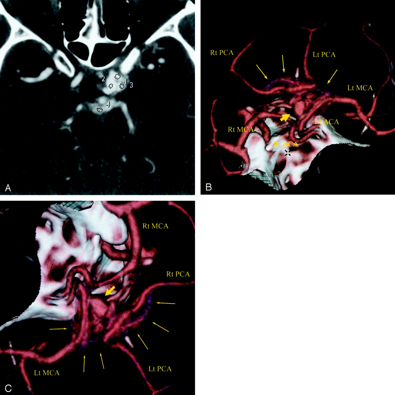
- Fig 1.
CTA of an actively bleeding aneurysm in a 35-year-old man.
A, Axial source image through the aneurysm. Region of interest 2 is in the aneurysm itself and measures 322 ± 14 (mean ± SD) HU. The other three regions of interest (1, 3, and 4) outline areas of extravasated contrast material outside the lumen of either the aneurysm or the blood vessels. The attenuations of these regions of interests are 300 ± 40, 275 ± 29, and 195 ± 15 HU, respectively. This is much higher than what is seen for blood, which is typically on the order of 80 HU. The patient did not receive contrast material before CTA was performed. This leads on to the inescapable conclusion that the only possible source of this attenuated material in the subarachnoid space is contrast material from an actively bleeding aneurysm. The attenuation measurements in the subarachnoid space (regions of interest 1, 3, and 4) are slightly below what was seen in the aneurysm and decrease as one moves further away form the aneurysm, most likely because of dilution of the contrast material with CSF.
B and C, 3D reconstruction of the CTA data. Large arrows depict the superior hypophyseal artery aneurysm. The small arrows show a nebulous area of increased attenuation in the subarachnoid space adjacent to the aneurysm that represents the actively extravasating contrast material from the bleeding aneurysm. The attenuation of this area is equal to or slightly below that of the contrast material in the vessels.


