Advertisement

Main menu

  • Home
  • Content
    • Current Issue
    • Accepted Manuscripts
    • Article Preview
    • Past Issue Archive
    • AJNR Case Collection
    • Case of the Week Archive
    • Classic Case Archive
    • Case of the Month Archive
  • Special Collections
    • Spinal CSF Leak Articles (Jan 2020-June 2024)
    • 2024 AJNR Journal Awards
    • Most Impactful AJNR Articles
  • Multimedia
    • AJNR Podcast
    • AJNR Scantastics
    • Video Articles
  • For Authors
    • Submit a Manuscript
    • Author Policies
    • Fast publishing of Accepted Manuscripts
    • Graphical Abstract Preparation
    • Manuscript Submission Guidelines
    • Imaging Protocol Submission
    • Submit a Case for the Case Collection
  • About Us
    • About AJNR
    • Editorial Board
  • More
    • Become a Reviewer/Academy of Reviewers
    • Subscribers
    • Permissions
    • Alerts
    • Feedback
    • Advertisers
    • ASNR Home
  • Other Publications
    • ajnr

User menu

  • Alerts
  • Log in

Search

  • Advanced search
American Journal of Neuroradiology

American Journal of Neuroradiology

ASHNR American Society of Functional Neuroradiology ASHNR American Society of Pediatric Neuroradiology ASSR
  • Alerts
  • Log in

Advanced Search

  • Home
  • Content
    • Current Issue
    • Accepted Manuscripts
    • Article Preview
    • Past Issue Archive
    • AJNR Case Collection
    • Case of the Week Archive
    • Classic Case Archive
    • Case of the Month Archive
  • Special Collections
    • Spinal CSF Leak Articles (Jan 2020-June 2024)
    • 2024 AJNR Journal Awards
    • Most Impactful AJNR Articles
  • Multimedia
    • AJNR Podcast
    • AJNR Scantastics
    • Video Articles
  • For Authors
    • Submit a Manuscript
    • Author Policies
    • Fast publishing of Accepted Manuscripts
    • Graphical Abstract Preparation
    • Manuscript Submission Guidelines
    • Imaging Protocol Submission
    • Submit a Case for the Case Collection
  • About Us
    • About AJNR
    • Editorial Board
  • More
    • Become a Reviewer/Academy of Reviewers
    • Subscribers
    • Permissions
    • Alerts
    • Feedback
    • Advertisers
    • ASNR Home
  • Follow AJNR on Twitter
  • Visit AJNR on Facebook
  • Follow AJNR on Instagram
  • Join AJNR on LinkedIn
  • RSS Feeds

Welcome to the new AJNR, Updated Hall of Fame, and more. Read the full announcements.


AJNR is seeking candidates for the position of Associate Section Editor, AJNR Case Collection. Read the full announcement.


OtherINTERVENTIONAL

Coil Embolization of an Aneurysm Associated with an Infraoptic Anterior Cerebral Artery in a Child

Sultan Al-Qahtani, Donatella Tampieri, Rolland Brassard, Denis Sirhan and Denis Mellanson
American Journal of Neuroradiology May 2003, 24 (5) 990-991;
Sultan Al-Qahtani
  • Find this author on Google Scholar
  • Find this author on PubMed
  • Search for this author on this site
Donatella Tampieri
  • Find this author on Google Scholar
  • Find this author on PubMed
  • Search for this author on this site
Rolland Brassard
  • Find this author on Google Scholar
  • Find this author on PubMed
  • Search for this author on this site
Denis Sirhan
  • Find this author on Google Scholar
  • Find this author on PubMed
  • Search for this author on this site
Denis Mellanson
  • Find this author on Google Scholar
  • Find this author on PubMed
  • Search for this author on this site
  • Article
  • Figures & Data
  • Info & Metrics
  • Responses
  • References
  • PDF
Loading

Abstract

Summary: We report an unusual case of a ruptured aneurysm from an infraoptic anterior cerebral artery in an 11-year-old child. To our knowledge, this is the second such case treated with Guglielmi detachable coils in the pediatric population that has been described in the literature, excluding that of a vein of Galen aneurysm that resulted in remarkable recovery. We conclude that the endovascular technique is a feasible treatment option for children with ruptured cerebral aneurysms, especially when more complex vascular anomalies are present.

The infraoptic anterior cerebral artery (ACA) is a rare congenital vascular anomaly that was first described by Robinson in 1959 (1). Many variations of this anomaly have subsequently been observed, and our search of the literature revealed a total of 21 reported cases. The origin and trajectory of the duplicated segment of the ACA are variable, and the infraoptic type has great significance because of its relationship with the optic nerve and frequent association with aneurysms.

This case illustrates an example of an infraoptic origin of a duplicated ACA associated with a ruptured aneurysm that was successfully treated with Guglielmi detachable coils (GDCs).

Case Report

A previously well 11-year-old boy was found by his mother unresponsive and gasping next to a playground slide. In the emergency room, the patient regained consciousness, vomited several times, and had several episodes of tonic clonic movements of the upper extremities.

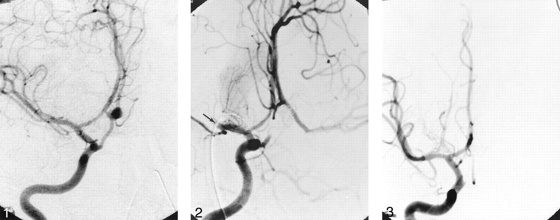
Patient history included long-standing intermittent headaches, always attributed to stress. On clinical examination, he was awake but agitated; his vital signs were normal, and his eyes were deviated to the left side. He had no sign of external injury. He had neck rigidity. Motor and sensory examination findings were normal. A non=contrast-enhanced CT scan of the head showed a diffuse subarachnoid hemorrhage, a small anterior interhemispheric hematoma, and mild hydrocephalus. The subarachnoid hemorrhage was grade III according to the Hunt and Hess classification system. Cerebral digital subtraction angiography (DSA) revealed an aneurysm located along an infraoptic ACA arising from the right internal carotid artery (ICA) near the origin of the ophthalmic artery (Fig 1); the origin of the left ACA was normal. In light of the location and morphology of the aneurysm, it was decided to proceed with endovascular treatment by using GDCs. During the embolization process, the patient received 2500 IU of heparin, and for the next 20 hours the patient received 500 IU/h. After placement of the third coil, during control DSA, a thrombus was noted in the distal M1 segment of the right middle cerebral artery (Fig 2). The clot was immediately dissolved by using 7 mg of intraarterial recombinant tissue plasminogen activator (Activase; Genetech, San Francisco, CA) injected locally, which achieved complete recanalization of the middle cerebral artery and its branches.

Fig 1.
  • Download figure
  • Open in new tab
  • Download powerpoint
Fig 1.

Right oblique ICA angiogram depicting the infraoptic ACA and associated aneurysm.

Fig 2.
  • Download figure
  • Open in new tab
  • Download powerpoint
Fig 2.

Frontal right ICA angiogram showing complete occlusion of the aneurysm and the infraoptic ACA. Note that the distal M1 segment of the right middle cerebral artery is occluded because of an acute thrombus (arrow).

After total coil placement in the aneurysm, the patient had an uneventful recovery and was discharged 10 days after the procedure. Control DSA was repeated after 3 months (Fig 3), and the examination showed complete obliteration of the aneurysm and the infraoptic ACA and normal middle cerebral artery circulation. The boy resumed his regular school and sports activities within a month of the procedure, and he successfully completed his school year. The 20-month clinical follow-up confirmed complete recovery.

Fig 3.
  • Download figure
  • Open in new tab
  • Download powerpoint
Fig 3.

Right ICA angiogram, demonstrating normal anterior cerebral circulation and complete obliteration of the aneurysm and the infraoptic ACA.

Discussion

Infraoptic ACA is a rare vascular anomaly, but it should be recognized by its unique angiographic appearance. This anomalous vessel usually arises from the intradural ICA at or near the origin of the ophthalmic artery and is rarely extradural in origin, according to Curtis (2). The artery then passes beneath the ipsilateral optic nerve and turns, ascending on the midline in front of the optic chiasma. Finally, the infraoptic ACA joins the distal A1 segment of the normal ACA or the anterior communicating artery (3). Twenty-one cases have been reported in the literature (1, 4–10). In 48% of the cases, there was an association with aneurysms: 10 patients presented with subarachnoid hemorrhage secondary to the associated aneurysm (10), and in one case, the subarachnoid hemorrhage was due to unknown cause (5).

In the cases described in the literature (1, 4–9), this anomaly occurs 75% of the time on the right side, 15% on the left side, and 10% bilaterally. Diagnosis was based on autopsy findings in four cases (1, 4), on angiographic and operative findings in nine cases (6, 9), on angiographic studies in seven cases (7, 8), and on angiographic, operative, and autopsy findings in one case (7).

The exact embryogenesis of this vascular anomaly is unclear, but the most logical explanation seems to be related to the persistence of the anatomic loop around the optic nerve of the primitive dorsal ophthalmic artery that, according to Robinson (1), normally disappears as the ophthalmic artery is formed. According to Bosma (7), the anomaly might represent the persistence of the primitive maxillary artery that supplies the optic stalk and cup and terminates at the prosencephalon during the early embryonic stage.

The aneurysms, which have a more proximal origin from the infraoptic ACA, are more difficult to treat surgically, because they are embedded within the cavernous sinus and are very close to several cranial nerves. According to Nutik and Dilenge (10), these aneurysms are difficult to treat surgically because of the inherent nature of the vessel and the aneurysm, which appear to have a thinner wall than normal.

Intracranial aneurysms in children are rare, comprising 0.5–2% of a large aneurysm series (11). Coil embolization of aneurysms in the pediatric population has been reported in only two cases; one by Cekirge et al (11), who reported a treatment of bilateral cavernous carotid artery aneurysms with mechanically detachable coils in a 4-year-old girl, and one by Firat et al (12), who reported a case in which GDCs were used to treat a partially thrombosed giant basilar artery aneurysm in an 11-year-old child who showed remarkable clinical improvement.

Conclusion

Vascular anomalies associated with intracranial aneurysms, even if rare, must be recognized to plan for appropriate treatment. Endovascular technique by using GDCs is a feasible treatment option in pediatric cerebral aneurysms, especially when an unusual vascular anomaly is coexisting, as in the case illustrated here.

References

  1. ↵
    Robinson LR. An unusual human anterior cerebral artery. J Anat 1959;93:131–133
  2. ↵
    Curtis A. Given P. Pearse Morris. Recognition and importance of an infraoptic anterior cerebral artery: case report. AJNR Am J Neuroradiol 2002;23:452–454
    Abstract/FREE Full Text
  3. ↵
    Piotre L, Mariusz M, Henryk M. The accessory anterior cerebral artery: case report and anatomic analysis of vascular anomaly. Surg Neurol 1997;48:171–174
    PubMed
  4. ↵
    McCormick WF. A unique anomaly of the intracranial arteries of man. Neurology 1969;19:77–80
    FREE Full Text
  5. ↵
    Handa J, Matsuda M, Koyama T, et al. Internal carotid aneurysm associated with multiple anomalies of cerebral arteries. Neuroradiology 1971;2:230–233
    PubMed
  6. ↵
    Genya O. Carotid-anterior cerebral artery anastomosis with aneurysm: case report and review of the literature. Neurosurgery 1988;23:654–658
    PubMed
  7. ↵
    Bosma NJ. Infra-optic course of anterior cerebral artery and low bifurcation of the internal carotid artery. Acta neurochir (Wien) 1977;38:305–312
  8. ↵
    Brismar J, Akerman R, Roberson G. Anomaly of anterior cerebral artery. Acta Radiol [Diagn] (Stockh) 1977;18:154–160
  9. ↵
    Ushioda T, Okumura K, Higuchi J, et al. A case of internal carotid-anterior artery anastomosis. Neuroradiology 1984;25:78–79
  10. ↵
    Nutik S, Dilenge D. Carotid-anterior cerebral artery anastomosis. J Neurosurg 1976;44:378–382
    PubMed
  11. ↵
    Cekirge S, Saatci I, Firat MM, et al. Bilateral cavernous carotid artery aneurysms in a 4-year-old child: endovascular treatment with mechanically detachable coils. Neuroradiology 1997;39:367–370
    PubMed
  12. ↵
    Firat MM, Cekirge S, Saatci I, Akalan N, Balkanci F. Guglielmi detachable coil treatment of a partially thrombosed giant basilar artery aneurysm in a child. Neuroradiology 2000;42:142–144
    PubMed
  • Received May 13, 2002.
  • Accepted after revision October 3, 2002.
  • Copyright © American Society of Neuroradiology
Back to top

In this issue

American Journal of Neuroradiology: 24 (5)
American Journal of Neuroradiology
Vol. 24, Issue 5
1 May 2003
  • Table of Contents
  • Index by author
Advertisement
Print
Download PDF
Email Article

Thank you for your interest in spreading the word on American Journal of Neuroradiology.

NOTE: We only request your email address so that the person you are recommending the page to knows that you wanted them to see it, and that it is not junk mail. We do not capture any email address.

Enter multiple addresses on separate lines or separate them with commas.
Coil Embolization of an Aneurysm Associated with an Infraoptic Anterior Cerebral Artery in a Child
(Your Name) has sent you a message from American Journal of Neuroradiology
(Your Name) thought you would like to see the American Journal of Neuroradiology web site.
CAPTCHA
This question is for testing whether or not you are a human visitor and to prevent automated spam submissions.
Cite this article
Sultan Al-Qahtani, Donatella Tampieri, Rolland Brassard, Denis Sirhan, Denis Mellanson
Coil Embolization of an Aneurysm Associated with an Infraoptic Anterior Cerebral Artery in a Child
American Journal of Neuroradiology May 2003, 24 (5) 990-991;

Citation Manager Formats

  • BibTeX
  • Bookends
  • EasyBib
  • EndNote (tagged)
  • EndNote 8 (xml)
  • Medlars
  • Mendeley
  • Papers
  • RefWorks Tagged
  • Ref Manager
  • RIS
  • Zotero
0 Responses
Respond to this article
Share
Bookmark this article
Coil Embolization of an Aneurysm Associated with an Infraoptic Anterior Cerebral Artery in a Child
Sultan Al-Qahtani, Donatella Tampieri, Rolland Brassard, Denis Sirhan, Denis Mellanson
American Journal of Neuroradiology May 2003, 24 (5) 990-991;
del.icio.us logo Twitter logo Facebook logo Mendeley logo
  • Tweet Widget
  • Facebook Like
  • Google Plus One

Jump to section

  • Article
    • Abstract
    • Case Report
    • Discussion
    • Conclusion
    • References
  • Figures & Data
  • Info & Metrics
  • Responses
  • References
  • PDF

Related Articles

  • No related articles found.
  • PubMed
  • Google Scholar

Cited By...

  • No citing articles found.
  • Crossref
  • Google Scholar

This article has not yet been cited by articles in journals that are participating in Crossref Cited-by Linking.

More in this TOC Section

  • SAVE vs. Solumbra Techniques for Thrombectomy
  • Contrast-Induced Encephalopathy after NeuroIR
  • CT Perfusion&Reperfusion in Acute Ischemic Stroke
Show more Interventional

Similar Articles

Advertisement

Indexed Content

  • Current Issue
  • Accepted Manuscripts
  • Article Preview
  • Past Issues
  • Editorials
  • Editors Choice
  • Fellow Journal Club
  • Letters to the Editor

Cases

  • Case Collection
  • Archive - Case of the Week
  • Archive - Case of the Month
  • Archive - Classic Case

Special Collections

  • Special Collections

Resources

  • News and Updates
  • Turn around Times
  • Submit a Manuscript
  • Author Policies
  • Manuscript Submission Guidelines
  • Evidence-Based Medicine Level Guide
  • Publishing Checklists
  • Graphical Abstract Preparation
  • Imaging Protocol Submission
  • Submit a Case
  • Become a Reviewer/Academy of Reviewers
  • Get Peer Review Credit from Publons

Multimedia

  • AJNR Podcast
  • AJNR SCANtastic
  • Video Articles

About Us

  • About AJNR
  • Editorial Board
  • Not an AJNR Subscriber? Join Now
  • Alerts
  • Feedback
  • Advertise with us
  • Librarian Resources
  • Permissions
  • Terms and Conditions

American Society of Neuroradiology

  • Not an ASNR Member? Join Now

© 2025 by the American Society of Neuroradiology All rights, including for text and data mining, AI training, and similar technologies, are reserved.
Print ISSN: 0195-6108 Online ISSN: 1936-959X

Powered by HighWire