Advertisement

Main menu

  • Home
  • Content
    • Current Issue
    • Accepted Manuscripts
    • Article Preview
    • Past Issue Archive
    • AJNR Case Collection
    • Case of the Week Archive
    • Classic Case Archive
    • Case of the Month Archive
  • Special Collections
    • Spinal CSF Leak Articles (Jan 2020-June 2024)
    • 2024 AJNR Journal Awards
    • Most Impactful AJNR Articles
  • Multimedia
    • AJNR Podcast
    • AJNR Scantastics
    • Video Articles
  • For Authors
    • Submit a Manuscript
    • Author Policies
    • Fast publishing of Accepted Manuscripts
    • Graphical Abstract Preparation
    • Manuscript Submission Guidelines
    • Imaging Protocol Submission
    • Submit a Case for the Case Collection
  • About Us
    • About AJNR
    • Editorial Board
  • More
    • Become a Reviewer/Academy of Reviewers
    • Subscribers
    • Permissions
    • Alerts
    • Feedback
    • Advertisers
    • ASNR Home
  • Other Publications
    • ajnr

User menu

  • Alerts
  • Log in

Search

  • Advanced search
American Journal of Neuroradiology

American Journal of Neuroradiology

ASHNR American Society of Functional Neuroradiology ASHNR American Society of Pediatric Neuroradiology ASSR
  • Alerts
  • Log in

Advanced Search

  • Home
  • Content
    • Current Issue
    • Accepted Manuscripts
    • Article Preview
    • Past Issue Archive
    • AJNR Case Collection
    • Case of the Week Archive
    • Classic Case Archive
    • Case of the Month Archive
  • Special Collections
    • Spinal CSF Leak Articles (Jan 2020-June 2024)
    • 2024 AJNR Journal Awards
    • Most Impactful AJNR Articles
  • Multimedia
    • AJNR Podcast
    • AJNR Scantastics
    • Video Articles
  • For Authors
    • Submit a Manuscript
    • Author Policies
    • Fast publishing of Accepted Manuscripts
    • Graphical Abstract Preparation
    • Manuscript Submission Guidelines
    • Imaging Protocol Submission
    • Submit a Case for the Case Collection
  • About Us
    • About AJNR
    • Editorial Board
  • More
    • Become a Reviewer/Academy of Reviewers
    • Subscribers
    • Permissions
    • Alerts
    • Feedback
    • Advertisers
    • ASNR Home
  • Follow AJNR on Twitter
  • Visit AJNR on Facebook
  • Follow AJNR on Instagram
  • Join AJNR on LinkedIn
  • RSS Feeds

Welcome to the new AJNR, Updated Hall of Fame, and more. Read the full announcements.


AJNR is seeking candidates for the position of Associate Section Editor, AJNR Case Collection. Read the full announcement.


OtherHEAD AND NECK

Non-Contrast-Enhanced CT Findings of High Attenuation within Metastatic Cervical Lymph Nodes in Patients with Stage I or II Tongue Carcinoma during a Follow-up Period

Takafumi Hayashi, Ray Tanaka, Shuhzou Taira, Jun-ichi Koyama, Kouji Katsura and Fukiko Kobayashi
American Journal of Neuroradiology August 2003, 24 (7) 1330-1333;
Takafumi Hayashi
aFrom the Division of Oral and Maxillofacial Radiology, Course for Oral Life Science, Niigata University Graduate School of Medical and Dental Sciences, Niigata, Japan
  • Find this author on Google Scholar
  • Find this author on PubMed
  • Search for this author on this site
Ray Tanaka
aFrom the Division of Oral and Maxillofacial Radiology, Course for Oral Life Science, Niigata University Graduate School of Medical and Dental Sciences, Niigata, Japan
  • Find this author on Google Scholar
  • Find this author on PubMed
  • Search for this author on this site
Shuhzou Taira
aFrom the Division of Oral and Maxillofacial Radiology, Course for Oral Life Science, Niigata University Graduate School of Medical and Dental Sciences, Niigata, Japan
  • Find this author on Google Scholar
  • Find this author on PubMed
  • Search for this author on this site
Jun-ichi Koyama
aFrom the Division of Oral and Maxillofacial Radiology, Course for Oral Life Science, Niigata University Graduate School of Medical and Dental Sciences, Niigata, Japan
  • Find this author on Google Scholar
  • Find this author on PubMed
  • Search for this author on this site
Kouji Katsura
aFrom the Division of Oral and Maxillofacial Radiology, Course for Oral Life Science, Niigata University Graduate School of Medical and Dental Sciences, Niigata, Japan
  • Find this author on Google Scholar
  • Find this author on PubMed
  • Search for this author on this site
Fukiko Kobayashi
aFrom the Division of Oral and Maxillofacial Radiology, Course for Oral Life Science, Niigata University Graduate School of Medical and Dental Sciences, Niigata, Japan
  • Find this author on Google Scholar
  • Find this author on PubMed
  • Search for this author on this site
  • Article
  • Figures & Data
  • Info & Metrics
  • Responses
  • References
  • PDF
Loading

Article Figures & Data

Figures

  • Fig 1
    • Download figure
    • Open in new tab
    • Download powerpoint
    Fig 1

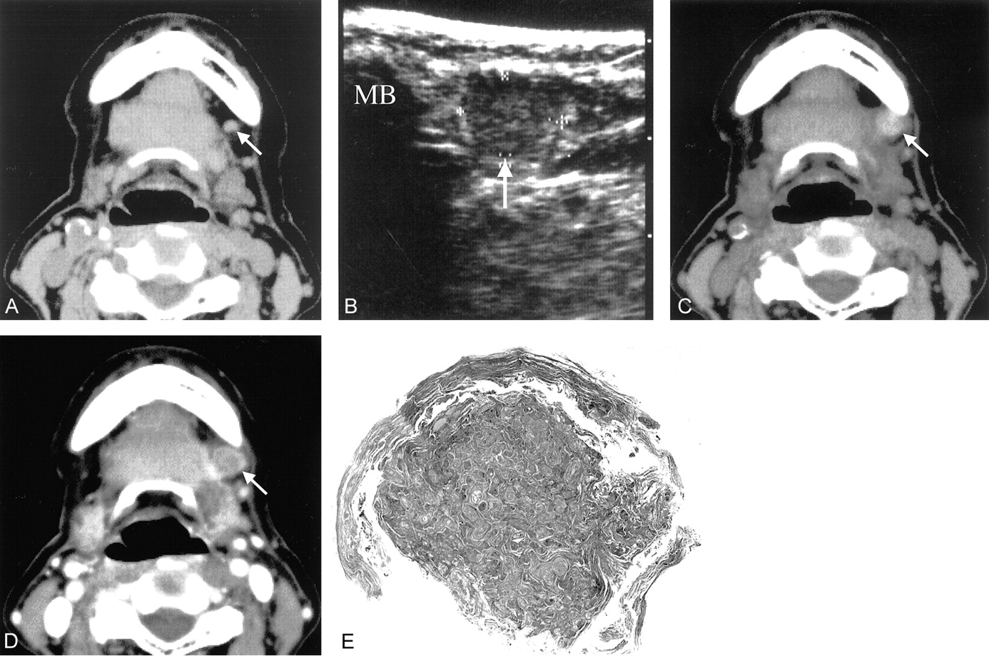
    Case 1, a 79-year-old woman with squamous cell carcinoma of the left side of her tongue.

    A, Non-contrast-enhanced CT image obtained at initial examination of a level I node (arrow); no hyperattenuation is evident.

    B, First follow-up sonogram at the level I node obtained 1 month after surgery of the primary tumor (transverse section). Note an enlarged lymph node (arrow) with heterogeneous internal echo. MB indicates mandible.

    C, Non-contrast-enhanced CT image at the level I node obtained 1 month after surgery of the primary tumor. Hyperattenuation entirely occupies the node (arrow). The size of the node has increased, and the minimal axial diameter of the node is 10 mm.

    D, Contrast-enhanced CT image obtained at the same level and time as that of A. Hyperattenuation within the node (arrow) is obscured after the administration of contrast medium.

    E, Photomicrograph (stain, hematoxyllin-eosin; original magnification, ×2.5) shows the level I node is entirely replaced by the area of marked keratinization (arrows). The non-contrast-enhanced CT finding of hyperattenuation is likely correlated with this marked keratinization.
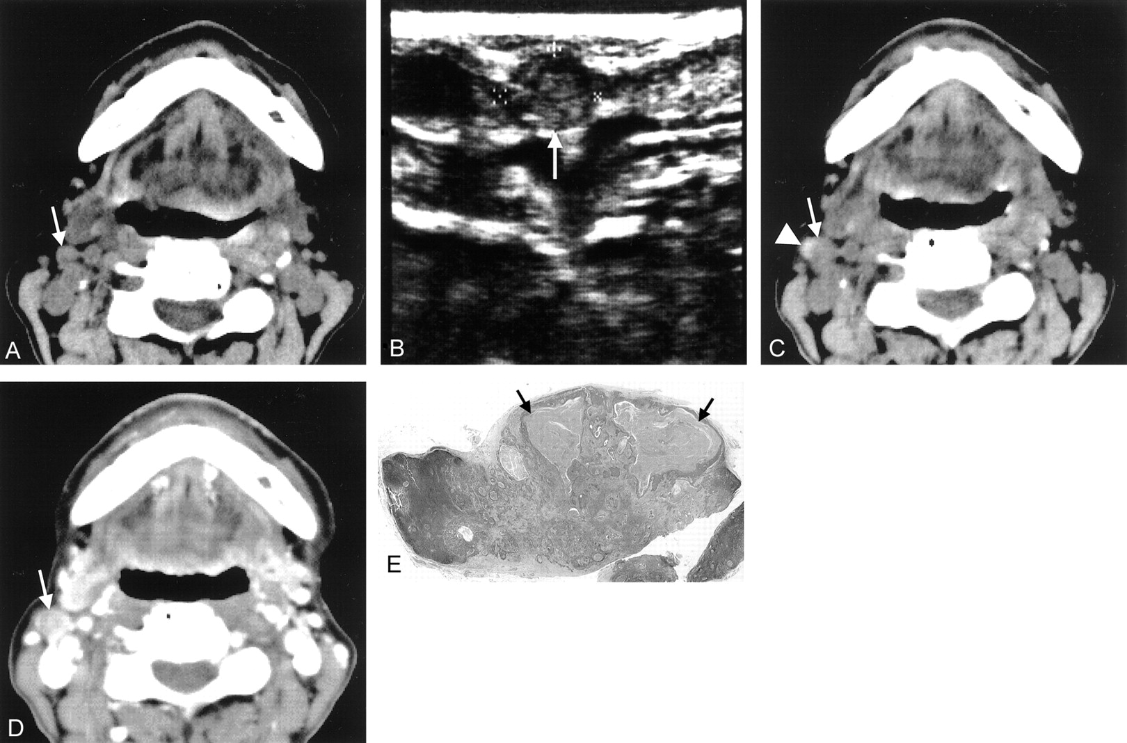
  • Fig 2
    • Download figure
    • Open in new tab
    • Download powerpoint
    Fig 2

    Case 2, an 80-year-old woman with squamous cell carcinoma of the right side of her tongue.

    A, Non-contrast-enhanced CT image at a level II node (arrow) obtained at initial examination; hyperattenuation is not evident.

    B, Ninth follow-up sonogram at the level II node obtained 7 months after surgery of the primary tumor (transverse section). Note an enlarged lymph node (arrow) with heterogeneous internal echo.

    C, Non-contrast-enhanced CT image at the level II node obtained 7 months after surgery of the primary tumor. Note the hyperattenuation (arrowhead) at a lateral margin of the node (arrow). The size of the node increases and the minimal axial diameter of the node is 9 mm.

    D, Contrast-enhanced CT image obtained at the same level and time as that of C. The level II node (arrow) enhances, and hyperattenuation is obscured after the administration of contrast medium.

    E, Photomicrograph (stain, hematoxyllin-eosin; original magnification, ×2) demonstrates the area of marked keratinization (arrows) within metastatic foci at the marginal portion of the largest one of level II node. The non-contrast-ehnahced CT finding of hyperattenuation appears to be correlated with this marked keratinization within metastatic foci.

PreviousNext
Back to top

In this issue

American Journal of Neuroradiology: 24 (7)
American Journal of Neuroradiology
Vol. 24, Issue 7
1 Aug 2003
  • Table of Contents
  • Index by author
Advertisement
Print
Download PDF
Email Article

Thank you for your interest in spreading the word on American Journal of Neuroradiology.

NOTE: We only request your email address so that the person you are recommending the page to knows that you wanted them to see it, and that it is not junk mail. We do not capture any email address.

Enter multiple addresses on separate lines or separate them with commas.
Non-Contrast-Enhanced CT Findings of High Attenuation within Metastatic Cervical Lymph Nodes in Patients with Stage I or II Tongue Carcinoma during a Follow-up Period
(Your Name) has sent you a message from American Journal of Neuroradiology
(Your Name) thought you would like to see the American Journal of Neuroradiology web site.
CAPTCHA
This question is for testing whether or not you are a human visitor and to prevent automated spam submissions.
Cite this article
Takafumi Hayashi, Ray Tanaka, Shuhzou Taira, Jun-ichi Koyama, Kouji Katsura, Fukiko Kobayashi
Non-Contrast-Enhanced CT Findings of High Attenuation within Metastatic Cervical Lymph Nodes in Patients with Stage I or II Tongue Carcinoma during a Follow-up Period
American Journal of Neuroradiology Aug 2003, 24 (7) 1330-1333;

Citation Manager Formats

  • BibTeX
  • Bookends
  • EasyBib
  • EndNote (tagged)
  • EndNote 8 (xml)
  • Medlars
  • Mendeley
  • Papers
  • RefWorks Tagged
  • Ref Manager
  • RIS
  • Zotero
0 Responses
Respond to this article
Share
Bookmark this article
Non-Contrast-Enhanced CT Findings of High Attenuation within Metastatic Cervical Lymph Nodes in Patients with Stage I or II Tongue Carcinoma during a Follow-up Period
Takafumi Hayashi, Ray Tanaka, Shuhzou Taira, Jun-ichi Koyama, Kouji Katsura, Fukiko Kobayashi
American Journal of Neuroradiology Aug 2003, 24 (7) 1330-1333;
del.icio.us logo Twitter logo Facebook logo Mendeley logo
  • Tweet Widget
  • Facebook Like
  • Google Plus One

Jump to section

  • Article
    • Abstract
    • CT Examination
    • Case Reports
    • Discussion
    • Conclusion
    • References
  • Figures & Data
  • Info & Metrics
  • Responses
  • References
  • PDF

Related Articles

  • No related articles found.
  • PubMed
  • Google Scholar

Cited By...

  • No citing articles found.
  • Crossref
  • Google Scholar

This article has not yet been cited by articles in journals that are participating in Crossref Cited-by Linking.

More in this TOC Section

  • Correlation of Apparent Diffusion Coefficient at 3T with Prognostic Parameters of Retinoblastoma
  • Parathyroid Lesions: Characterization with Dual-Phase Arterial and Venous Enhanced CT of the Neck
  • Efficacy of Diffusion-Weighted Imaging for the Differentiation between Lymphomas and Carcinomas of the Nasopharynx and Oropharynx: Correlations of Apparent Diffusion Coefficients and Histologic Features
Show more Head and Neck

Similar Articles

Advertisement

Indexed Content

  • Current Issue
  • Accepted Manuscripts
  • Article Preview
  • Past Issues
  • Editorials
  • Editors Choice
  • Fellow Journal Club
  • Letters to the Editor

Cases

  • Case Collection
  • Archive - Case of the Week
  • Archive - Case of the Month
  • Archive - Classic Case

Special Collections

  • Special Collections

Resources

  • News and Updates
  • Turn around Times
  • Submit a Manuscript
  • Author Policies
  • Manuscript Submission Guidelines
  • Evidence-Based Medicine Level Guide
  • Publishing Checklists
  • Graphical Abstract Preparation
  • Imaging Protocol Submission
  • Submit a Case
  • Become a Reviewer/Academy of Reviewers
  • Get Peer Review Credit from Publons

Multimedia

  • AJNR Podcast
  • AJNR SCANtastic
  • Video Articles

About Us

  • About AJNR
  • Editorial Board
  • Not an AJNR Subscriber? Join Now
  • Alerts
  • Feedback
  • Advertise with us
  • Librarian Resources
  • Permissions
  • Terms and Conditions

American Society of Neuroradiology

  • Not an ASNR Member? Join Now

© 2025 by the American Society of Neuroradiology All rights, including for text and data mining, AI training, and similar technologies, are reserved.
Print ISSN: 0195-6108 Online ISSN: 1936-959X

Powered by HighWire