Advertisement

Main menu

  • Home
  • Content
    • Current Issue
    • Accepted Manuscripts
    • Article Preview
    • Past Issue Archive
    • AJNR Case Collection
    • Case of the Week Archive
    • Classic Case Archive
    • Case of the Month Archive
  • Special Collections
    • Spinal CSF Leak Articles (Jan 2020-June 2024)
    • 2024 AJNR Journal Awards
    • Most Impactful AJNR Articles
  • Multimedia
    • AJNR Podcast
    • AJNR Scantastics
    • Video Articles
  • For Authors
    • Submit a Manuscript
    • Author Policies
    • Fast publishing of Accepted Manuscripts
    • Graphical Abstract Preparation
    • Manuscript Submission Guidelines
    • Imaging Protocol Submission
    • Submit a Case for the Case Collection
  • About Us
    • About AJNR
    • Editorial Board
  • More
    • Become a Reviewer/Academy of Reviewers
    • Subscribers
    • Permissions
    • Alerts
    • Feedback
    • Advertisers
    • ASNR Home
  • Other Publications
    • ajnr

User menu

  • Alerts
  • Log in

Search

  • Advanced search
American Journal of Neuroradiology

American Journal of Neuroradiology

ASHNR American Society of Functional Neuroradiology ASHNR American Society of Pediatric Neuroradiology ASSR
  • Alerts
  • Log in

Advanced Search

  • Home
  • Content
    • Current Issue
    • Accepted Manuscripts
    • Article Preview
    • Past Issue Archive
    • AJNR Case Collection
    • Case of the Week Archive
    • Classic Case Archive
    • Case of the Month Archive
  • Special Collections
    • Spinal CSF Leak Articles (Jan 2020-June 2024)
    • 2024 AJNR Journal Awards
    • Most Impactful AJNR Articles
  • Multimedia
    • AJNR Podcast
    • AJNR Scantastics
    • Video Articles
  • For Authors
    • Submit a Manuscript
    • Author Policies
    • Fast publishing of Accepted Manuscripts
    • Graphical Abstract Preparation
    • Manuscript Submission Guidelines
    • Imaging Protocol Submission
    • Submit a Case for the Case Collection
  • About Us
    • About AJNR
    • Editorial Board
  • More
    • Become a Reviewer/Academy of Reviewers
    • Subscribers
    • Permissions
    • Alerts
    • Feedback
    • Advertisers
    • ASNR Home
  • Follow AJNR on Twitter
  • Visit AJNR on Facebook
  • Follow AJNR on Instagram
  • Join AJNR on LinkedIn
  • RSS Feeds

Welcome to the new AJNR, Updated Hall of Fame, and more. Read the full announcements.


AJNR is seeking candidates for the position of Associate Section Editor, AJNR Case Collection. Read the full announcement.


Getting new auth cookie, if you see this message a lot, tell someone!
OtherBRAIN

Peripheral Third Cranial Nerve Enhancement in Multiple Sclerosis

M. Tariq Bhatti, Ilona M. Schmalfuss, Lorna S. Williams and Ronald G. Quisling
American Journal of Neuroradiology August 2003, 24 (7) 1390-1395;
M. Tariq Bhatti
aDepartment of Ophthalmology, University of Florida College of Medicine, Gainesville, FL
bDepartment of Neurology, University of Florida College of Medicine, Gainesville, FL
  • Find this author on Google Scholar
  • Find this author on PubMed
  • Search for this author on this site
Ilona M. Schmalfuss
cDepartment of Radiology, University of Florida College of Medicine, Gainesville, FL
  • Find this author on Google Scholar
  • Find this author on PubMed
  • Search for this author on this site
Lorna S. Williams
dDepartment of Radiology, JFK Medical Center, Atlantis, FL
  • Find this author on Google Scholar
  • Find this author on PubMed
  • Search for this author on this site
Ronald G. Quisling
cDepartment of Radiology, University of Florida College of Medicine, Gainesville, FL
  • Find this author on Google Scholar
  • Find this author on PubMed
  • Search for this author on this site
  • Article
  • Figures & Data
  • Info & Metrics
  • Responses
  • References
  • PDF
Loading

Article Figures & Data

Figures

  • Tables
  • Fig 1.
    • Download figure
    • Open in new tab
    • Download powerpoint
    Fig 1.

    Coronal gadolinium-enhanced T1-weighted images (TR/TE, 600/14) through the cerebral peduncle and cisternal course of cranial nerve III (A) demonstrate marked enhancement and subtle enlargement of cranial nerve III on the left side (white arrows in A). Notice the normal appearance of cranial nerve III on the right (arrowheads in A). There is also subtle enhancement at the cranial nerve III exit zone on the left side (black arrows in A and B), which is better appreciated on the axial gadolinium-enhanced T1-weighted image (B) performed with the same imaging parameters.

  • Fig 2.
    • Download figure
    • Open in new tab
    • Download powerpoint
    Fig 2.

    Axial fluid-attenuated inversion recovery (FLAIR) image (TR/TE, 9500/110) through the midbrain (A) demonstrates a subtle area of increased signal intensity (arrows) along the medial margin of the cerebral peduncle on the left side. No enhancement is seen on the axial gadolinium-enhanced T1-weighted image (600/17) (B) in this region. The coronal gadolinium-enhanced T1-weighted images (600/14) through the cisternal course of cranial nerve III (C) demonstrate subtle enhancement of the cranial nerve III on the left side (arrows in C). Notice the normal appearance of cranial nerve III on the right (arrowheads in C).
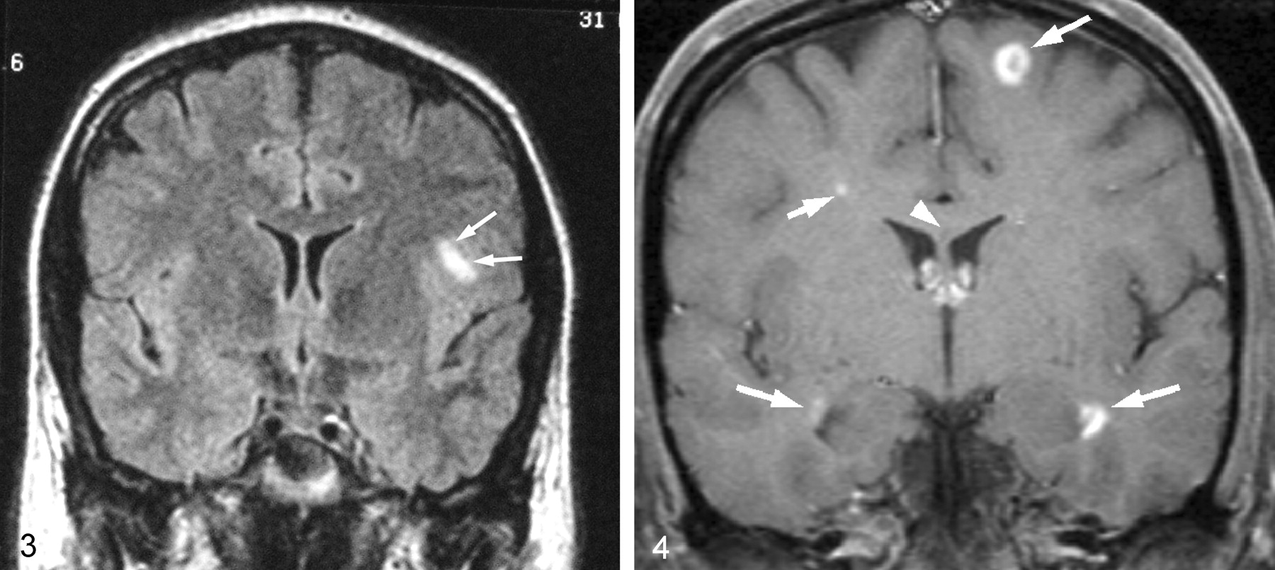
  • Fig 3.
    • Download figure
    • Open in new tab
    • Download powerpoint
    Fig 3.

    Coronal FLAIR-weighted image (TR/TE, 10,002/168) obtained at the level of the anterior horns of the lateral ventricles shows a focal area of increased signal intensity (arrows) in the subcortical white matter of the inferior frontal lobe on the left side.

  • Fig 4.
    • Download figure
    • Open in new tab
    • Download powerpoint
    Fig 4.

    Coronal gadolinium-enhanced T1-weighted image (TRTE, 882/14) obtained at the level of the anterior horns of the lateral ventricles shows multiple enhancing white matter lesions (arrows). There is also a nonenhancing lesion in the corpus callosum in midline (arrowhead).

Tables

  • Figures
  • Third CN palsy and underlying lesions

    Anatomic Site3. CN Dysfunction FeaturesAssociated Clinical FindingsEtiologiesContrast Enhancement of 3.CN or Its Nuclei
    Midbrain:
     Nuclear(+/−) Bilateral pupil involvementSupranuclear ocular motility deficitsInfectious:
    (+/−) Bilateral ptosis Lyme diseaseUsually present
    Incomplete paresis (isolated extraocular muscle) SyphilisVariable
    Inflammatory:
    Contralateral superior rectus muscle paresisMultiple sclerosisVariable
     SarcoidosisPresent
     Autoimmune vasculitisVariable
    Neoplastic:
     Primary brain tumorVariable
     MetastasisPresent
     LymphomaPresent
    TraumaticUsually none
    Vascular:
     IschemiaVariable
     Cavernous angiomaUsually none
     Arteriovenous malformation (AVM)Enhancement of the AVM only
    FascicularComplete or incomplete (divisional) paresisCerebellar Ataxia: Nothnagel’sSame as above
    Contralateral dyskinesia: Benedickt’s
    (+/−) Pupil involvementContralateral hemiparesis: Weber’s
    Subarachnoid spaceComplete or incomplete (divisional) paresisMultiple cranial neuropathiesInfectious:
    Meningeal irritation Bacterial, viral or fungal meningitisPresent
    (+/−) Pupil involvementMental status changes SyphilisVariable
    Raised intracranial pressure Lyme diseaseUsually present
     HIVUsually present
    Inflammatory:
     Multiple sclerosisVariable
     SarcoidosisPresent
     Inflammatory demyelinating polyneuropathyVariable
    Neoplastic:
     LymphomaPresent
     LeukemiaPresent
     Meningeal carcinomatosisPresent
     SchwannomaPresent
    TraumaticUsually none
    Vascular:
     IschemiaVariable
     AneurysmEnhancement of the Aneurysm only
    Other:
     Ophthalmoplegic migrainePresent
    Cavernous sinus/superior orbital fissureComplete or incomplete (divisional) paresisMultiple cranial neuropathiesInfectious:
    Visual Loss Bacterial, viral or fungal meningitisPresent
    (+/−) Pupil involvementProptosis SyphilisVariable
     Lyme diseaseUsually present
     HIVUsually present
    Inflammatory:
     Tolosa-Hunt syndromePresent
     SarcoidosisPresent
    Neoplastic:
     LymphomaPresent
     LeukemiaPresent
     MetastasisUsually present
     SchwannomaPresent
     Pituitary macroadenomaPresent
     Cavernous sinus meningiomaPresent
     CraniopharyngiomaVariable
    TraumaticUsually none
    Vascular:
     IschemiaVariable
     AneurysmEnhancement of the aneurysm only
     Pituitary gland apoplexyUsually none
    Other:
     Ophthalmoplegic migrainePresent
PreviousNext
Back to top

In this issue

American Journal of Neuroradiology: 24 (7)
American Journal of Neuroradiology
Vol. 24, Issue 7
1 Aug 2003
  • Table of Contents
  • Index by author
Advertisement
Print
Download PDF
Email Article

Thank you for your interest in spreading the word on American Journal of Neuroradiology.

NOTE: We only request your email address so that the person you are recommending the page to knows that you wanted them to see it, and that it is not junk mail. We do not capture any email address.

Enter multiple addresses on separate lines or separate them with commas.
Peripheral Third Cranial Nerve Enhancement in Multiple Sclerosis
(Your Name) has sent you a message from American Journal of Neuroradiology
(Your Name) thought you would like to see the American Journal of Neuroradiology web site.
CAPTCHA
This question is for testing whether or not you are a human visitor and to prevent automated spam submissions.
Cite this article
M. Tariq Bhatti, Ilona M. Schmalfuss, Lorna S. Williams, Ronald G. Quisling
Peripheral Third Cranial Nerve Enhancement in Multiple Sclerosis
American Journal of Neuroradiology Aug 2003, 24 (7) 1390-1395;

Citation Manager Formats

  • BibTeX
  • Bookends
  • EasyBib
  • EndNote (tagged)
  • EndNote 8 (xml)
  • Medlars
  • Mendeley
  • Papers
  • RefWorks Tagged
  • Ref Manager
  • RIS
  • Zotero
0 Responses
Respond to this article
Share
Bookmark this article
Peripheral Third Cranial Nerve Enhancement in Multiple Sclerosis
M. Tariq Bhatti, Ilona M. Schmalfuss, Lorna S. Williams, Ronald G. Quisling
American Journal of Neuroradiology Aug 2003, 24 (7) 1390-1395;
del.icio.us logo Twitter logo Facebook logo Mendeley logo
  • Tweet Widget
  • Facebook Like
  • Google Plus One

Jump to section

  • Article
    • Abstract
    • Case Report
    • Discussion
    • Conclusion
    • Footnotes
    • References
  • Figures & Data
  • Info & Metrics
  • Responses
  • References
  • PDF

Related Articles

  • No related articles found.
  • PubMed
  • Google Scholar

Cited By...

  • Isolated third nerve palsies as the initial manifestation of multiple sclerosis
  • Crossref
  • Google Scholar

This article has not yet been cited by articles in journals that are participating in Crossref Cited-by Linking.

More in this TOC Section

  • Statin Therapy Does Not Affect the Radiographic and Clinical Profile of Patients with TIA and Minor Stroke
  • Optimal MRI Sequence for Identifying Occlusion Location in Acute Stroke: Which Value of Time-Resolved Contrast-Enhanced MRA?
  • SWI or T2*: Which MRI Sequence to Use in the Detection of Cerebral Microbleeds? The Karolinska Imaging Dementia Study
Show more Brain

Similar Articles

Advertisement

Indexed Content

  • Current Issue
  • Accepted Manuscripts
  • Article Preview
  • Past Issues
  • Editorials
  • Editors Choice
  • Fellow Journal Club
  • Letters to the Editor

Cases

  • Case Collection
  • Archive - Case of the Week
  • Archive - Case of the Month
  • Archive - Classic Case

Special Collections

  • Special Collections

Resources

  • News and Updates
  • Turn around Times
  • Submit a Manuscript
  • Author Policies
  • Manuscript Submission Guidelines
  • Evidence-Based Medicine Level Guide
  • Publishing Checklists
  • Graphical Abstract Preparation
  • Imaging Protocol Submission
  • Submit a Case
  • Become a Reviewer/Academy of Reviewers
  • Get Peer Review Credit from Publons

Multimedia

  • AJNR Podcast
  • AJNR SCANtastic
  • Video Articles

About Us

  • About AJNR
  • Editorial Board
  • Not an AJNR Subscriber? Join Now
  • Alerts
  • Feedback
  • Advertise with us
  • Librarian Resources
  • Permissions
  • Terms and Conditions

American Society of Neuroradiology

  • Not an ASNR Member? Join Now

© 2025 by the American Society of Neuroradiology All rights, including for text and data mining, AI training, and similar technologies, are reserved.
Print ISSN: 0195-6108 Online ISSN: 1936-959X

Powered by HighWire