
Article Figures & Data
Figures
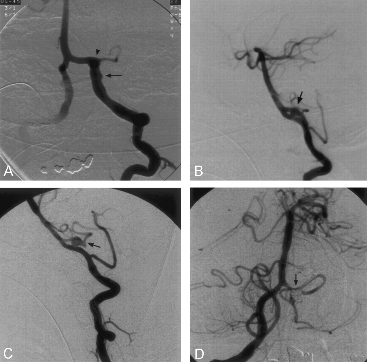
- Fig 1.
Patient 25.
A, Anteroposterior and B, lateral diagnostic angiograms demonstrate a dissecting aneurysm of the dominant left vertebral artery involving the supra-PICA segment and proximal basilar artery (arrow). Arrowhead indicates the pseudoaneurysm.
C, Lateral view of the left internal carotid artery injection shows good collateral circulation to the basilar artery (arrow) through the posterior communicating artery after proximal occlusion of the left vertebral artery. Arrowhead indicates the tip of the distal balloon.
D, Two-year follow-up left internal carotid artery angiogram in the lateral projection shows interval thrombosis of the pseudoaneurysm and dissected segment of the left vertebral artery. The basilar artery (arrow) appears less irregular.
- Fig 2.
Patient 27.
A, Anteroposterior angiogram of the left vertebral artery shows irregular narrowing of the supra-PICA segment (arrow) and a pseudoaneurysm at the vertebrobasilar junction (arrowhead). The anterior spinal artery and left PICA are not included in the abnormal segment.
B, Anteroposterior angiogram of the right vertebral artery shows a codominant vessel with full delineation of the vertebrobasilar junction pseudoaneurysm (arrowhead). The dissection extends in the basilar artery to include the AICA origins (arrow).
C, Posttreatment angiogram of the right vertebral artery shows coil occlusion of the vertebrobasilar junction pseudoaneurysm (arrowhead).
D, Posttreatment angiogram of the left vertebral artery shows proximal occlusion of the supra-PICA segment of the vessel with preservation of flow in the anterior spinal artery (arrow) and PICA.
- Fig 3.
Patient 11.
A, Anteroposterior angiogram of the left vertebral artery demonstrates irregularity of the lumen (arrow) and a 4–5-mm pseudoaneurysm (arrowhead) near the PICA origin.
B, Lateral angiogram of the left vertebral artery shows the small pseudoaneurysm (arrow).
C, Lateral angiogram of the right vertebral artery at the time of rehemorrhage 2 weeks after proximal occlusion of the left vertebral artery shows that the pseudoaneurysm (arrow) has not enlarged and the left PICA remains patent.
D, Anteroposterior angiogram of the right vertebral artery after delivery of coils across the vertebrobasilar junction into the pseudoaneurysm shows the lesion is occluded (arrow), with the PICA filling antegrade
- Fig 4.
Patient 22.
A, Anteroposterior angiogram of the right vertebral artery shows a dissection of this codominant vertebral artery (black arrow) and psuedoaneurysm (arrowhead). Note the fenestration in the basilar artery (white arrow).
B and C, Follow-up angiograms of the right (B) and left (C) vertebral arteries, respectively, show no residual aneurysm.
D, Follow-up angiogram 1 year later shows recurrence of the dissecting aneurysm (arrow) proximally in the left vertebral artery to involve the PICA. The coil mass has compacted (arrowhead).


Tables
Patient No./Age (y)/Sex Symptom Hunt Hess Grade Type Site Dominant Vessel BTO Treatment Rankin Score 1/50/F SAH 1 Infra LVA LVA + Proximal occlusion Unknown 2/66/F SAH 4 Infra RVA LVA − Proximal occlusion 0 3/49/M Stroke 0 Infra RVA Co + Proximal occlusion 0 4/49/M Stroke 0 Infra RVA LVA − Trapping 0 5/53/M SAH 1 Infra RVA Co − Trapping 0 6/61/F SAH 3 Infra RVA Co − Trapping 0 7/51/M SAH 5 Infra RVA Co − Trapping 5 8/54/M SAH + stroke 4 No PICA RVA Co − Trapping 4 9/49/M SAH 3 No PICA R > L RVA + Trapping 5 10/48/F SAH + stroke 3 No PICA LVA Co + Trapping 0 11/54/M SAH + stroke 3 PICA LVA Co − Proximal occlusion + trapping 2 12/53/M SAH + stroke 4 PICA RVA LVA − Proximal occlusion 5 13/38/F Headache 3 PICA RVA RVA − Clip 3 14/79/F Headache 1 Supra RVA Co + Proximal occlusion 0 15/55/M SAH 4 Supra LVA Co − Proximal occlusion Unknown 16/17/M Stroke 0 Supra RVA Co − Proximal occlusion 0 17/49/M SAH 4 Supra RVA RVA − Proximal occlusion Unknown 18/66/M SAH 1 Supra RVA LVA − Proximal occlusion 5 19/59/M SAH 3 Supra RVA LVA − Proximal occlusion 5 20/44/M TIA 0 Supra LVA RVA − Trapping 0 21/45/M SAH 3 Supra LVA RVA − Trapping 2 22/29/F SAH 3 Supra RVA Co − Trapping 0 23/43/F SAH 1 Supra LVA Co − Trapping 1 24/47/F SAH 3 Supra LVA RVA − Trapping 3 25/41/M SAH 3 VBJ LVA, BA LVA + Proximal occlusion 0 26/71/M SAH 1 VBJ RVA, LVA, BA RVA + Proximal occlusion 0 27/45/M SAH 3 VBJ LVA, BA Co − Proximal occlusion 0 28/45/F SAH 1 BA BA LVA − Clip 2 Tr = trauma.
Note.—TIA indicates transient ischemic attack; VBJ, vertebrobasilar junction; BA, basilar artery; L, left; R, right; VA, vertebral artery; BTO, balloon test occlusion; Co, codominant.
Patient No./Age (y)/Sex Symptom Hunt Hess Grade Type Site Dominant Vessel History Rankin Score 1/42/F SAH 5 Infra RVA Co Rapid decline 5 2/48/M SAH 4 > 5 Infra RVA Co Rapid decline 5 3/87/F SAH 3 No PICA RVA Co Family decision 3 4/47/M SAH + stroke 3 PICA LVA RVA Thrombosed Unknown 5/70/M SAH 4 PICA LVA LVA Stable 0 6/61/M Trauma 3 Supra LVA LVA Carotid cavernous fistula 5 7/44/F SAH 2 VBJ RVA, LVA, BA Co Thrombosed 0 Note.—VBJ indicates vertebrobasilar junction; BA, basilar artery; L, left; R, right; VA, vertebral artery; Co, codominant.
In this issue


Jump to section
Related Articles
Cited By...
- Endovascular sacrifice of the proximal posterior inferior cerebellar artery for treatment of ruptured intracranial aneurysms
- Endovascular parent vessel sacrifice in ruptured dissecting vertebral and posterior inferior cerebellar artery aneurysms: clinical outcomes and review of the literature
- Republished: Monotherapy with stenting in subarachnoid hemorrhage (SAH) after middle cerebral artery dissection
- Elderly patients with intracranial aneurysms have higher quality of life after coil embolization: a decision analysis
- Deconstructive and Reconstructive Techniques in Treatment of Vertebrobasilar Dissecting Aneurysms: A Systematic Review and Meta-Analysis
- Monotherapy with stenting in subarachnoid hemorrhage (SAH) after middle cerebral artery dissection
- Natural Course of Dissecting Vertebrobasilar Artery Aneurysms without Stroke
- Safety of Unilateral Endovascular Occlusion of the Cervical Segment of the Vertebral Artery without Antecedent Balloon Test Occlusion
- Multiple overlapping stents as monotherapy in the treatment of 'blister' pseudoaneurysms arising from the supraclinoid internal carotid artery: a single institution series and review of the literature
- Subarachnoid haemorrhage with bilateral intracranial vertebral artery dissecting aneurysms treated by staged endovascular stenting
- Pipeline Embolization Device in Aneurysmal Subarachnoid Hemorrhage
- Novel use of Onyx for treatment of intracranial vertebral artery dissection
- Incidence and Risk Factors of Recurrence After Endovascular Treatment of Intracranial Vertebrobasilar Dissecting Aneurysms
- Reconstructive Endovascular Treatment of Intracranial Fusiform Aneurysms: A 1-Stage Procedure with Stent and Balloon
- Endovascular Strategies for Vertebrobasilar Dissecting Aneurysms
- Clinical and Angiographic Follow-Up of Stent-Only Therapy for Acute Intracranial Vertebrobasilar Dissecting Aneurysms
- Stent-Assisted Reconstructive Endovascular Repair of Cranial Fusiform Atherosclerotic and Dissecting Aneurysms: Long-Term Clinical and Angiographic Follow-Up
- Mechanically-induced proximal arterial occlusion and stent-within-a-stent technique for the treatment of bilateral vertebral artery dissecting aneurysms.