
Article Figures & Data
Figures
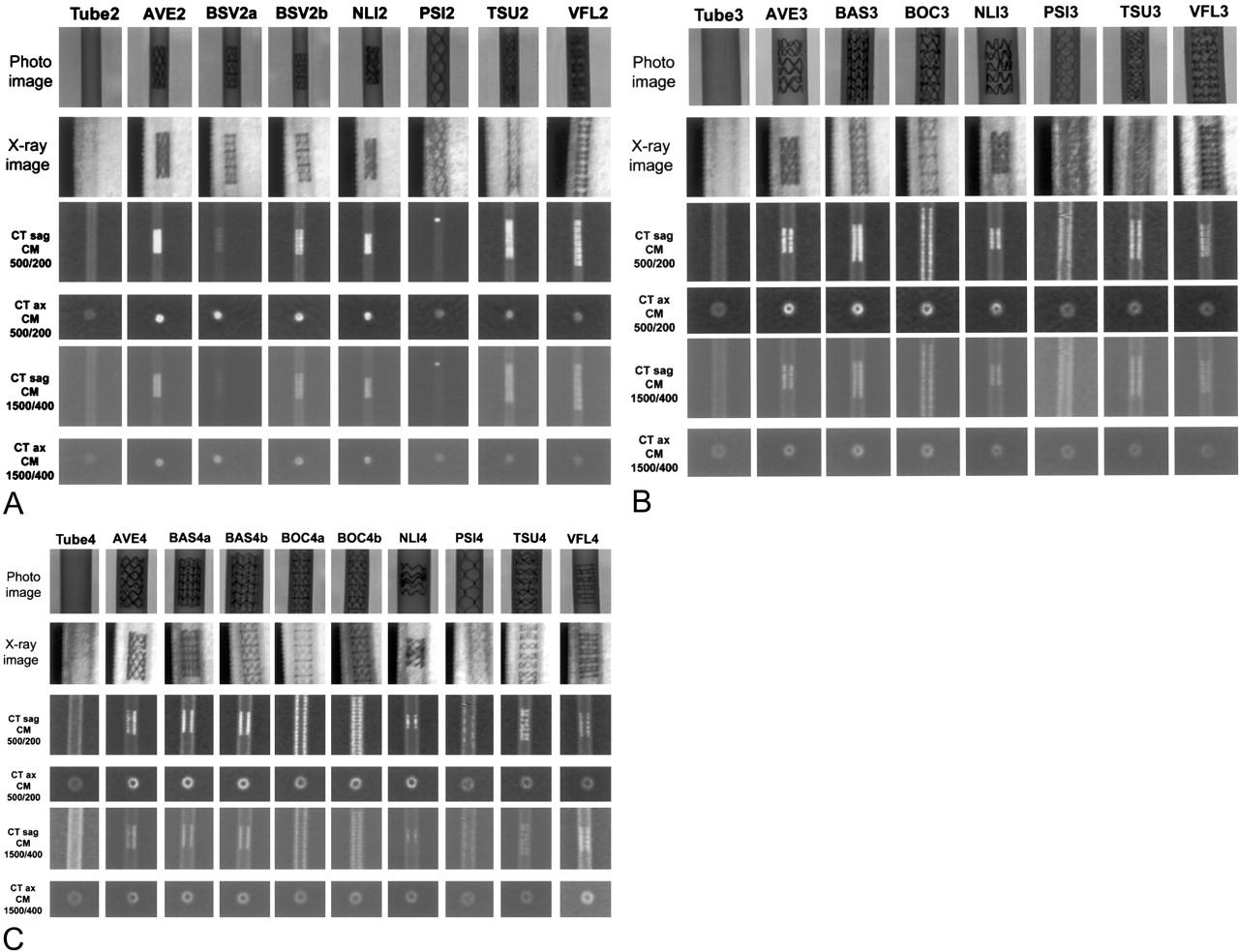
- Fig 1.
(a) Imaging features of the stents. CT sag indicates sagittal CT image; CT ax, axial CT image; CM, stent filled with diluted contrast medium. The numbers separated by the slash indicate the window width and window level (in Hounsfield units).
A, 2.0-mm stents.
B, 3.0-mm stents.
C, 4.0-mm stents.
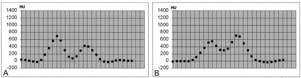
- Fig 2.
Attenuation profile of the BOC3 stent. The difference between two vertical lines represents a distance of 0.5 mm.
A, BOC3 filled with only NaCl solution only. PNHU<200(NaCl) = 3.
B, BOC3 filled with diluted contrast medium. PNHU<200(CM) = 0.
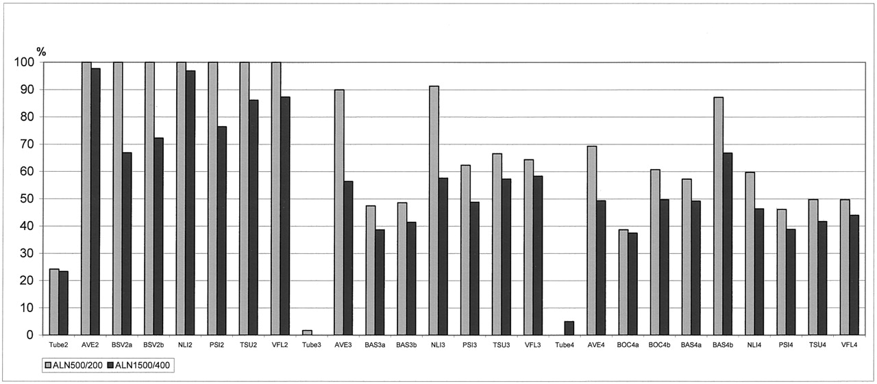
- Fig 3.
ALN of the stents and tubes when filled with diluted contrast medium. ALN1500/400 was 0% for T3, and ALN500/200 was 0% for T4.
- Fig 4.
DIFFHU<200 of the stents. DIFFHU<200 was zero for all 2.0-mm stents.


Tables
Properties of the analyzed stents
Stent or Tube Trade Name Manufacturer Material Stent Design Nominal Stent Length, mm Diameter, mm Strut Thickness, mm Strut Width, mm Stent Free Area, % Nominal Dxray* 2.0 mm AVE2 AVE S660 Medtronic, Minneapolis, MN SS 316 L Modular 9 2.5 2.17 0.128 0.153 80 BSV2a BioDivYsio SV Abbott Vascular Devices, Mervue, UK SS 316 L Modular 10 2.0 1.96 0.091 0.08 85–90 BSV2b BioDivYsio SV Abbott Vascular Devices, Mervue, UK SS 316 L Modular 10 2.5 2.34 0.091 0.08 85–90 NLI2 Neurolink Guidant, Indianapolis, IN SS 316 L MC 8 2.0 2.14 0.094 0.127 85 PSI2 PSI-Stent Dendron, Bochum, Germany Nitinol SHCA 20 2.0 1.91 0.06 0.06 85 TSU2 Tsunami Terumo, Tokyo, Japan SS 316 L MC 15 2.0 2.17 0.08 0.09 75 VFL2 V-Flex Plus Cook, Bloomington, IN SS 316 L MC 16 2.5 1.97 0.08 0.08 >85 3.0 mm AVE3 AVE S670 Medtronic, Minneapolis, MN SS 316 L Modular 9 3.0 2.98 0.128 0.154 77–83 BAS3 BioDivYsio AS Abbott Vascular Devices, Mervue, UK SS 316 L MC-modular 15 3.0 3.04 0.091 0.08 NA BOC3 BioDivYsio OC Abbott Vascular Devices, Mervue, UK SS 316 L MC-modular 28 3.0 3.24 0.091 0.08 88–91 NLI3 Neurolink Guidant, Indianapolis, IN SS 316 L MC 8 3.0 3.26 0.094 0.127 87 PSI3 PSI-Stent Dendron, Bochum, Germany Nitinol SHCA 20 3.0 2.83 0.06 0.06 85 TSU3 Tsunami Terumo, Tokyo, Japan SS 316 L MC 15 3.0 3.04 0.08 0.09 83 VFL3 V-Flex Plus Cook Bloomington, IN SS 316 L MC 12 3.0 3.04 0.08 0.08 >85 4.0 mm AVE4 AVE S670 Medtronic, Minneapolis, MN SS 316 L MC-modular 12 4.0 3.91 0.128 0.154 77–83 BAS4a BioDivYsio AS Abbott Vascular Devices, Mervue, UK SS 316 L MC-modular 15 4.0 4.13 0.091 0.08 75–81 BAS4b BioDivYsio AS Abbott Vascular Devices, Mervue, UK SS 316 L MC-modular 11 4.0 3.91 0.091 0.08 75–81 BOC4a BioDivYsio OC Abbott Vascular Devices, Mervue, UK SS 316 L MC-modular 28 3.5 4.10 0.091 0.08 88–91 BOC4b BioDivYsio OC Abbott Vascular Devices, Mervue, UK SS 316 L MC-modular 28 4.0 4.07 0.091 0.08 88–91 NLI4 Neurolink Guidant, Indianapolis, IN SS 316 L MC 8 4.0 4.13 0.094 0.127 88 PSI4 PSI-Stent Dendron, Bochum, Germany Nitinol SHCA 20 4.0 3.62 0.06 0.06 85 TSU4 Tsunami Terumo, Tokyo, Japan SS 316 L MC 15 4.0 3.91 0.08 0.09 88 VFL4 V-Flex Plus Cook, Bloomington, IN SS 316 L MC 12 3.5 4.13 0.08 0.08 >85 Tube T2 Tube 2 mm NeoLab, Heidelberg, Germany Silicone NA NA 2.0 NA NA NA NA T3 Tube 3 mm NeoLab, Heidelberg, Germany Silicone NA NA 3.0 NA NA NA NA T4 Tube 4 mm NeoLab, Heidelberg, Germany Silicone NA NA 4.0 NA NA NA NA Note.—MC indicates multicellular; NA, not applicable; SHCA, semihelical coil array; and SS, stainless steel.
* The diameter of each stent after balloon inflation was calculated on the basis of a radiopaque reference scale that was simultaneously x-ray imaged with the stented tube.


